Özet
Akut yan ağrısı, hematüri veya böbrek işlevinde bozulma bulunan; nefrotik sendromu, malignitesi ya da hiperkoagülabilitesi olan hastada renal ven trombozu düşünülür. Kontrastlı BT, renal ven içindeki trombüsü doğrudan gösterir ve trombüsün ana venden inferior kaval ven'e uzanımını belirler; aynı zamanda etkilenen böbreğin parankimal kontrastlanmasını değerlendirmeye olanak tanır. Venöz çıkışın tıkanması böbrekte ödem ve venöz konjesyona neden olabilir; ayrıca eşlik eden pulmoner emboli riski klinik açıdan önemlidir. Tümör trombüsünün tümör dışı trombüsten ayırt edilmemesi, malignitenin evrelenmesini ve radyoloji raporunu değiştireceği için tedavi planlamasını doğrudan etkiler.
Faz ve pencere
- Portal tanısal
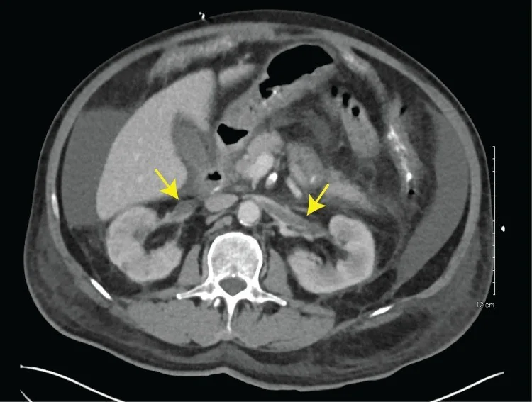
- Venöz/nefrografik kontrastlı görüntülerde renal ven içinde kontrastla çevrili düşük atenüasyonlu dolum defekti ve akut olguda genişlemiş ven görülür; böbrek büyümesi, nefrogramın azalması veya heterojenliği, renal sinüs ve perirenal ödem eşlik edebilir. Trombüsün ana renal venden inferior kaval vene (VKİ) doğru uzanımı, çok düzlümlü yeniden yapılandırmalarla birlikte değerlendirilir.
Önerilen pencereler: Batın (G 400 / M 50), Anjiyo (G 600 / M 150), Yumuşak doku (G 400 / M 40).
BT bulguları
- Renal ven dolum defekti — kontrastlı lümen içinde santral veya eksantrik düşük atenüasyonlu materyal.
- Akut ven genişlemesi — trombüs içeren renal venin karşı tarafa göre dolgun görünmesi.
- Nefrogram asimetrisi — etkilenen böbrekte azalmış ya da heterojen parankimal kontrastlanma.
- Böbrek büyümesi ve ödem — akut venöz konjesyonla birlikte parankim hacminde artış ve sinüs çevresinde bulanıklık.
- Perirenal ödem — renal fasya içinde yağ planlarının silinmesi veya çizgilenmesi.
- VKI uzanımı — renal ven trombüsünün ana kaval lümene süreklilik göstermesi.
- Tümör trombüsü olasılığı — renal kitleyle devamlılık gösteren ve kontrastlanan intravasküler yumuşak doku.
Normalde
Normalde ana renal venler böbrek hilusundan VKİ'ye kadar kontrastla homojen dolar ve lümenlerinde dolum defekti bulunmaz. İki böbreğin boyutunu, parankim kontrastlanmasını ve renal sinüs çevresi yağ planlarını yan yana kıyaslayın; sol renal venin seyri daha uzundur ve normal asimetri tek başına tromboz değildir.
Normal BT ile kıyasla
Aynı bölgenin, kaynak veri setinde patoloji içermediği belirtilen bir BT incelemesini kesit kesit kaydırın; organ sınırlarını açarak anatomiyi hasta görüntüsüyle karşılaştırın. Küçük rastlantısal bulgular tümüyle dışlanmamıştır.
Kaynak: TotalSegmentator v2.01 veri seti (Wasserthal ve ark., Radiology: Artificial Intelligence 2023), CC BY 4.0, vaka s0541; organ sınırları veri setinin otomatik segmentasyonundan, birleştirilerek sadeleştirildi. Görüntüler 2,5 mm kesit aralığına yeniden örneklendi.
Ayırıcı tanı
- Tümör trombüsü
- renal hücreli karsinom kitlesiyle devamlılık ve trombüs içinde kontrastlanma görülür.
- Bland trombüs
- lümen içi materyal belirgin kontrastlanmaz; komşu solid renal kitle beklenmez.
- Renal enfarkt
- ven lümeni açık olabilir; parankimde kama biçimli kontrastlanmayan alanlar öne çıkar.
- Sol renal ven aortomezenterik segmentte daralır; tipik olarak trombüs yokken, sekonder RVT gelişmişse lümende trombüs görülebilir.
- Akım karışım artefaktı
- VKİ'de katmanlaşan kontrast, farklı faz veya düzlemde gerçek trombüs gibi görünebilir.
Tuzaklar
- Tümör ve bland trombüs birlikte bulunabilir; böbrek kitlesini ve trombüsün kontrastlanmasını ayrı ayrı inceleyin.
- Erken kontrast zamanlamasında venöz opasifikasyon yetersiz olabilir; yalancı dolum defektini başka düzlemde ve daha iyi opasifiye kesitlerde doğrulayın.
- Kronik trombozda belirgin ven genişlemesi olmayabilir; küçük kollateral kanalları ve böbrek boyut kaybını da arayın.
Kendini dene
Nefrotik sendromlu hastada böbrek büyümüş, nefrogram azalmış ve renal vende geniş dolum defekti var. Tanı nedir?
Cevabı göster
Renal ven trombozu. Genişlemiş renal ven içindeki dolum defekti ve eşlik eden konjestif nefrogram renal ven trombozunu destekler. Arter anevrizması arteriyel sakküler genişlemedir; piyelonefrit parankim/ürotelyum inflamasyonu yapar; karsinom renal parankimde kitle olarak görülür.
Renal kitleye bitişik ven içi doku kontrastlanıyor ve VKİ'ye uzanıyor. En olası intravasküler içerik hangisidir?
Cevabı göster
Tümör trombüsü. Bland trombüs damar duvarı sınırları içinde kalır ve tümör trombüsüne göre farklı kontrastlanma gösterir; karışım artefaktı anatomik devamlılık oluşturamaz; Nutcracker fenomeni ise lümen içi doku yapmaz.
Kaynaklar
Bu sayfadaki 36 cümle ve 2 quiz sorusu aşağıdaki kaynaklardan alıntılarla tek tek karşılaştırıldı; 6 cümle bu karşılaştırmada düzeltildi (2026-10-08).
- Renal Vein Thrombosis: A Narrative Reviewpmc.ncbi.nlm.nih.gov
- Imaging of renal emergencies: Review of infectious, hemorrhagic, vascular, and traumatic etiologiespmc.ncbi.nlm.nih.gov
- Tumor in the veins: an abdominal perspective with an emphasis on CT and MR imagingpmc.ncbi.nlm.nih.gov
- Imaging Complications of Renal Transplantation.pubmed.ncbi.nlm.nih.gov
- Transplant Renal Vein Thrombosis.pubmed.ncbi.nlm.nih.gov
- Bilateral renal lymphangiectasia mimicking renal cystic disease and literature review.pubmed.ncbi.nlm.nih.gov
- Contrast enhanced ultrasound of renal masses. A reappraisal of EFSUMB recommendations and possible emerging applications.pubmed.ncbi.nlm.nih.gov
- Clinical Scenarios in Acute Kidney Injury-Parenchymal Acute Kidney Injury - Vascular Diseases.pubmed.ncbi.nlm.nih.gov
- Thromboembolic complications in nephrotic syndrome: imaging spectrum.pubmed.ncbi.nlm.nih.gov
- Intra-abdominal Venous Thromboses and Their Management.pubmed.ncbi.nlm.nih.gov
- Infrahepatic inferior vena cava agenesis with bilateral renal vein thrombosis.pubmed.ncbi.nlm.nih.gov
- Wunderlich Syndrome: Comprehensive Review of Diagnosis and Management.pubmed.ncbi.nlm.nih.gov
- Thrombophlebitis hiding under a KILT - case report on 40 years long-term follow-up of neonatal renal vein thrombosis.pubmed.ncbi.nlm.nih.gov
- Malignant Solitary Fibrous Tumor of the Renal Vein Presenting as a Giant Renal Artery Aneurysm: A Case Report and Review of Literature.pubmed.ncbi.nlm.nih.gov
- Antiphospholipid syndrome presenting as isolated renal vein thrombosis: a case report and review of the literature.pubmed.ncbi.nlm.nih.gov
- A young Asian woman with thrombosis in the right renal vein and inferior vena cava: A rare case report and literature review.pubmed.ncbi.nlm.nih.gov
- Venous Thromboembolism in Neonates, Children, and Adolescents: A Comprehensive Narrative Review of Risk Factors, Diagnosis, Treatment, and Prevention.pubmed.ncbi.nlm.nih.gov
- Rapid renal salvage in acute renal vein thrombosis secondary to membranous nephropathy using large-bore mechanical thrombectomy.pubmed.ncbi.nlm.nih.gov
- Renal vein thrombosis as a reversible vascular renal complication of CLOVES syndrome: a case report.pubmed.ncbi.nlm.nih.gov
- Renal vein thrombosis with inferior vena cava extension in PLA2R-positive membranous nephropathy: A radiological mimic of acute pyelonephritis.pubmed.ncbi.nlm.nih.gov
- Neonatal Thromboembolic Disease: Clinical Spectrum and Emerging Evidence for Diagnostic and Therapeutic Strategies Addressing Unmet Needs.pubmed.ncbi.nlm.nih.gov
- When Cancer Clots: An Extensive Radiologic Analysis of Cancer-Associated Thromboembolism.pubmed.ncbi.nlm.nih.gov
- Perioperative Point-of-Care Ultrasound Utilization in Abdominal Organ Transplantation. Part II: Postoperative Care.pubmed.ncbi.nlm.nih.gov
- Endovascular Treatment of Hemoptysis: Does Bronchial Artery Embolization Fit All?pubmed.ncbi.nlm.nih.gov
- Giant Clear Cell Renal Cell Carcinoma With Continuous Tumor Thrombus Extension Into the Left Renal, Gonadal, Ascending Lumbar, and Hemiazygos Veins: A Case Report.pubmed.ncbi.nlm.nih.gov
- Can Artificial Intelligence Really Help? A Practicing Radiologist's Simplified Guide to AI, with a Critical Appraisal of the Use of AI in Cancer-Associated Thromboembolism.pubmed.ncbi.nlm.nih.gov
- Multimodality imaging of renal lymphoma and its mimics.pubmed.ncbi.nlm.nih.gov
- Development and Proof-of-Concept Evaluation of a Structured Reporting Template for Emergency Radiology Using Synthetic Cases.pubmed.ncbi.nlm.nih.gov
- Aggressive Sporadic Renal Angiomyolipoma With Tumor Thrombus Extending Into the Inferior Vena Cava, Right Atrium, and Right Ventricle: A Multidisciplinary Case Report.pubmed.ncbi.nlm.nih.gov
- Intravascular ultrasound-guided retrieval of a fish bone embedded in the inferior vena cava: a case report.pubmed.ncbi.nlm.nih.gov
- Percutaneous Mechanical Thrombectomy of a Superior Vena Cava and Right Atrial Thrombus Complicating a Malfunctioning Tunneled Dialysis Catheter.pubmed.ncbi.nlm.nih.gov
- Value of Multimodality Imaging in the Early Diagnosis of Intravascular and Intracardiac Leiomyomatosis.pubmed.ncbi.nlm.nih.gov
- A Case Report of an Incidental Finding of a Duplicated Inferior Vena Cava in a Patient With Persistent Lower-Extremity Swelling.pubmed.ncbi.nlm.nih.gov
- Recurrent transplant renal vein thrombosis caused by May-Thurner syndrome: a case report and review of literature.pubmed.ncbi.nlm.nih.gov
- Endovenous reconstruction of congenital iliocaval anomalies.pubmed.ncbi.nlm.nih.gov
- Quantitative renal magnetic resonance imaging: magnetic resonance urography.pubmed.ncbi.nlm.nih.gov
- Tuberculosis revisted: classic imaging findings in childhood.pubmed.ncbi.nlm.nih.gov
- CT and MRI in Urinary Tract Infections: A Spectrum of Different Imaging Findings.pubmed.ncbi.nlm.nih.gov
- Multimodal imaging features to diagnose abdominal complications of sickle cell disease.pubmed.ncbi.nlm.nih.gov
- Computed tomography of renal emergencies: A comprehensive diagnostic guide for radiology residents.pubmed.ncbi.nlm.nih.gov
- Characteristics of the Hantaan virus complicated with SARS-CoV2 infection: A case series report.pubmed.ncbi.nlm.nih.gov
- Understanding CT Resolution Timeline in Acute Pyelonephritis: A Framework for Clinical Practice.pubmed.ncbi.nlm.nih.gov
- A novel strategy for temporary caval flow diversion and embolic protection during resection of advanced renal cell carcinoma.pubmed.ncbi.nlm.nih.gov
- Intravascular extension of uterine sarcoma: A rare presentation mimicking venous thromboembolism.pubmed.ncbi.nlm.nih.gov
- Intracardiac Neoplastic Extension of a Malignant Solitary Fibrous Tumor Mimicking a Giant Left Atrial Thrombus.pubmed.ncbi.nlm.nih.gov
- Contrast-Enhanced CT Finding of Hot Spot Sign Secondary to Supra Hilar Mass Causing Superior Vena Cava Syndrome: A Case Report.pubmed.ncbi.nlm.nih.gov
- Balloon-assisted coil embolization for high-flow renal arteriovenous fistula with inferior vena cava dilatation: A report of two cases.pubmed.ncbi.nlm.nih.gov
- Double-J stent traversing the inferior vena cava into the right atrium: a rare cause of bacteremia after chronic stent retention.pubmed.ncbi.nlm.nih.gov
- Arterial-Venous Balance in the Human Renal Vasculature: Renal-Aortic and Renal-Caval Ratios and Factors on Contrast-Enhanced Computed Tomography.pubmed.ncbi.nlm.nih.gov
- Pediatric Varicocele as the Presenting Sign of Nutcracker Syndrome: An Ultrasound Case Report.pubmed.ncbi.nlm.nih.gov
- Bilateral Renal Infarction Associated With Left Renal Artery Thrombosis and Predominant Left Renal Infarction: A Case Report.pubmed.ncbi.nlm.nih.gov
- Long-term follow-up monitoring with multimodal ultrasound in a case of suspected renal angiomyolipoma with renal vein/inferior vena cava thrombus: a case report.pubmed.ncbi.nlm.nih.gov
- Complicated renal angiomyolipoma with renal vein fat embolism mimicking lipid-rich renal cell carcinoma: A case report.pubmed.ncbi.nlm.nih.gov
- Primary Renal Ewing Sarcoma Presenting With Divergent Radiological Phenotypes: A Radiology-Focused Case Series Featuring the Mimicking of Renal Cell Carcinoma and Urinary Tract Urothelial Carcinoma.pubmed.ncbi.nlm.nih.gov
- Adult Hepatic Infarction for Internists: A Practical Diagnostic and Management Pathway to Avoid Misdiagnosis, Unnecessary Drainage, and Delayed Vascular Recognition.pubmed.ncbi.nlm.nih.gov
- Paediatric cranial ultrasound: abnormalities of the brain in term neonates and young infants.pubmed.ncbi.nlm.nih.gov
- Infarcts and ischemia in the abdomen: an imaging perspective with an emphasis on cross-sectional imaging findings.pubmed.ncbi.nlm.nih.gov
- Use of the Wrapsody™ Cell-Impermeable Endoprosthesis for the treatment of pelvic congestion syndrome due to nutcracker syndrome.pubmed.ncbi.nlm.nih.gov
- Nutcracker syndrome in 2026: a nephrologist-oriented diagnosis and management.pubmed.ncbi.nlm.nih.gov
- Spinal venous hypertension secondary to nutcracker syndrome-a previously unreported cause of congestive myelopathy.pubmed.ncbi.nlm.nih.gov
- Pseudothrombosis due to blood-contrast level in the infrarenal vena cava.pubmed.ncbi.nlm.nih.gov
- Case Report: En bloc resection of inferior vena cava and renal cell carcinoma with level IV tumor thrombus associated with tumor thrombus embolization to the pulmonary arteries: presence of blood vessels inside the tumor thrombus.pubmed.ncbi.nlm.nih.gov
- The State of the Art in Combination Locoregional and Systemic Treatment Strategies for Hepatocellular Carcinoma: Recent Advancements and Future Horizons.pubmed.ncbi.nlm.nih.gov
- Spectral CT: Current Liver Applications.pubmed.ncbi.nlm.nih.gov
- Intracranial venous thrombosis imaging by anatomical location: an educational review with mimics and diagnostic pitfalls.pubmed.ncbi.nlm.nih.gov
- Acute Traumatic Aortic Injury: What the Radiologist Needs to Know.pubmed.ncbi.nlm.nih.gov
- Optimal approach to performing and reporting computed tomography angiography for suspected acute pulmonary embolism: a clinical consensus statement of the ESC Working Group on Pulmonary Circulation & Right Ventricular Function, the Fleischner Society, the Association for Acute CardioVascular Care (ACVC) and the European Association of Cardiovascular Imaging (EACVI) of the ESC, endorsed by European Respiratory Society (ERS), Asian Society of Thoracic Radiology (ASTR), European Society of Thoracic Imaging (ESTI), and Society of Thoracic Radiology (STR).pubmed.ncbi.nlm.nih.gov
- Cross-sectional imaging in an inferior vena cava agenesis associated with antiphospholipid syndrome, lower limb, and portomesenteric deep vein thrombosis and renal insufficiency in a young patient: A rare presentation.pubmed.ncbi.nlm.nih.gov
- Kidney and inferior vena cava abnormalities with leg thrombosis (KILT) syndrome presenting as extensive iliofemoral deep vein thrombosis in a young patient.pubmed.ncbi.nlm.nih.gov
- Extensive calcified inferior vena cava and renal vein thrombosis in a preterm infant: A case report.pubmed.ncbi.nlm.nih.gov
- Transplant Indications in Polycystic Liver and Kidney Diseasepubmed.ncbi.nlm.nih.gov
- Impaired lymphatic drainage in a child with hydrops fetalis and a novel variant in the PROX1 gene: case report and review of literature.pubmed.ncbi.nlm.nih.gov
- ARID1A-Deficient and 11q13-Amplified Metastatic Pancreatic Cancer Initially Presenting as Retroperitoneal Fibrosis in a Patient with Familial CHEK2 Variant.pubmed.ncbi.nlm.nih.gov
- Cerebral blood flow alterations in non-auditory tinnitus: implications for cerebral venous congestion pathophysiology.pubmed.ncbi.nlm.nih.gov
- Non-Thrombotic Filling Defects in Cerebral Veins and Sinuses: When Normal Structures Mimic a Disease.pubmed.ncbi.nlm.nih.gov
- Hypersensitive detection of single millimeter vascular emboli from adhesive in vivo.pubmed.ncbi.nlm.nih.gov
- Case report - AML of left kidney with fatty embolus Involving the renal vein and IVC.pubmed.ncbi.nlm.nih.gov
- The Central Role of Imaging in Renal Cell Carcinoma: A Comprehensive Review of Tumor Aggressiveness, Histology, and Radiomics.pubmed.ncbi.nlm.nih.gov
- Right adrenal metastasis from left apical non-small cell lung carcinoma with contiguous tumor thrombus extending through the inferior vena cava into the right atrium.pubmed.ncbi.nlm.nih.gov
- American Society of Nephrology Quiz and Questionnaire 2012: glomerulonephritis.pubmed.ncbi.nlm.nih.gov
- Minimal-change nephrotic syndrome in a hematopoietic stem-cell transplant recipient.pubmed.ncbi.nlm.nih.gov
- A Huge High-Flow Aneurysmal Renal Arteriovenous Malformation Treated With Endovascular Transcatheter Embolization.pubmed.ncbi.nlm.nih.gov
- Multidetector CT imaging of complications after laparoscopic nephron-sparing surgery.pubmed.ncbi.nlm.nih.gov
- International Society of Paediatric Surgical Oncology (IPSO) Surgical Practice Guidelines.pubmed.ncbi.nlm.nih.gov
- 2025 Heart Disease and Stroke Statistics: A Report of US and Global Data From the American Heart Association.pubmed.ncbi.nlm.nih.gov
- A Step-by-Step Practical Approach to Imaging Diagnosis and Interventional Radiologic Therapy in Vascular Malformations.pubmed.ncbi.nlm.nih.gov
- High-grade leiomyosarcoma of the common femoral vein masquerading as a femoral artery aneurysm in an elderly patient: A diagnostic challenge.pubmed.ncbi.nlm.nih.gov
- Hughes-Stovin Syndrome: An Experience of Management of 3 Cases From a Tertiary Health Care Centre.pubmed.ncbi.nlm.nih.gov
- Solitary Fibrous Tumor of Internal Jugular Vein: An Extremely Rare Entity with Review of Literature.pubmed.ncbi.nlm.nih.gov
- Systematic Review of Renal Infarction Clinical Presentation, Causes, and Outcomes.pubmed.ncbi.nlm.nih.gov
- Machine Perfusion as a Temporal and Resuscitative Bridge in Emergent Renal Autotransplantation: A Novel Case Report.pubmed.ncbi.nlm.nih.gov
- Renal cell carcinoma with local tumor extension and pulmonary tumor embolism captured on hybrid CT and F18-PSMA PET.pubmed.ncbi.nlm.nih.gov
- Imaging of Acute Abdominopelvic Pain in Pregnancy and Puerperium-Part II: Non-Obstetric Complications.pubmed.ncbi.nlm.nih.gov
- MDCT of abdominopelvic oncologic emergencies.pubmed.ncbi.nlm.nih.gov
- Long-term safety and effectiveness of the "OptEase" vena cava filter.pubmed.ncbi.nlm.nih.gov
Eğitim amaçlıdır; hasta başında tanı ve tedavi kararının yerine geçmez. İçerik yapay zekâ destekli bir süreçle güncel literatüre göre cümle cümle kaynaklarla karşılaştırıldı, ancak uzman radyolog incelemesinden geçmedi. Klinik kararlarda güncel kılavuzları, kurum protokollerini ve radyoloji raporunu esas alın.