Özet
Reaktif lenfadenopati, enfeksiyon veya inflamasyona drene olan nodların bağışıklık yanıtı sırasında belirginleşmesidir. BT, nodların dağılımını ve olası primer odağı aynı anatomik alanda gösterir; ancak boyut ve kontrastlanma paterni tek başına reaktif nodu metastazdan güvenle ayırmaz. Bölgesel dağılım, çevre dokudaki inflamasyon ve uygun klinik bağlam benign açıklamayı güçlendirirken, atipik istasyonlar veya gerilemeyen nodlar yeniden değerlendirilmelidir. Nodları doğrudan primer odağa bağlamadan okumak metastatik hastalığı olduğundan fazla evreleyebilir; gerilemeyen şüpheli nodları reaktif saymak ise maligniteyi geciktirebilir.
Faz ve pencere
- Venöz tanısal
- Kontrastlı boyun BT'sinde nodların homojen yumuşak doku kontrastlanması, nodal zincire göre dağılımı ve çevre yağlı dokudaki inflamatuvar değişiklikler görülür; bu görünüm reaktif etiyolojiyi tek başına kanıtlamaz.
Önerilen pencereler: Yumuşak doku (G 400 / M 40), Mediasten (G 350 / M 50), Batın (G 400 / M 50).
BT bulguları
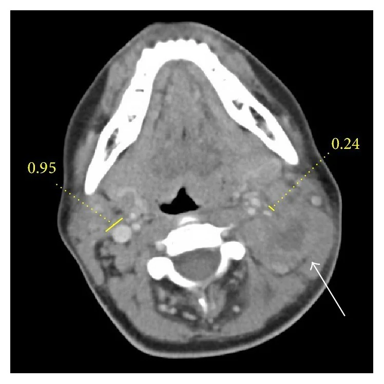
- Diş, tonsil veya farenks enfeksiyonuna drene olan üst servikal ve jugulodigastrik nodlarda bölgesel belirginleşme.
- Kontrastlı baş-boyun BT'sinde, enfeksiyon kaynaklı reaktif lenfadenopatiler çevresinde yumuşak doku ödemine ve inflamatuar değişikliklere rastlanır.
- Birden çok küçük nodun aynı drenaj zincirinde bulunması; ayrı anatomik istasyonlara yayılma yoksa bölgesel yanıtı destekler.
- Nod içinde nekroz veya sıvı atenüasyonu yerine çoğunlukla homojen yumuşak doku atenüasyonu görülür; merkezi düşük atenüasyon ya da nekroz metastaz veya apse için endişe vericidir.
- Reaktif nodlarda kısa/uzun eksen oranının <0.5 kalması, oval şekil ve hiperalkojenik hilumun korunması; metastazda beklenen hilum kaybı, yuvarlaklaşma ve eksantrik kortikal kalınlaşmadan ayrılır.
- Önceki incelemede nodların küçülmesi veya kaybolması reaktif süreci destekleyen zamansal değişimdir.
Normalde
Normal boyun görüntülerinde uzun ekseni kısa eksenden büyük, oval/kidney şeklinde ve hiperalkojenik kapak içeren küçük lenf nodları çevre yumuşak doku planları içinde belirgindir. Enfeksiyonlu örnekte aynı düzeyde nod sayısı ve çapı artarken tonsil çevresinde yumuşak doku ödemi ve yağ dokuda çizgilenme görülebilir; tek taraflı asimetriyi karşı tarafla kıyasla.
Normal BT ile kıyasla
Aynı bölgenin, kaynak veri setinde patoloji içermediği belirtilen bir BT incelemesini kesit kesit kaydırın; organ sınırlarını açarak anatomiyi hasta görüntüsüyle karşılaştırın. Küçük rastlantısal bulgular tümüyle dışlanmamıştır.
Kaynak: TotalSegmentator v2.01 veri seti (Wasserthal ve ark., Radiology: Artificial Intelligence 2023), CC BY 4.0, vaka s0622; organ sınırları veri setinin otomatik segmentasyonundan, birleştirilerek sadeleştirildi. Görüntüler 2,5 mm kesit aralığına yeniden örneklendi.
Ayırıcı tanı
- Nodal metastaz
- beklenen drenaj istasyonunda yuvarlaklaşma, kistik/nekrotik merkez veya düzensiz ekstranodal uzanım reaktif örüntüden ayrılır.
- Lenfoma
- birden fazla nodal bölgeyi tutan homojen nodal kitleler ve nodların birbirine yaslanarak konglomera oluşturması görülebilir.
- Bakteriyel nodal apse
- merkezde sıvılaşma, çevresel kontrastlanma ve komşu yağ dokuda belirgin inflamatuvar reaksiyon gösterir.
- Tüberküloz lenfadeniti
- merkezî düşük atenüasyon ve periferik kontrastlanma, bazen bitişik nodların konglomerasyonu ile seyreder.
Tuzaklar
- BT'de yağlı hilusun korunması her nodda güvenilir biçimde seçilmez; ultrason bulgusunu kontrastlı BT'de varmış gibi raporlama.
- Reaktif nodlar da büyüyebilir ve malign nodlar küçük kalabilir; boyut, klinik odak ve seri değişim birlikte ele alınmalıdır.
- Yalnızca aynı bölgedeki nod sayısının artışı malignite kanıtı değildir; diş, tonsil, cilt ve hava yolu enfeksiyonu odağını ara.
Kendini dene
Tonsillitli hastada aynı taraftaki üst servikal zincirde homojen oval nodlar ve komşu yağda hafif çizgilenme var. En olası yorum nedir?
Cevabı göster
Reaktif lenfadenopati. Nodların tonsil enfeksiyonunun drenaj zincirinde bulunması ve komşu inflamasyon bölgesel reaktif yanıtı destekler. Kistik metastazda sıvı merkezli nod, lenfomada çok istasyonlu kitle paterni, tüberkülozda ise nekrotik nodlar beklenebilir.
Enfeksiyon bulguları geriledikten sonra aynı zincirdeki nodların küçülmesi en çok hangi süreci destekler?
Cevabı göster
Reaktif yanıt. Primer inflamasyonla birlikte gerileyen nodlar geçici immün yanıtla uyumludur. Lenfoma çoğunlukla enfeksiyon odağından bağımsız kalıcı nodal yük oluşturur; ekstranodal yayılım ve apse de küçülen homojen nodları açıklamaz.
İlgili konular
Kaynaklar
Bu sayfadaki 32 cümle ve 2 quiz sorusu aşağıdaki kaynaklardan alıntılarla tek tek karşılaştırıldı; 4 cümle bu karşılaştırmada düzeltildi (2026-10-09).
- Oncologic Imaging of the Lymphatic System: Current Perspective with Multi-Modality Imaging and New Horizonpmc.ncbi.nlm.nih.gov
- Cervical: Lymphadenopathypmc.ncbi.nlm.nih.gov
- Peripheral Lymphadenopathy: Approach and Diagnostic Toolspmc.ncbi.nlm.nih.gov
- Pelvic Inflammatory Disease: Multimodality Imaging Approach with Clinical-Pathologic Correlation.pubmed.ncbi.nlm.nih.gov
- Evaluation and management of neck masses in children.pubmed.ncbi.nlm.nih.gov
- Lymphadenopathy post-COVID-19 vaccination with increased FDG uptake may be falsely attributed to oncological disorders: A systematic review.pubmed.ncbi.nlm.nih.gov
- Management of Unilateral Axillary Lymphadenopathy Detected on Breast MRI in the Era of COVID-19 Vaccination.pubmed.ncbi.nlm.nih.gov
- Disseminated Kaposi Sarcoma.pubmed.ncbi.nlm.nih.gov
- Lymphadenopathy after the third Covid-19 vaccine.pubmed.ncbi.nlm.nih.gov
- COVID-19 Vaccination-Associated Lymphadenopathy on FDG PET/CT: Distinctive Features in Adenovirus-Vectored Vaccine.pubmed.ncbi.nlm.nih.gov
- Frequency of Ipsilateral Axillary Lymphadenopathy After the Inactivated COVID-19 Vaccine.pubmed.ncbi.nlm.nih.gov
- COVID-19 vaccination and breast cancer surgery timing.pubmed.ncbi.nlm.nih.gov
- Axillary Lymphadenopathy Associated With COVID-19 Vaccination: Updates and Recommendations.pubmed.ncbi.nlm.nih.gov
- Lymphocyte-Rich Classical Hodgkin Lymphoma. A Case with Difficulty in Distinguishing from Nodular Lymphocyte-Predominant Hodgkin Lymphoma.pubmed.ncbi.nlm.nih.gov
- Laparoscopic retroperitoneal/mesenteric lymph node sampling: a safe and effective technique.pubmed.ncbi.nlm.nih.gov
- Dasatinib associated lymphadenopathy in a chronic myeloid leukemia patient: A case report.pubmed.ncbi.nlm.nih.gov
- The incidence and duration of COVID-19 vaccine-related reactive lymphadenopathy on 18 F-FDG PET-CT.pubmed.ncbi.nlm.nih.gov
- Unusual Hemodynamic Presentation of Pediatric Kikuchi-Fujimoto Disease: Persistent Sinus Bradycardia and Vasopressor-Dependent Hypotension.pubmed.ncbi.nlm.nih.gov
- EBUS-guided Cryobiopsy-Proven Anthracotic Mediastinal Lymph Node Mimicking Malignancy on FDG PET-CT: A Case Report.pubmed.ncbi.nlm.nih.gov
- Histiocytic necrotizing lymphadenitis with central nervous system involvement in children: An analysis of clinical features.pubmed.ncbi.nlm.nih.gov
- Metastatic Carotid Body Paraganglioma Following Three Decades of Indolent Growth: A Case Report.pubmed.ncbi.nlm.nih.gov
- Gut microbiota profiles in reactive and metastatic lymphadenopathy in colorectal cancer: a tumor location-stratified analysis.pubmed.ncbi.nlm.nih.gov
- Multiparametric MRI Assessment of Cervical Lymphadenopathy: Combined Diagnostic Performance of Morphological Features and Apparent Diffusion Coefficient.pubmed.ncbi.nlm.nih.gov
- A diagnostic pitfall on cervical ultrasound: Lipoplastic lymphadenopathy mimicking malignancy.pubmed.ncbi.nlm.nih.gov
- A Fibroblast Activation Protein Inhibitor (FAPI)-Avid Gastric Antral Mass Mimicking Advanced Gastric Cancer: A Case of Presumed Inflamed Gastric Ectopic Pancreas.pubmed.ncbi.nlm.nih.gov
- Simultaneous DOTATATE uptake in three non-neuroendocrine pathologies: Follicular lymphoma, Crohn's disease, and vertebral hemangioma.pubmed.ncbi.nlm.nih.gov
- Low-grade Central Osteosarcoma with High-grade Transformation in the Distal Tibia: A Rare Case Report.pubmed.ncbi.nlm.nih.gov
- Disseminated Melanoma With Rare Multiorgan Visceral Involvement in a Young Adult: A Case Report.pubmed.ncbi.nlm.nih.gov
- HIV Complicated with Talaromyces Marneffei Multisystem Infection: A Case Report and Literature Review.pubmed.ncbi.nlm.nih.gov
- Tuberculosis revisted: classic imaging findings in childhood.pubmed.ncbi.nlm.nih.gov
- Imaging of human papilloma virus associated oropharyngeal squamous cell carcinoma and its impact on diagnosis, prognostication, and response assessment.pubmed.ncbi.nlm.nih.gov
- Lemierre Syndrome Following Dental Extraction: A Case Report.pubmed.ncbi.nlm.nih.gov
- CT and MRI Key Features of Benign Tumors and Tumor-like Lesions of the Tongue: A Pictorial Review.pubmed.ncbi.nlm.nih.gov
- Vector-Borne Zoonotic Lymphadenitis-The Causative Agents, Epidemiology, Diagnostic Approach, and Therapeutic Possibilities-An Overview.pubmed.ncbi.nlm.nih.gov
- Computed Tomography Imaging Characteristics in the Diagnosis and Assessment of Cellulitis in Patients with Leg Swelling.pubmed.ncbi.nlm.nih.gov
- Acute abdomen revealing abdominal tuberculosis complicated by a proximal jejunal perforation: Case report.pubmed.ncbi.nlm.nih.gov
- Spectrum of [ 18 F]FDG-PET/CT Findings in Benign Lymph Node Pathology.pubmed.ncbi.nlm.nih.gov
- Sequential imaging approach with lymphoscintigraphy and intraoperative indocyanine green lymphography for refractory chylous ascites in an infant: A case report.pubmed.ncbi.nlm.nih.gov
- COVID-19 Vaccination and Pulmonary Nodules: A Narrative Review of Causality, Detection Bias and Thoracic Imaging Pitfalls.pubmed.ncbi.nlm.nih.gov
- Treatment-Resistant Femoral Lymph Node Abscess Caused by Cat Scratch Disease: A Case Confirmed by Polymerase Chain Reaction.pubmed.ncbi.nlm.nih.gov
- Seeing Angiogenesis: Multiparametric Ultrasound as a Non-Invasive Window into the Vascularization Status of Cervical Lymph Nodes in Head and Neck Squamous Cell Carcinoma.pubmed.ncbi.nlm.nih.gov
- Necrotizing (Abscessing) Lymphadenopathy and the Diagnostic Value of Contrast-Enhanced Ultrasound (CEUS): A Review with Clinical Vignettes.pubmed.ncbi.nlm.nih.gov
- Lymph Node Assessment with Multiparametric Ultrasound: Normal Values, Morphologic Patterns, and Diagnostic Algorithms.pubmed.ncbi.nlm.nih.gov
- Follicular-Patterned Lesions of the Thyroid Gland.pubmed.ncbi.nlm.nih.gov
- Tips and Tricks in Thoracic Radiology for Beginners: A Findings-Based Approach.pubmed.ncbi.nlm.nih.gov
- Nonproliferative and Proliferative Lesions of the Rat and Mouse Hematolymphoid System.pubmed.ncbi.nlm.nih.gov
- Longitudinal Mediastinal Lymph Node Dynamics Associate With Mortality and Fibrosis Progression in Fibrotic Interstitial Lung Disease.pubmed.ncbi.nlm.nih.gov
- Diagnostic value of high-frame-rate contrast-enhanced ultrasound and nomogram model in lymphomas.pubmed.ncbi.nlm.nih.gov
- Surgical Parapharyngeal Space Tumor Analysis with Case Series Study.pubmed.ncbi.nlm.nih.gov
- Imaging for Pediatric Head and Neck Emergencies: Critical Insights.pubmed.ncbi.nlm.nih.gov
- ESR Essentials: acute infections of the head and neck-practice recommendations by the European Society of Head and Neck Radiology.pubmed.ncbi.nlm.nih.gov
- Lymph Node Abscessation Secondary to Neoplasia in Two Dogs.pubmed.ncbi.nlm.nih.gov
- Malignant lymph nodes of the head and neck: imaging pearls, pitfalls, and practical approach to reporting in the era of extranodal extension.pubmed.ncbi.nlm.nih.gov
- Lymphadenopathy in Concurrent Head and Neck Malignancies.pubmed.ncbi.nlm.nih.gov
- Hidden in plain sight: Extrapulmonary manifestations of tuberculosis in Asia.pubmed.ncbi.nlm.nih.gov
- Cat at home? Cat scratch disease with atypical presentations and aggressive radiological findings mimicking sarcoma, a potential diagnostic pitfall.pubmed.ncbi.nlm.nih.gov
- Cat Scratch Disease-A Benign Disease with Thymic Hyperplasia Mimicking Lymphoma.pubmed.ncbi.nlm.nih.gov
- The silent pandemic in South Africa: Extra-pulmonary tuberculosis from head to heel.pubmed.ncbi.nlm.nih.gov
- Endoscopic ultrasound in mediastinal tuberculosis.pubmed.ncbi.nlm.nih.gov
- Spectral Precision: The Added Value of Dual-Energy CT for Axillary Lymph Node Characterization in Breast Cancer.pubmed.ncbi.nlm.nih.gov
- Computer-assisted and image-guided surgery for surgical lymph-nodal staging of gynecologic cancer in the era of digital and robotic surgery: a review of current evidence.pubmed.ncbi.nlm.nih.gov
- Perioperative CALLY dynamics after curative gastrectomy for gastric adenocarcinoma: associations with nodal tumor burden, textbook surgical outcome, and disease-free survival.pubmed.ncbi.nlm.nih.gov
- Standardizing pCR/MPR Assessment in NSCLC After Neoadjuvant Therapy: Challenges and Perspectives.pubmed.ncbi.nlm.nih.gov
- Intraoperative Ex Vivo Shear-Wave Elastography of Sentinel Lymph Nodes in Endometrial Cancer and Other Gynaecological Malignancies.pubmed.ncbi.nlm.nih.gov
- Same-Slide Spatial Multiomics Integration with IN-DEPTH Reveals Tumor Virus-Linked Spatial Reorganization of the Tumor Microenvironment.pubmed.ncbi.nlm.nih.gov
- Special Issue: EACR 2026 Congress: Innovative Cancer Science, 8-11 June 2026.pubmed.ncbi.nlm.nih.gov
- Recent advancements in artificial intelligence-powered cancer prediction from oral microbiome.pubmed.ncbi.nlm.nih.gov
- Concomitant Pulmonary and Tonsillar Tuberculosis.pubmed.ncbi.nlm.nih.gov
- Tonsillar Post-transplant Lymphoproliferative Disorder Presenting With Hemoptysis After Heart Transplantation: A Case Report and Review of the Literature.pubmed.ncbi.nlm.nih.gov
- Case Report: Synchronous lingual osseous choristoma and suprahyoid thyroglossal duct cyst: insights into the embryological thyroglossal-hyoid axis.pubmed.ncbi.nlm.nih.gov
- Incidentally Discovered Ovarian Cancer Combined With Obturator Nerve Schwannoma: A Rare Case Report.pubmed.ncbi.nlm.nih.gov
- The Central Role of Imaging in Renal Cell Carcinoma: A Comprehensive Review of Tumor Aggressiveness, Histology, and Radiomics.pubmed.ncbi.nlm.nih.gov
- Parapharyngeal Schwannoma: Limitations Regarding Diagnosis and Surgical Intervention-A Case Report and Focused Literature Review.pubmed.ncbi.nlm.nih.gov
- Excision of a Giant Bilobed Second Branchial Cleft Cyst: A Case Report.pubmed.ncbi.nlm.nih.gov
- Pre-treatment and post-treatment nasopharyngeal carcinoma imaging: imaging updates, pearls and pitfalls.pubmed.ncbi.nlm.nih.gov
- Human biomarker navigator.pubmed.ncbi.nlm.nih.gov
- New insights into imaging of pulmonary metastases from extra-thoracic neoplasms.pubmed.ncbi.nlm.nih.gov
- China Anti-Cancer Association Guidelines for the diagnosis, treatment, and follow-up of thymic epithelial tumors (2023).pubmed.ncbi.nlm.nih.gov
- Kikuchi-Fujimoto Disease in Pregnancy: A Rare Case With Atypical Presentation and Potential Viral Trigger.pubmed.ncbi.nlm.nih.gov
- Corticosteroid-responsive portal lymphadenopathy mimicking malignant progression induced by furmonertinib in EGFR-mutated NSCLC: a case report.pubmed.ncbi.nlm.nih.gov
- Migratory Pulmonary Infiltrates Mimicking Malignancy, Tuberculosis, and Sarcoidosis in an Apparently Immunocompetent Adult: A Case of Tissue-Proven Invasive Fungal Disease Due to a Non-<i>marneffei</i><i>Penicillium</i> Species.pubmed.ncbi.nlm.nih.gov
- Sustained Regression of Parotid Mucosa-Associated Lymphoid Tissue (MALT) Lymphoma After Nutritional and Nutraceutical Intervention Without Chemoimmunotherapy: A Case Report.pubmed.ncbi.nlm.nih.gov
- Differentiating lymphoma progression from benign lesions: features of incident [<sup>18</sup>F]FDG-avid foci on interim and end-of-treatment PET/CT.pubmed.ncbi.nlm.nih.gov
- Neurolymphomatosis: A Comprehensive Review of Clinical and Imaging Features.pubmed.ncbi.nlm.nih.gov
- Evaluation of Lymphadenopathy in Urgent Care: A Case of Hodgkin Lymphoma.pubmed.ncbi.nlm.nih.gov
- The Diagnostic Utility of Endobronchial Ultrasound-Guided Fine-Needle Aspiration Cytology (EBUS-FNAC) for Thoracic Lymphadenopathy: A Correlation Study With Radiological Findings.pubmed.ncbi.nlm.nih.gov
- An Aggressive Neoplasm with Mixed Epithelial and Sarcoma-like Features, of Uncertain Primary Origin, Presenting as an Upper Lip Mass: A Case Report.pubmed.ncbi.nlm.nih.gov
- Australian clinical practice guideline: diagnosis and treatment of idiopathic multicentric Castleman disease.pubmed.ncbi.nlm.nih.gov
- Cervical lymph node metastases in thoracic esophageal cancer: a systematic review.pubmed.ncbi.nlm.nih.gov
- Isolated Parotitis Following a Cheek Slap: A Case Report and Clinical Implications.pubmed.ncbi.nlm.nih.gov
- Role of magnetic resonance in bowel pathologies: Beyond inflammatory bowel disease.pubmed.ncbi.nlm.nih.gov
- A Case Report of Extrapulmonary Tuberculosis: A Potpourri of Imaging Manifestations.pubmed.ncbi.nlm.nih.gov
Eğitim amaçlıdır; hasta başında tanı ve tedavi kararının yerine geçmez. İçerik yapay zekâ destekli bir süreçle güncel literatüre göre cümle cümle kaynaklarla karşılaştırıldı, ancak uzman radyolog incelemesinden geçmedi. Klinik kararlarda güncel kılavuzları, kurum protokollerini ve radyoloji raporunu esas alın.