Özet
Sarkoidoz, çoğunlukla genç ve orta yaş erişkinlerde akciğer ve intratorasik lenf nodlarını birlikte etkileyen sistemik granülomatöz hastalıktır. Toraks BT nodal dağılımı, simetriyi, kalsifikasyon biçimini ve eşlik eden perilenfatik akciğer nodüllerini akciğer grafisinden daha ayrıntılı gösterir; tipik görünüm tanıyı destekler, ancak enfeksiyon ve lenfomayı tek başına dışlamaz. Simetrik bilateral hiler ve sağ paratrakeal büyüme tipik olsa da tek taraflı, nekrotik veya asimetrik nodlar alternatif tanı aramayı gerektirir. Atipik dağılımın ya da akciğer parankimindeki eşlik eden hastalığın gözden kaçması, enfeksiyon veya malignitenin yanlışlıkla sarkoidoz olarak yorumlanmasına yol açabilir.
Faz ve pencere
- Kontrastsız tanısal
- İnce kesit toraks BT'de bilateral hiler ve mediastinal nodal büyümenin simetrisi, istasyon dağılımı ve nod içi kalsifikasyon; akciğer penceresinde fissürler, plevra ve bronkovasküler demetler boyunca perilenfatik mikronodüller değerlendirilir.
Önerilen pencereler: Mediasten (G 350 / M 50), Akciğer (G 1500 / M -600).
BT bulguları
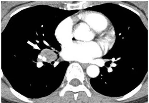
- Bilateral hiler nodların ve sağ alt paratrakeal istasyon 4R'nin düzgün sınırlı, çoğunlukla simetrik büyümesi
- Subkarinal istasyon 7 ve bilateral interlober/hiler istasyonların eşlik eden nodal tutulumu
- Nodların genellikle homojen yumuşak doku yoğunluğunda olması; belirgin merkezi nekrozun tipik olmaması
- Kronik hastalıkta bilateral, noktasal, kaba veya periferik kabuk biçimli nodal kalsifikasyon
- Fissür, plevra ve bronkovasküler demet boyunca dağılmış perilenfatik mikronodüllerin eşlik etmesi
- Üst ve orta akciğer zonlarında nodül kümelenmesi veya bronkovasküler interstisyel kalınlaşma
- Nodal büyüme olmadan parankimal tutulum ya da asimetrik nodlar gibi atipik örüntülerin bulunabilmesi
- Nodların bronşa basısı veya komşu damarı yer değiştirmesi; ileri fibrotik hastalıkta hiler mimaride çekilme
Normalde
Normal mediastinal ve hiler lenf nodları çevre yağ dokusu içinde ayrı seçilir ve ana bronşları ya da pulmoner arterleri belirgin biçimde itmez; akciğer fissürleri ve bronkovasküler demetler boyunca rastgele dağılmış nodüller bulunmaz. Sağ paratrakeal zincir ile iki hiler bölgenin kontur ve hacmini karşılaştır; nodların simetrik büyümesine, kalsifikasyonuna ve aynı kesitte eşlik eden perilenfatik nodüllere bak.
Normal BT ile kıyasla
Aynı bölgenin, kaynak veri setinde patoloji içermediği belirtilen bir BT incelemesini kesit kesit kaydırın; organ sınırlarını açarak anatomiyi hasta görüntüsüyle karşılaştırın. Küçük rastlantısal bulgular tümüyle dışlanmamıştır.
Kaynak: TotalSegmentator v2.01 veri seti (Wasserthal ve ark., Radiology: Artificial Intelligence 2023), CC BY 4.0, vaka s1278; organ sınırları veri setinin otomatik segmentasyonundan, birleştirilerek sadeleştirildi. Görüntüler 2,5 mm kesit aralığına yeniden örneklendi.
Ayırıcı tanı
- Tüberküloz lenfadeniti
- asimetrik veya tek taraflı nodlar, merkezi düşük atenüasyon ve çevresel kontrastlanan halka, matlaşma ve perinodal yağ değişikliği TB'yi destekler.
- Lenfoma
- hacimli, birleşen veya belirgin asimetrik nodal kitleler ve eşlik eden ekstratorasik hastalık kuşkuyu artırır; nadiren simetrik hiler görünüm de olabilir.
- Silikozis
- üst loblarda, çoğunlukla santrallobüler ve subplevral yerleşimli küçük nodüller ve bazen yumurta kabuğu biçimli nodal kalsifikasyon görülebilir; mesleki silika maruziyeti öyküsü ve üst lob ağırlıklı nodüler fibrozis ayırıcıdır.
- Histoplazmoz
- akut formlarında bilateral küçük nodüller (<3 mm) ve lenfadenopati; kronik formlarında üst loblarda kavitasyon ve nodüler fibrozis görülür; geçirilmiş enfeksiyonlarda nodal kalsifikasyon izlenebilir.
- Metastatik lenfadenopati
- bilinen primer tümör, tek taraflı veya istasyon dışı nodlar ve eşlik eden akciğer/organ lezyonları sarkoidozdan uzaklaştırır.
Tuzaklar
- Bilateral hiler lenfadenopati tek başına sarkoidoz tanısı değildir; TB ve lenfoma da nodal büyüme yapabildiğinden asimetri, nekroz ve klinik bağlamı kontrol et.
- Nodal nekroz sarkoidozda seyrek görülebilir; özellikle belirgin rim tarzı kontrastlanma varsa TB, mantar enfeksiyonu veya malignite için ek kanıt ara.
- Yumurta kabuğu kalsifikasyonu sarkoidoza özgü değildir; silikozis ve başka kronik granülomatöz süreçlerle birlikte de görülebilir.
- Kontrastlı BT, hiler lenf nodlarının komşu pulmoner damarlarla ilişkisini ve daraltma etkisini değerlendirmek açısından faydalıdır.
Kendini dene
Toraks BT'de simetrik bilateral hiler ve sağ paratrakeal nodlara, üst-orta zonlarda fissür ve bronkovasküler demetler boyunca perilenfatik mikronodüller eşlik ediyor; silika maruziyeti öyküsü yok. En olası tanı nedir?
Cevabı göster
Sarkoidoz. Simetrik hiler ve sağ paratrakeal lenfadenopatiyle birlikte fissür ve bronkovasküler demetleri izleyen perilenfatik nodüller sarkoidozu destekler. Silikozis için mesleki silika maruziyeti ve üst lob ağırlıklı nodüler fibrozis aranır; TB ve lenfoma bu dağılımın daha az tipik taklitçileridir.
Sarkoidoz düşünülen hastada tek taraflı mediastinal nodlarda düşük dansiteli merkez ve kontrastlanan çevresel halka var. Hangi taklitçi özellikle araştırılmalıdır?
Cevabı göster
Tüberküloz lenfadeniti. Merkezi nekroz ve çevresel kontrastlanma, özellikle asimetrik nodlarda tüberküloz lenfadenitini düşündürür; bu görünüm patognomonik değildir. Timik, vasküler ve plevral lezyonlar mediastinal lenf nodlarından farklı yapılara ait olabilir.
İlgili konular
Kaynaklar
Bu sayfadaki 37 cümle ve 2 quiz sorusu aşağıdaki kaynaklardan alıntılarla tek tek karşılaştırıldı; 3 cümle bu karşılaştırmada düzeltildi (2026-10-09).
- Imaging of Pulmonary Sarcoidosis—A Reviewpmc.ncbi.nlm.nih.gov
- Diagnosis and Detection of Sarcoidosis. An Official American Thoracic Society Clinical Practice Guidelinepmc.ncbi.nlm.nih.gov
- Differential diagnosis of pulmonary sarcoidosis: a reviewpmc.ncbi.nlm.nih.gov
- Carcinoma Breast Presenting with Concurrent Extensive Lymph Nodal Sarcoidosis.pubmed.ncbi.nlm.nih.gov
- Emerging phenotypes of sarcoidosis based on 18F-FDG PET/CT: a hierarchical cluster analysis.pubmed.ncbi.nlm.nih.gov
- Common variable immunodeficiency presenting as sarcoidosis in a 9-year-old child.pubmed.ncbi.nlm.nih.gov
- Fatal clinical outcome in a patient with sarcoidosis-lymphoma syndrome.pubmed.ncbi.nlm.nih.gov
- Clinical impact of liquid-based cytology test on diagnostic yields from transbronchial needle aspiration.pubmed.ncbi.nlm.nih.gov
- Fibrosing mediastinitis mimicking sarcoidosis.pubmed.ncbi.nlm.nih.gov
- Peritoneal sarcoidosis: A case report.pubmed.ncbi.nlm.nih.gov
- [Squamous cell carcinoma (SCC)--nature and usage in patients with cervical carcinoma].pubmed.ncbi.nlm.nih.gov
- Restrictive lung defects: parenchymal, chest wall and neuromuscular.pubmed.ncbi.nlm.nih.gov
- The "cluster of black pearls" sign of sarcoid lymphadenopathy: a new sign on thin-section contrast-enhanced multidetector CT.pubmed.ncbi.nlm.nih.gov
- Evaluation of ¹⁸F-fluorodeoxyglucose uptake in enlarged mediastinal lymph nodes in patients with lung cancer.pubmed.ncbi.nlm.nih.gov
- Hodgkin's Lymphoma Presenting as Multiple Cavitary Lung Lesions.pubmed.ncbi.nlm.nih.gov
- (18)F-FDG and (18)F-FLT PET/CT imaging in the characterization of mediastinal lymph nodes.pubmed.ncbi.nlm.nih.gov
- A Rare Presentation of Sarcoidosis: A Case of Acute Onset Bilateral Pulmonary Infiltrates and Skin Lesions.pubmed.ncbi.nlm.nih.gov
- Carotid Artery Dissection as Initial Manifestation of Overlapping Sarcoidosis and Antiphospholipid Syndrome in a Young Woman.pubmed.ncbi.nlm.nih.gov
- Tattoos causing local and even generalized lymphadenopathies including sarcoidosis.pubmed.ncbi.nlm.nih.gov
- Symptomatic diffuse pulmonary meningotheliomatosis with persistent diffusing-capacity impairment.pubmed.ncbi.nlm.nih.gov
- Sarcoidosis Presenting With Severe Hypercalcemia, Renal Dysfunction, and Bilateral Submandibular Swelling: A Case Report.pubmed.ncbi.nlm.nih.gov
- Multimodality Imaging for the Diagnosis and Evaluation of Pulmonary Sarcoidosis.pubmed.ncbi.nlm.nih.gov
- Breaking Symmetry: Unilateral Sarcoidosis Presenting as a Diagnostic Challenge.pubmed.ncbi.nlm.nih.gov
- CT Imaging Features of Pulmonary Sarcoidosis: Typical and Atypical Radiological Features and Their Differential Diagnosis.pubmed.ncbi.nlm.nih.gov
- Differential diagnosis of pulmonary sarcoidosis: a review.pubmed.ncbi.nlm.nih.gov
- Paucibacillary Pleural Tuberculosis Presenting as Subpleural Nodules: A Diagnostic Challenge With Negative PCR and Smear Tests.pubmed.ncbi.nlm.nih.gov
- Pulmonary cryptococcosis with cavitation in an immunocompetent child: a case report and literature review.pubmed.ncbi.nlm.nih.gov
- Development and Internal Validation of the Yuvarajan Sarcoidosis Diagnostic Score (YSDS): A Retrospective Cohort Study.pubmed.ncbi.nlm.nih.gov
- Calcified Lung Nodules: A Diagnostic Challenge in Clinical Daily Practice.pubmed.ncbi.nlm.nih.gov
- Korean Guidelines for the Diagnosis and Management of Interstitial Lung Diseases: Sarcoidosis.pubmed.ncbi.nlm.nih.gov
- Mediastinal Lymphadenopathy: Associations Between Computed Tomography Characteristics and Final Etiological Diagnoses in 200 Patients.pubmed.ncbi.nlm.nih.gov
- Scoping out sarcoidosis: the evolving role of bronchoscopy.pubmed.ncbi.nlm.nih.gov
- Predictors of malignant lymph node involvement in patients with mediastinal lymphadenopathy and previous cancer: A cohort study.pubmed.ncbi.nlm.nih.gov
- Typical and atypical manifestations of intrathoracic sarcoidosis.pubmed.ncbi.nlm.nih.gov
- PET-CT for assessing mediastinal lymph node involvement in patients with suspected resectable non-small cell lung cancer.pubmed.ncbi.nlm.nih.gov
- AWMF S2k Guideline: Diagnosis and Assessment of Lung Disease Caused by Occupational Silica Dust Exposure (Silicosis) - Disease No. 4101 as per Occupational Diseases Ordinance (BKV) Annex 1.pubmed.ncbi.nlm.nih.gov
- Non-metastatic causes of multiple pulmonary nodules.pubmed.ncbi.nlm.nih.gov
- New insights into imaging of pulmonary metastases from extra-thoracic neoplasms.pubmed.ncbi.nlm.nih.gov
- Bilateral giant progressive massive fibrosis mimicking lung cancer in a rubber factory worker: A rare presentation.pubmed.ncbi.nlm.nih.gov
- Silicosis, Sarcoidosis, and Silicosarcoidosis Are Overlapping Diagnoses and Difficult to Differentiate.pubmed.ncbi.nlm.nih.gov
- TNF Inhibitor-Induced Sarcoidosis-Like Lesions in Inflammatory Bowel Disease.pubmed.ncbi.nlm.nih.gov
- Noninfectious Granulomatous Lung Disease: Radiological Findings and Differential Diagnosis.pubmed.ncbi.nlm.nih.gov
- Childhood interstitial lung diseases: current understanding of the classification and imaging findings.pubmed.ncbi.nlm.nih.gov
- Atypical Fibrotic-Mimicking Pulmonary Sarcoidosis in a Young Female: The Basal Conundrum.pubmed.ncbi.nlm.nih.gov
- Bone involvement in sarcoidosis: Insights from a multicentre Italian cohort.pubmed.ncbi.nlm.nih.gov
- CT Features of Granulomatous-Lymphocytic Interstitial Lung Disease (GLILD): The "Kebab Sign" as a Marker to Support Differential Diagnosis.pubmed.ncbi.nlm.nih.gov
- Fibrotic Pulmonary Sarcoidosis-From Pathogenesis to Management.pubmed.ncbi.nlm.nih.gov
- Tips and Tricks in Thoracic Radiology for Beginners: A Findings-Based Approach.pubmed.ncbi.nlm.nih.gov
- Imaging of pulmonary hypertension: an update.pubmed.ncbi.nlm.nih.gov
- Lung imaging methods: indications, strengths and limitations.pubmed.ncbi.nlm.nih.gov
- Commentary: an eye on PET quantification.pubmed.ncbi.nlm.nih.gov
- Patterns of injury and violence in Yaoundé Cameroon: an analysis of hospital data.pubmed.ncbi.nlm.nih.gov
- Extrapulmonary tuberculosıs: an old but resurgent problem.pubmed.ncbi.nlm.nih.gov
- High-affinity alphavbeta3 integrin targeted optical probe as a new imaging biomarker for early atherosclerosis: initial studies in Watanabe rabbits.pubmed.ncbi.nlm.nih.gov
- Imaging of Retrosternal Space Lesions - A Pictorial Review.pubmed.ncbi.nlm.nih.gov
- Hidden danger: silicosis in a dental technician-an overlooked occupational risk.pubmed.ncbi.nlm.nih.gov
- Broncholithiasis: Diagnostic and Therapeutic Insights From a Contemporary Case Series of Five Patients.pubmed.ncbi.nlm.nih.gov
- Radiological features of chronic pulmonary histoplasmosis: Easily mistaken for tuberculosis.pubmed.ncbi.nlm.nih.gov
- Unilateral hilar sarcoidosis with anemia and low T3 syndrome: a case report.pubmed.ncbi.nlm.nih.gov
- Emerging and re-emerging infectious disease in otorhinolaryngology.pubmed.ncbi.nlm.nih.gov
- Spontaneous bacteremia and spontaneous bacterial peritonitis share similar prognosis in patients with cirrhosis: a cohort study.pubmed.ncbi.nlm.nih.gov
- EBUS-guided Cryobiopsy-Proven Anthracotic Mediastinal Lymph Node Mimicking Malignancy on FDG PET-CT: A Case Report.pubmed.ncbi.nlm.nih.gov
- Multisystem Sarcoidosis Presenting as Severe Hypercalcemic Crisis and Generalized Lymphadenopathy Mimicking Hematological Malignancy: A Case Report.pubmed.ncbi.nlm.nih.gov
- Cisplatin-induced sarcoidosis-like reaction after primary chemoradiotherapy for locally advanced cervical cancer.pubmed.ncbi.nlm.nih.gov
- Unusual Presentation of Tuberculosis: Diagnostic Challenges in Isolated Axillary Tuberculous Lymphadenitis.pubmed.ncbi.nlm.nih.gov
- Nasopharyngeal tuberculosis mimicking malignancy on MRI: A rare clinical entity in an immunocompetent adult.pubmed.ncbi.nlm.nih.gov
- A case report of tuberculous fibrosing mediastinitis: focusing on the diagnostic value of three-dimensional reconstruction from pulmonary contrast-enhanced CT and bronchoscopic features.pubmed.ncbi.nlm.nih.gov
- Three-Dimensional Bronchovascular Modelling in Sublobar Pulmonary Resection: A Tool for Personalised Thoracic Surgery.pubmed.ncbi.nlm.nih.gov
- Artificial Intelligence in Pulmonary Endoscopy: Current Evidence, Limitations, and Future Directions.pubmed.ncbi.nlm.nih.gov
- State-of-the-Art Mediastinal Staging in Non-Small-Cell Lung Cancer: Integration of Combined Endosonographic Techniques with Updated IASLC TNM 9th Classification.pubmed.ncbi.nlm.nih.gov
- Right-first bilateral transcervical single-port robotic esophagectomy: a cadaveric feasibility study toward a safer extrapleural strategy.pubmed.ncbi.nlm.nih.gov
- Aggressive Progression of High Programmed Death-Ligand 1 (PD-L1) Non-small Cell Lung Cancer Presenting as Life-Threatening Esophageal Obstruction: A Case of Food Impaction Secondary to Subcarinal Lymph Node Compression.pubmed.ncbi.nlm.nih.gov
- Unlocking the Diagnostic Challenge of Tuberculosis and Sarcoidosis Intrathoracic Lymphadenopathy: Potential Role of HMGB1 and miRNA-221 as Diagnostic Tools.pubmed.ncbi.nlm.nih.gov
- Dilemma of diagnosing thoracic sarcoidosis in tuberculosis endemic regions: An imaging-based approach. Part 1.pubmed.ncbi.nlm.nih.gov
- Mimickers of chronic thromboembolic pulmonary hypertension on imaging tests: a review.pubmed.ncbi.nlm.nih.gov
- Systemic Sarcoidosis With Neurosarcoidosis and Obstructive Hydrocephalus: A Radiologic-Pathologic Case Report.pubmed.ncbi.nlm.nih.gov
- Cardiac Sarcoidosis: A Practical Guide for Cardiologists.pubmed.ncbi.nlm.nih.gov
- High prevalence of coxsackievirus B antibodies in patients with cardiac sarcoidosis: a retrospective cohort study.pubmed.ncbi.nlm.nih.gov
- Arrhythmias in Cardiac Sarcoidosis: Pathophysiology, Diagnostic Strategies, Risk Stratification for Sudden Cardiac Death, and Contemporary Management-A Narrative Review.pubmed.ncbi.nlm.nih.gov
- Primary Lymphoproliferative Lung Diseases: Imaging and Multidisciplinary Approach.pubmed.ncbi.nlm.nih.gov
- Radiological and pathological analysis of the galaxy sign in patients with pulmonary mucosa-associated lymphoid tissue (MALT) lymphoma.pubmed.ncbi.nlm.nih.gov
- Coenzyme Q10 alleviates silicotic fibrosis in rats through the TGF-β1/Smad pathway.pubmed.ncbi.nlm.nih.gov
- Presumed Infected Bronchogenic Cyst Presenting as Acute Pericarditis: A Case Report.pubmed.ncbi.nlm.nih.gov
- Ocular TB as the only manifestation of TB: diagnostic uncertainty and treatment thresholds.pubmed.ncbi.nlm.nih.gov
- Diagnostic Imaging of Extrapulmonary Tuberculosis Across Organ Systems.pubmed.ncbi.nlm.nih.gov
- The application of grayscale quantitative analysis in the differential diagnosis of necrosis between cervical lymph node metastases in nasopharyngeal carcinoma and tuberculous lymphadenitis.pubmed.ncbi.nlm.nih.gov
- Ultrasound-Guided Needle Aspiration Biopsy for Superficial Cervical Lymphadenopathy: Diagnostic Yield Beyond Tuberculosis.pubmed.ncbi.nlm.nih.gov
- Obstructive Jaundice due to a Periportal Tuberculous Mass Complicated by Anti-Tuberculous Drug-Induced Liver Injury: A Case Report From a Low-Resource Setting.pubmed.ncbi.nlm.nih.gov
- Necrotizing (Abscessing) Lymphadenopathy and the Diagnostic Value of Contrast-Enhanced Ultrasound (CEUS): A Review with Clinical Vignettes.pubmed.ncbi.nlm.nih.gov
- An Uncommon Case of Recurring Pulmonary, Lymphatic, and Skeletal Tuberculosis Accompanied by Myositis: Is It Associated with Particular Resistance?pubmed.ncbi.nlm.nih.gov
- Pathology Competencies in Medical Education and Educational Cases: Update 2023.pubmed.ncbi.nlm.nih.gov
- Pictorial essay of radiological features of benign intrathoracic masses.pubmed.ncbi.nlm.nih.gov
- A comprehensive review of endobronchial ultrasound-guided transbronchial needle aspiration (EBUS-TBNA): staging, techniques, and future directions.pubmed.ncbi.nlm.nih.gov
- Multisystem FDG-avid sarcoidosis with osseous involvement mimicking disseminated metastatic malignancy.pubmed.ncbi.nlm.nih.gov
- Dual-tracer <sup>18</sup>F-FDG and <sup>18</sup>F-FLT PET/CT for differentiating metastatic lymph nodes from sarcoid involvement in a patient with vulvar squamous cell carcinoma and coexisting pulmonary sarcoidosis.pubmed.ncbi.nlm.nih.gov
- Immunological biomarkers predict pulmonary and nodal, but not cardiac positron emission tomography-assessed sarcoidosis activity.pubmed.ncbi.nlm.nih.gov
- International Harmonization of Nomenclature and Diagnostic Criteria (INHAND): Non-proliferative and Proliferative Lesions of the Non-human Primate ( M. fascicularis ).pubmed.ncbi.nlm.nih.gov
- Diagnosis and Management of Sarcoidosis-like Reaction in Adjuvant Immunotherapy: A Comprehensive Review and Clinical Implications.pubmed.ncbi.nlm.nih.gov
- Clinical and computed tomography (CT) features of nodular pulmonary histoplasmosis.pubmed.ncbi.nlm.nih.gov
- Pulmonary sarcoidosis complicated with pulmonary cryptococcosis: a case report.pubmed.ncbi.nlm.nih.gov
Eğitim amaçlıdır; hasta başında tanı ve tedavi kararının yerine geçmez. İçerik yapay zekâ destekli bir süreçle güncel literatüre göre cümle cümle kaynaklarla karşılaştırıldı, ancak uzman radyolog incelemesinden geçmedi. Klinik kararlarda güncel kılavuzları, kurum protokollerini ve radyoloji raporunu esas alın.