Özet
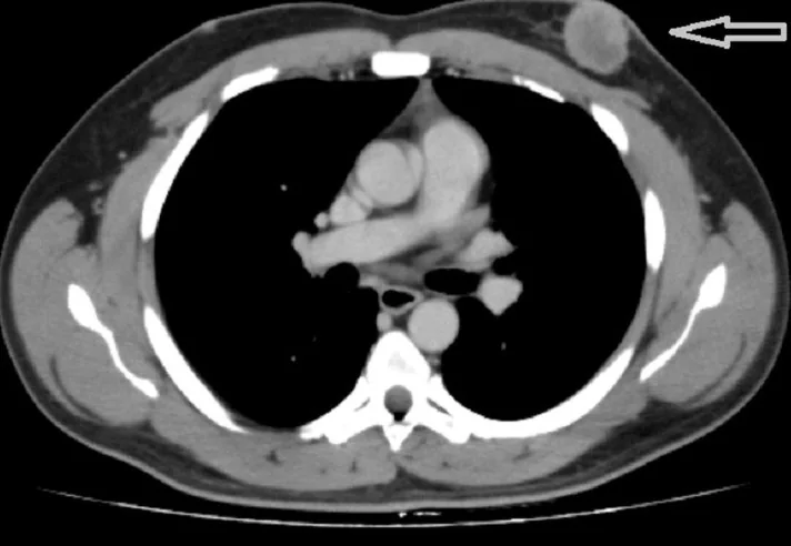
Primer meme kanseri, meme taraması veya tanısal meme incelemesinde saptanabileceği gibi başka nedenle çekilen toraks BT'sinde de ilk kez fark edilebilir. BT'de irregüler-spiküle veya lobüle kitle, asimetrik belirgin kontrastlanma, cilt-trabeküler değişiklik ve bölgesel nodlar şüpheyi artırır; BT'nin meme için tarama ya da kesin tanı yöntemi olmadığı unutulmamalıdır. Toraks BT'sinin güçlü katkısı tümörün göğüs duvarı ve toraks içi nodal istasyonlarla ilişkisini aynı incelemede göstermesidir. Primer odağın veya uzanımın raporlanmaması tanı ve evreleme sürecini geciktirebilir; yalnız nod boyutuna bakarak nodal metastaz varsaymak yanlış evrelemeye yol açabilir.
Faz ve pencere
- Portal tanısal
- İntravenöz kontrastlı toraks BT'de çevre fibroglandüler dokuya göre belirgin veya heterojen kontrastlanan, irregüler/lobüle konturlu kitle ya da kitle dışı asimetrik kontrastlanma seçilebilir; aynı incelemede cilt, pektoralis, kaburga ve aksiller ve internal mammary-supraklaviküler nodal alanlar incelenir.
Önerilen pencereler: Yumuşak doku (G 400 / M 40), Mediasten (G 350 / M 50).
BT bulguları
- Spiküle meme kitlesi — parankim içinde merkezden çevreye uzanan ince çizgisel çıkıntıları olan düzensiz yumuşak doku odağı.
- Lobüle veya irregüler kontur — düzgün oval sınıra göre asimetrik ve girintili çıkıntılı kitle biçimi.
- Lezyonun çevre parankimden farklı yoğunluk kazanması — malign kitlenin Hounsfield birimi genellikle yüksektir (ortalama ~49 HU), 20 HU altı ise çoğunlukla kistlere işaret eder.
- Asimetrik kitle dışı doku artışı — belirgin kapsüllü kitle oluşturmadan tek bölgede parankim kontrastlanması ve mimari bozulma.
- Cilt ve trabeküler kalınlaşma — primer odağa bakan ciltte kalınlaşma/çekinti veya meme içi çizgisel bağ dokusunda belirginleşme.
- Meme başı retraksiyonu — meme başının içe çekilmesidir; meme Paget hastalığı olgularının mamografilerinde bildirilmiştir.
- Pektoralis ve göğüs duvarı ilişkisi — retromamari yağ planı kaybı temas veya invazyon kuşkusu doğurur ancak BT bu ayrımı güvenilir biçimde yapamaz; patolojik doğrulama gereklidir.
- Bölgesel lenfadenopati — aksiller, subpektoral, supraklaviküler veya internal mammary zincirde asimetrik nod; tek başına boyut metastazı kanıtlamaz.
Normalde
Normal aynı düzeyde meme parankimi çevre yağla karışık, taraflar arasında değişken yoğunlukta olabilir; tek odakta spiküler çıkıntı, yeni cilt çekintisi veya retromamari yağ planı kaybı beklenmez. Karşı memede meme başı yönünü, cilt ve trabeküler çizgileri ve pektoralis ön sınırını eşleştirin; aksiller ve internal mammary nodları yalnız boyutla değil, asimetri ve şekil özellikleriyle kıyaslayın.
Normal BT ile kıyasla
Aynı bölgenin, kaynak veri setinde patoloji içermediği belirtilen bir BT incelemesini kesit kesit kaydırın; organ sınırlarını açarak anatomiyi hasta görüntüsüyle karşılaştırın. Küçük rastlantısal bulgular tümüyle dışlanmamıştır.
Kaynak: TotalSegmentator v2.01 veri seti (Wasserthal ve ark., Radiology: Artificial Intelligence 2023), CC BY 4.0, vaka s1278; organ sınırları veri setinin otomatik segmentasyonundan, birleştirilerek sadeleştirildi. Görüntüler 2,5 mm kesit aralığına yeniden örneklendi.
Ayırıcı tanı
- Düzgün sınırlı kitleler
- malign kitlerde düzgün sınır %33 oranında iken, benign kitlerin %93'ünde düzgün sınır bulunur; BT bulguları tek başına tüm benignlikleri kanıtlamayabilir.
- Birincil meme difüz büyük B-hücreli lenfoma (PB-DLBCL)
- klinik ve radyolojik olarak meme karsinomuna benzer, kesin tanı için dokuya bakılması (core biopsy) ve immünfenotipleme gereklidir.
- Meme metastazı
- bilinen başka primer tümör, birden fazla odak veya eş zamanlı başka organ metastazları ile düşünülür; primer meme kanseri morfolojisiyle örtüşebilir.
- Mastit veya apse
- cilt ve trabeküler ödem, yaygın inflamatuvar değişiklik ya da çevresel kontrastlanan koleksiyon olabilir; klinik enfeksiyon ve koleksiyon içeriğiyle ayrılır.
- Postoperatif değişiklikler
- akut inflamatuvar değişimlerden kronik fibrozis, yağ nekrozu ve mimari bozulmaya kadar zamanla evrilen karakteristik morfolojik değişiklikler gösterir.
- İnflamatuvar meme kanseri
- katı bir kitle olmaksızın tek memede diffüz şişlik ve lenfadenopati ile kendini gösterebilir, kesin tanı için biyopsi şarttır.
Tuzaklar
- Enfiltratif büyüme paterni (örneğin invaziv lobüler karsinom) — konvansiyonel görüntüleme ve klinik muayene doğruluğunu sıkça sınırlar, düzensiz kitle dışı lezyon olarak izlenebilir.
- Pektoralis majorla temas veya yağ planı kaybı invazyon şüphesi yaratasa da BT tek başına kesin ayırım yapamaz; klinik bağlam ve ileri görüntüleme/biyopsi ile değerlendirilmelidir.
- Aksiller veya subpektoral nodun büyük olması metastazı kanıtlamaz; bilateral dağılım, yağlı hilus, kortikal şekil ve klinik bağlam birlikte düşünülmelidir.
- Küçük mamografik mikrokalsifikasyonlar rutin toraks BT çözünürlüğünde görünmeyebilir; BT'de kalsifikasyon saptanmaması DCIS'i veya mamografik şüpheyi dışlamaz.
Kendini dene
Göğüs BT'sinde kanserli meme nodüllerinin konturu kaynakta en sık hangi özelliklerle tarif edilmiştir?
Cevabı göster
Lobüle veya spiküle. Kaynakta kanserli nodüllerin sıklıkla lobüle veya spiküle olduğu bildirilmiştir.
Bu çalışmada kontrastlı ve kontrastsız toraks BT'nin meme kanseri saptamadaki duyarlılığı nasıl karşılaştırılmıştır?
Cevabı göster
Kontrastlı BT'de %85,71, kontrastsız BT'de %80,00. Çalışmada meme kanseri saptama duyarlılığı kontrastlı toraks BT'de %85,71, kontrastsız BT'de %80,00 olarak bildirilmiştir.
İlgili konular
Kaynaklar
Bu sayfadaki 34 cümle ve 2 quiz sorusu aşağıdaki kaynaklardan alıntılarla otomatik olarak karşılaştırıldı; 11 cümle bu karşılaştırmada düzeltildi (2026-10-09).
- Chest CT for Breast Cancer Diagnosispmc.ncbi.nlm.nih.gov
- From Dyspnea to Suspected Breast Cancer: A Post-traumatic Hematoma as a Diagnostic Pitfall.pubmed.ncbi.nlm.nih.gov
- Incidental Detection of a Small, Non-spiculated Breast Cancer During Preoperative Evaluation for an Unruptured Cerebral Aneurysm With Gradual Enlargement Over 16 Years: A Case Report.pubmed.ncbi.nlm.nih.gov
- Incidental Malignancies in the Emergency Department: Missed Diagnoses, Radiological Difficulty, and a Risk-Based Framework for Follow-Up.pubmed.ncbi.nlm.nih.gov
- Incidental Breast Findings on Chest CT: Rates of Report and Follow-Up.pubmed.ncbi.nlm.nih.gov
- Beyond the lung: recognizing incidental breast lesions on chest computed tomography.pubmed.ncbi.nlm.nih.gov
- Extranodal marginal zone lymphoma presenting as a skin-based breast finding detected on screening mammography.pubmed.ncbi.nlm.nih.gov
- Combination of vinorelbine capsules and the small molecule anti-angiogenic drug apatinib for first-line treatment of advanced triple-negative breast cancer with schizophrenia after failure of anthracycline and taxane therapy: A case report.pubmed.ncbi.nlm.nih.gov
- Case Report: Metachronous bilateral primary and secondary angiosarcoma of the breast.pubmed.ncbi.nlm.nih.gov
- An exceptionally late and severe radiation recall dermatitis with chest-wall ulceration 32 years after breast radiotherapy: a case report and literature review.pubmed.ncbi.nlm.nih.gov
- Prediction of axillary lymph node metastasis in breast cancer using preoperative CT Hounsfield units: A retrospective study.pubmed.ncbi.nlm.nih.gov
- The role of <sup>68</sup>Ga-FAPi PET/CT in Staging and Restaging of Breast Cancer Subtypes with Limited <sup>18</sup>F-FDG Uptake.pubmed.ncbi.nlm.nih.gov
- Navigating lymphatic drainage uncertainty in abdominal ectopic breast cancer: a novel application of indocyanine green fluorescence-a case report and literature review.pubmed.ncbi.nlm.nih.gov
- Small intestinal metastasis of triple-negative breast cancer mimicking lymphoma: imaging findings and case report.pubmed.ncbi.nlm.nih.gov
- Diverse Radiologic Presentations of Paget's Disease of the Breast: From Subtle to Striking Findings.pubmed.ncbi.nlm.nih.gov
- Unexpected Link Between Myasthenia Gravis and Lung Adenocarcinoma: A Case Report.pubmed.ncbi.nlm.nih.gov
- Primary Small Cell Neuroendocrine Carcinoma of the Breast in a 75-Year-Old Woman With Aggressive Recurrence Despite Multimodal Therapy: A Case Report and Literature Review.pubmed.ncbi.nlm.nih.gov
- Impact of Contrast-Enhanced Mammography on Breast Imaging Reporting and Data System (BI-RADS) Reclassification: Correlation With Histopathology and Clinical Outcomes.pubmed.ncbi.nlm.nih.gov
- The pseudocirrhotic liver: Hepar lobatum carcinomatosum in a patient with triple-negative breast cancer.pubmed.ncbi.nlm.nih.gov
- Primary splenic angiosarcoma presenting with multiple bone destruction as the initial manifestation: a case report and narrative review.pubmed.ncbi.nlm.nih.gov
- Seminal vesicle mixed epithelial-stromal tumor delayed local recurrence treated by salvage robotic Re-excision in a previously operated pelvis: A case report.pubmed.ncbi.nlm.nih.gov
- Chronic Expanding Hematoma of the Postmastectomy Axilla: A Pictorial Review of Long-Term Multimodality Imaging.pubmed.ncbi.nlm.nih.gov
- Background Parenchymal Enhancement on Contrast-Enhanced Mammography: Determinants, Technical Considerations, and Emerging Role as a Breast Cancer Risk Biomarker.pubmed.ncbi.nlm.nih.gov
- Fluid-filled breast: A unique clinical presentation of invasive micropapillary carcinoma.pubmed.ncbi.nlm.nih.gov
- Breast cancer cryoablation with radiologic-pathologic correlation.pubmed.ncbi.nlm.nih.gov
- Secondary breast involvement by peripheral T-cell lymphoma, not otherwise specified.pubmed.ncbi.nlm.nih.gov
- Clinical and radiological features of primary breast sarcoma: a 14-year retrospective case series from a single institution.pubmed.ncbi.nlm.nih.gov
- Primary breast diffuse large B-cell lymphoma mimicking breast carcinoma: A case report and literature review.pubmed.ncbi.nlm.nih.gov
- A deceptive fungating breast mass: giant fibroadenoma with multiple epidermal inclusion cysts mimicking malignancy.pubmed.ncbi.nlm.nih.gov
- Single-incision robotic nipple-sparing mastectomy with immediate breast reconstruction: a comprehensive surgical technique and strategy.pubmed.ncbi.nlm.nih.gov
- Aesthetic breast surgery: putting in context-a narrative review.pubmed.ncbi.nlm.nih.gov
- Extensive Axillary Nodal Metastasis Despite a Clinically Negative Axilla in Invasive Lobular Carcinoma: A Case Report.pubmed.ncbi.nlm.nih.gov
- Accessory Breast Carcinoma Masquerading as a Sebaceous Cyst in a Breast Cancer Survivor: A Diagnostic Pitfall in Axillary Assessment.pubmed.ncbi.nlm.nih.gov
- Standard imaging-guided chest wall perforator flap reconstruction without intraoperative handheld Doppler for granulomatous lobular mastitis: a simplified approach for resource-limited settings.pubmed.ncbi.nlm.nih.gov
- The inframammary fold: Structure, clinical considerations, and reconstructive techniques.pubmed.ncbi.nlm.nih.gov
- Radiologic Axillary Response After Neoadjuvant Chemotherapy in cN2 Breast Cancer: Decision Support for Selective Axillary De-Escalation.pubmed.ncbi.nlm.nih.gov
- Diagnostic Performance of ⁶⁸Ga-FAPI PET/CT, ¹⁸F-FDG PET/CT, and Breast MRI for Assessing Tumor Extent and Axillary Metastasis in Breast Cancer: A Histopathology-Referenced Retrospective Study.pubmed.ncbi.nlm.nih.gov
- ETV6-NTRK3 Fusion-Positive Late-Recurrent Parotid Carcinoma With Contralateral Axillary Metastasis and Early Response to Entrectinib: A Case Report.pubmed.ncbi.nlm.nih.gov
- Primary Mammary Angiosarcoma in a Postmenopausal Woman: A Case Report.pubmed.ncbi.nlm.nih.gov
- Breast cystic echinococcosis with ultrasonographic "cerebral gyrus sign": a case report.pubmed.ncbi.nlm.nih.gov
- Isolated bilateral breast myeloid sarcoma: A rare case report.pubmed.ncbi.nlm.nih.gov
- Multimodality imaging features of primary mammary angiosarcoma: a case report with delayed osseous metastases.pubmed.ncbi.nlm.nih.gov
- Progressive Liver Metastasis of Breast Cancer on Endocrine Therapy Showing Favorable Response to Additive Alternate-Day S-1 Therapy: A Case Report.pubmed.ncbi.nlm.nih.gov
- Breast cancer after polyacrylamide hydrogel augmentation: A clinicopathological analysis of 5 cases and literature review.pubmed.ncbi.nlm.nih.gov
- Imaging Evaluation and Management of Breast Symptoms in the Pediatric Population.pubmed.ncbi.nlm.nih.gov
- Breast imaging of infectious disease.pubmed.ncbi.nlm.nih.gov
- Medical Imaging of Inflammations and Infections of Breast Implants.pubmed.ncbi.nlm.nih.gov
- Beyond the Beam: Multimodal Imaging and Surveillance of Post-Radiotherapy Changes in the Breast.pubmed.ncbi.nlm.nih.gov
- Surgical Management of Posttraumatic Breast Bisection Following Seat Belt Injury: A Series of 3 Cases and Technique Description.pubmed.ncbi.nlm.nih.gov
- Fibromatosis of the breast: a case report and literature review.pubmed.ncbi.nlm.nih.gov
- Breast Carcinoma En Cuirasse: Proposal of a Novel Diagnostic Criterion.pubmed.ncbi.nlm.nih.gov
- Triple-Negative Inflammatory Metaplastic Breast Cancer Presenting as a Non-Mass Lesion.pubmed.ncbi.nlm.nih.gov
- Synchronous Bilateral Breast Cancer and Thymoma: A Rare Case of Multiple Primary Malignancies.pubmed.ncbi.nlm.nih.gov
- Invasive Lobular Carcinoma of the Breast With Imaging Features Resembling Mucosa-Associated Lymphoid Tissue Lymphoma: A Case Report.pubmed.ncbi.nlm.nih.gov
- Bilateral-Asymmetry-Aware Multiple-Instance Learning for Breast Cancer Classification on Chest CT.pubmed.ncbi.nlm.nih.gov
- Post-mastectomy radiation therapy in Poland syndrome with left breast cancer: A case report.pubmed.ncbi.nlm.nih.gov
- Management of Breast Phyllodes Tumors: A Grade-Stratified Surgical and Reconstructive Strategy.pubmed.ncbi.nlm.nih.gov
- The Role and Feasibility of Automated Breast Ultrasound in the Evaluation of Male Breast Disease: Workflow Efficiency and Imaging Spectrum.pubmed.ncbi.nlm.nih.gov
- Breast and Contralateral Axillary Lymph Node Metastases After Ovarian Cancer Surgery: A Case Report.pubmed.ncbi.nlm.nih.gov
- Enlargement of a Preexisting Pulmonary Nodule Mimicking Metastasis during Neoadjuvant Chemotherapy with Pembrolizumab for Triple-Negative Breast Cancer.pubmed.ncbi.nlm.nih.gov
- Radiomic Characterization of Breast Tissue from Breast CT Images Obtained with Synchrotron Beams.pubmed.ncbi.nlm.nih.gov
- Contrast-enhanced mammography in the setting of neoadjuvant therapy from a multidisciplinary perspective.pubmed.ncbi.nlm.nih.gov
- Key Points of Endoscopic Minimally Invasive Surgery for Gynecomastia Based on Refined Anatomy: A Case Series.pubmed.ncbi.nlm.nih.gov
- Expansion of bone marrow adipocytes in obese mice leads to PD-L1-driven bone marrow immunosuppression and osteoclastogenesis.pubmed.ncbi.nlm.nih.gov
- A multidisciplinary view of mastectomy and breast reconstruction: Understanding the challenges.pubmed.ncbi.nlm.nih.gov
- Extensive migration of injected free liquid silicone for breast augmentation with related major complications.pubmed.ncbi.nlm.nih.gov
- Simultaneous Occurrence of Breast Cancer Metastases and Cervical Cancer Metastases in the Same Lymph Node.pubmed.ncbi.nlm.nih.gov
- Axillary De-Escalation After Neoadjuvant Therapy in cN2/N3 HER2-Positive and Triple-Negative Breast Cancer: Navigating Biological Promise and Clinical Uncertainty.pubmed.ncbi.nlm.nih.gov
- Predictors and prevalence of lateral supraclavicular lymph node metastases on [<sup>18</sup>F]FDG PET/CT in patients with locally advanced, or multifocal invasive ductal carcinoma of breast: potential impact on adjuvant radiotherapy targets.pubmed.ncbi.nlm.nih.gov
- Adenoid cystic carcinoma of the breast: report of the diagnosis and management of two cases.pubmed.ncbi.nlm.nih.gov
- Case Report: Mammary Paget's disease with multifocal microinvasive carcinoma and extensive lymph node metastasis: therapeutic challenges and insights from a case of stage pT1mic pN3c cM0.pubmed.ncbi.nlm.nih.gov
- Surgical treatment of giant phyllodes tumors of the breast: a series of rare cases.pubmed.ncbi.nlm.nih.gov
- Gastric and peritoneal metastases from breast carcinoma with a mixed ductal-lobular phenotype and aberrant E-cadherin: a case report.pubmed.ncbi.nlm.nih.gov
- Clinicoradiologicial aspects of secretory carcinoma breast: A rare pediatric breast malignancy.pubmed.ncbi.nlm.nih.gov
- Patterns of injury and violence in Yaoundé Cameroon: an analysis of hospital data.pubmed.ncbi.nlm.nih.gov
- Why we still miss breast cancers: strategies for improving mammography interpretation.pubmed.ncbi.nlm.nih.gov
- Recent strategies in diagnosis, screening, prevention, and treatment of breast cancer in young women.pubmed.ncbi.nlm.nih.gov
- Pancreatic Tail Leiomyosarcoma With Hepatic Metastases: Diagnostic and Therapeutic Challenges: A Case Report.pubmed.ncbi.nlm.nih.gov
- Parallel Battles: Managing Synchronous Cervical and Triple-Negative Breast Cancers.pubmed.ncbi.nlm.nih.gov
- Brachial Plexopathies: A Comprehensive Radiologic Method Integrating Ultrasound and MRI.pubmed.ncbi.nlm.nih.gov
- Extensive dystrophic calcinosis in dermatomyositis with concurrent breast carcinoma: an imaging pitfall.pubmed.ncbi.nlm.nih.gov
- Case report of invasive ductal carcinoma of the breast in a Pakistani male aged 55.pubmed.ncbi.nlm.nih.gov
- Total laparoscopic radical hysterectomy for cervical cancer in prolapsed uterus.pubmed.ncbi.nlm.nih.gov
- Proceedings of the 15th International Congress on Circumpolar Health.pubmed.ncbi.nlm.nih.gov
- Capsular Contracture in Implant-Based Breast Reconstruction: A Comprehensive Narrative Review of Pathophysiology, Risk Factors, and Contemporary Controversies.pubmed.ncbi.nlm.nih.gov
- Case Report: A rare case of thyroid metastasis from breast cancer.pubmed.ncbi.nlm.nih.gov
- Malignant phyllodes tumor of the breast with short-term multiple recurrences and systemic distant metastasis: a case report and literature review.pubmed.ncbi.nlm.nih.gov
- Late breast cancer recurrence concurrent with silicone breast implant rupture 17 years after mastectomy and immediate implant-based reconstruction in a 53-year-old woman: a case report.pubmed.ncbi.nlm.nih.gov
- Recurrent Radiation-Induced Angiosarcoma of the Breast Without Distant Metastasis: A Case Report.pubmed.ncbi.nlm.nih.gov
- The Gems and Pitfalls of Inflammatory Breast Disease: An Illustrated Review of Mastitis. What the Radiologist Should Know.pubmed.ncbi.nlm.nih.gov
- Primary adenoid cystic carcinoma of the breast: a clinicopathological case series.pubmed.ncbi.nlm.nih.gov
- Adenoid Cystic Carcinoma of the Male Breast: An Exceptionally Rare Presentation.pubmed.ncbi.nlm.nih.gov
- Primary breast diffuse large B-cell lymphoma mimicking breast carcinoma: a case report.pubmed.ncbi.nlm.nih.gov
Eğitim amaçlıdır; hasta başında tanı ve tedavi kararının yerine geçmez, tıbbi tavsiye değildir. Metinler yapay zekâ desteğiyle yazıldı ve otomatik bir adımda kaynak alıntılarıyla karşılaştırıldı; bu bir tıbbi doğrulama değildir ve içerik uzman radyolog incelemesinden geçmedi. Klinik kararlarda güncel kılavuzları, kurum protokollerini ve radyoloji raporunu esas alın.