Özet
Toraks BT’de görülen meme kalsifikasyonları çoğu kez benign involüsyon, yağ nekrozu veya damar duvarı değişikliğine bağlıdır; bazen de kitle ya da duktal hastalığa eşlik eder. BT, makrokalsifikasyonları ve eşlik eden kitleyi gösterebilir; küçük şüpheli kalsifikasyon kümelerini dışlamak veya BI-RADS sınıflaması yapmak için tasarlanmamıştır. Düzensiz yumuşak doku kitlesi ya da kuşkulu kontrastlanma ile birlikte görülen kalsifikasyon, yalnız görünüşüne dayanarak benign sayılmamalıdır. Şüpheli odak gözden kaçarsa meme kanserine yönelik tanısal inceleme gecikebilir.
Faz ve pencere
- Kontrastsız tanısal
- Kalsiyum yüksek atenüasyonlu odaklar olarak seçilir; kaba, kümeli veya tübüler biçim ayırt edilebilir. İnce duktal mikrokalsifikasyonlar rutin toraks BT çözünürlüğünde güvenilir biçimde değerlendirilemez.
Önerilen pencereler: Yumuşak doku (G 400 / M 40), Kemik (G 1800 / M 400).
BT bulguları
- Yuvarlak ya da lobüle sınırlı nodül içinde birleşik iri kalsifikasyonlar ('patlamış mısır' görünümü), involüsyon gösteren fibroadenomla uyumludur.
- Yağ yoğunluklu bir lezyonun çevresinde ince, düzgün kalsifiye bir halka görünüp görülmesi yağ nekrozu veya yağ kistini düşündürür.
- Toraks BT'nin uzaysal çözünürlüğünün altında kalan mikrokalsifikasyon kümeleri güvenilir biçimde ayırt edilemez; tüm görünen kırıntılı kalsifikasyonların morfolojisi ve dağılımı açısından değerlendirmesi mamografiye bırakılır.
- Kalsifikasyonla birlikte düzensiz veya spiküle yumuşak doku kitlesi varsa malignite olasılığı açık kalır.
- Damar boyunca devam eden çift çizgi ya da kıvrımlı tübüler kalsifikasyon, parankimal kümelenmeden ayrılır.
Normalde
Normal meme parankiminde belirgin yüksek atenüasyonlu kalsiyum odakları veya kütle oluşturan lezyonlar gözlenmez; cilt, cilt altı yağ ve glandüler dokunun sınırları net biçimde ayırt edilir. Glandüler doku, cilt altı yağ ve pektoral kas arasındaki anatomik sınırlar ayırt edilir; şüpheli odakların hangi katmanda bulunduğunun ardışık aksiyel kesitlerle incelenmesi, normal yapıların patoloji sanılmasını önler.
Normal BT ile kıyasla
Aynı bölgenin, kaynak veri setinde patoloji içermediği belirtilen bir BT incelemesini kesit kesit kaydırın; organ sınırlarını açarak anatomiyi hasta görüntüsüyle karşılaştırın. Küçük rastlantısal bulgular tümüyle dışlanmamıştır.
Kaynak: TotalSegmentator v2.01 veri seti (Wasserthal ve ark., Radiology: Artificial Intelligence 2023), CC BY 4.0, vaka s1278; organ sınırları veri setinin otomatik segmentasyonundan, birleştirilerek sadeleştirildi. Görüntüler 2,5 mm kesit aralığına yeniden örneklendi.
Ayırıcı tanı
- İnvolüsyon gösteren fibroadenom
- yuvarlak/lobüle kitle içinde iri birleşik kalsifikasyon.
- Yağ nekrozu
- yağ yoğunluklu bir kaviteyi çevreleyen ince kalsifiye halka veya düzensiz distrofik kalsifikasyon.
- Meme arter kalsifikasyonu
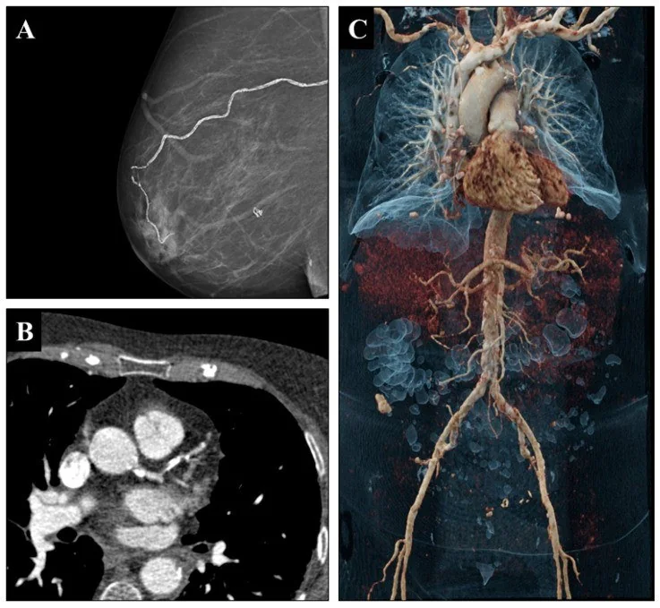
- dallanan damar yatağı boyunca çizgisel ve tübüler uzanım.
- Duktal karsinoma in situ
- BT’de mikrokalsifikasyonlar silik kalabilir; eşlik eden asimetrik doku veya kitle kuşkuludur.
Tuzaklar
- Toraks BT'de görülmeyen mikrokalsifikasyonların mamografide de mevcut olmayacağı varsayılmamalıdır; BT, primer meme taramasında mamografinin yerini tutmaz.
- Kaba kalsifikasyon çoğunlukla benign görünse de eşlik eden lobüle veya düzensiz kitleyi ayrıca değerlendir.
- Toraks BT çözünürlüğünün altında kalan mikrokalsifikasyonlar güvenilir biçimde seçilemez; görünen tüm kırıntılı odağın damar duvarı, yağ nekrozu veya involüsyonel fibroadenom gibi benign bir yapıya ait olduğu akla gelmelidir.
Kendini dene
Toraks BT’de meme içinde iri, birleşmiş kalsifikasyonlar düzgün sınırlı nodülü dolduruyor. En olası açıklama nedir?
Cevabı göster
İnvolüsyonel fibroadenom. Nodül içindeki kaba, birleşik kalsifikasyon involüsyon gösteren fibroadenom görünümüdür. Duktal kalsifikasyonlar daha ince olabilir; arter kalsiyumu tübüler seyreder, yağ nekrozu ise yağlı kavite/rim ile ilişkilidir.
Bir kalsifikasyon odağına komşu düzensiz kontrastlanan meme kitlesi de seçiliyor. BT yorumu nasıl olmalıdır?
Cevabı göster
Şüpheli kitle eşlik ediyor. Kalsifikasyon tek başına benignlik sağlamaz; düzensiz eşlikçi kitle meme görüntülemesiyle değerlendirilmelidir. Normal lobül kitle oluşturmaz, damar kalsifikasyonu ise damar boyunca uzanır.
İlgili konular
Kaynaklar
Bu sayfadaki 33 cümle ve 2 quiz sorusu aşağıdaki kaynaklardan alıntılarla tek tek karşılaştırıldı; 8 cümle bu karşılaştırmada düzeltildi (2026-10-08).
- Beyond the lung: recognizing incidental breast lesions on chest computed tomographypmc.ncbi.nlm.nih.gov
- Intramammary Findings on CT of the Chest – a Review of Normal Anatomy and Possible Findingspmc.ncbi.nlm.nih.gov
- Imaging Evaluation and Management of Breast Symptoms in the Pediatric Population.pubmed.ncbi.nlm.nih.gov
- Case Report: A rare case of serous cystadenocarcinoma of the ovary with a benign teratoma in the other ovary.pubmed.ncbi.nlm.nih.gov
- Adipose Tissue Heterogeneity: Depot-Specific Location and Functional Specialization in Obesity-Related Disease.pubmed.ncbi.nlm.nih.gov
- Artificial Intelligence-Powered Mammography: Navigating the Landscape of Deep Learning for Breast Cancer Detection.pubmed.ncbi.nlm.nih.gov
- Breast Cancer Calcifications: Identification Using a Novel Segmentation Approach.pubmed.ncbi.nlm.nih.gov
- Diverse Radiologic Presentations of Paget's Disease of the Breast: From Subtle to Striking Findings.pubmed.ncbi.nlm.nih.gov
- Extensive dystrophic calcinosis in dermatomyositis with concurrent breast carcinoma: an imaging pitfall.pubmed.ncbi.nlm.nih.gov
- Incidental Breast Findings on Chest CT: Rates of Report and Follow-Up.pubmed.ncbi.nlm.nih.gov
- Multimodality imaging features of primary mammary angiosarcoma: a case report with delayed osseous metastases.pubmed.ncbi.nlm.nih.gov
- Why we still miss breast cancers: strategies for improving mammography interpretation.pubmed.ncbi.nlm.nih.gov
- Viewpoint on the Consequences and Mitigation of Cognitive Bias in the Radiological Interpretation of Breast Cancer Imaging Using Artificial Intelligence.pubmed.ncbi.nlm.nih.gov
- Contrast-enhanced mammography in the setting of neoadjuvant therapy from a multidisciplinary perspective.pubmed.ncbi.nlm.nih.gov
- Radiation dose comparison of photon-counting breast CT and mammography: A phantom study.pubmed.ncbi.nlm.nih.gov
- Machine Learning-Based Classification of BI-RADS 4 and BI-RADS 5 Microcalcifications in Mammography Combined with DCE-MRI for Malignant-Benign Discrimination.pubmed.ncbi.nlm.nih.gov
- Beyond the lung: recognizing incidental breast lesions on chest computed tomography.pubmed.ncbi.nlm.nih.gov
- Primary breast osteosarcoma: A case report highlighting the importance of radiologic-pathologic correlation.pubmed.ncbi.nlm.nih.gov
- Breast calcifications following electrical defibrillation: An unusual mammographic appearance.pubmed.ncbi.nlm.nih.gov
- Beyond the Beam: Multimodal Imaging and Surveillance of Post-Radiotherapy Changes in the Breast.pubmed.ncbi.nlm.nih.gov
- Imaging features of lacrimal gland disease.pubmed.ncbi.nlm.nih.gov
- Submucosal laryngeal lesions: A puzzling diagnostic conundrum.pubmed.ncbi.nlm.nih.gov
- Characterization of Breast Microcalcifications Using Dual-Energy CBCT: Impact of Detector Configuration on Imaging Performance-A Simulation Study.pubmed.ncbi.nlm.nih.gov
- Male accessory breast mucinous carcinoma: a case report and literature review.pubmed.ncbi.nlm.nih.gov
- Artificial intelligence in the radiologic assessment of ductal carcinoma <i>in situ</i>: a systematic review.pubmed.ncbi.nlm.nih.gov
- From Dyspnea to Suspected Breast Cancer: A Post-traumatic Hematoma as a Diagnostic Pitfall.pubmed.ncbi.nlm.nih.gov
- Systemic Vascular Calcification After Lung Transplantation: The Role of Mammographic Breast Arterial Calcifications.pubmed.ncbi.nlm.nih.gov
- Cancer predisposition syndromes: an imaging review.pubmed.ncbi.nlm.nih.gov
- Mammographic Vascular Microcalcifications as a Surrogate Parameter for Coronary Heart Disease: Correlation to Cardiac Computer Tomography and Proposal of a Classification Score.pubmed.ncbi.nlm.nih.gov
- Combined heart and liver transplantation for homozygous familial hypercholesterolaemia: 35-year post-operative outcomes.pubmed.ncbi.nlm.nih.gov
- Understanding Photon-Counting CT: Physics, Detector Technology, and Image Reconstructions.pubmed.ncbi.nlm.nih.gov
- Chinese expert consensus on imaging examination and diagnosis of nasal cavity and paranasal sinus tumors.pubmed.ncbi.nlm.nih.gov
- LNMSNet: a multi-task deep learning network for pulmonary nodules segmentation and malignancy classification.pubmed.ncbi.nlm.nih.gov
- Diagnostic Imaging Features of Mammary Gland Tumors in Dogs and Cats.pubmed.ncbi.nlm.nih.gov
- Silent Intruders: A Dual Case Report of Retained Foreign Bodies in the Breasts.pubmed.ncbi.nlm.nih.gov
- Case Report: Primary gastric extraskeletal osteosarcoma and literature review.pubmed.ncbi.nlm.nih.gov
- Pseudoepitheliomatous Hyperplasia of the Medial Thigh with Dystrophic Calcification and Metaplastic Ossification: A Case Report.pubmed.ncbi.nlm.nih.gov
- Immediate DIEP flap breast reconstruction following neoadjuvant therapy: Histopathological assessment of vascular integrity in internal mammary and deep inferior epigastric arteries.pubmed.ncbi.nlm.nih.gov
- Incidental Coronary Artery Calcification on Simulation Breast Radiotherapy Imaging and Risk of Subsequent Ischemic Heart Disease:A Missed Opportunity?pubmed.ncbi.nlm.nih.gov
- Coronary artery calcification in thoracic cancer radiotherapy: A scoping review.pubmed.ncbi.nlm.nih.gov
- Complete disappearance of ductal carcinoma <i>in situ</i> after neoadjuvant therapy in HER2-positive breast cancer: a case report.pubmed.ncbi.nlm.nih.gov
- Congenital unilateral athelia associated with mammary Paget's disease, high-grade ductal carcinoma <i>in situ</i> and microinvasive breast cancer: a case report.pubmed.ncbi.nlm.nih.gov
- Diagnosis of Ductal Carcinoma in Situ in Breast Biopsies for a Radiological Mass Lesion: Did We Miss Something?pubmed.ncbi.nlm.nih.gov
- A Heterogeneous Phenotypic Presentation of a PALB2 Mutation Carrier With Multiple Metachronous Primary Malignancies: A Case Report.pubmed.ncbi.nlm.nih.gov
- Chronic Expanding Hematoma of the Postmastectomy Axilla: A Pictorial Review of Long-Term Multimodality Imaging.pubmed.ncbi.nlm.nih.gov
- Multimodal imaging in neuro-ophthalmology.pubmed.ncbi.nlm.nih.gov
- Photon-Counting CT: Technology, Current and Potential Future Clinical Applications, and Overview of Approved Systems and Those in Various Stages of Research and Development.pubmed.ncbi.nlm.nih.gov
- Retrospective Review Is the Best View: A Primer for Residents and Radiologists on Missed Findings in Chest Radiography.pubmed.ncbi.nlm.nih.gov
- The PRINCE-BC trial: precision regional irradiation of nodal stations for clinical evaluation of high-risk pN0 breast cancer - a phase III, open-label, randomized controlled trial.pubmed.ncbi.nlm.nih.gov
- The inframammary fold: Structure, clinical considerations, and reconstructive techniques.pubmed.ncbi.nlm.nih.gov
- Unsupervised Anomaly Detection in Medical Imaging: A Survey of Methods, Challenges, and Future Directions.pubmed.ncbi.nlm.nih.gov
- From Innovation to Application: Can Emerging Imaging Techniques Transform Breast Cancer Diagnosis?pubmed.ncbi.nlm.nih.gov
- Pericardial Diseases: International Position Statement on New Concepts and Advances in Multimodality Cardiac Imaging.pubmed.ncbi.nlm.nih.gov
- ESUR female pelvis group approach to cystic female pelvic lesions.pubmed.ncbi.nlm.nih.gov
- Pediatric endocrine disorders: a review of intra-abdominal findings and appropriate imaging.pubmed.ncbi.nlm.nih.gov
- Review of the Imaging Modalities in the Gynecological Neoplasms During Pregnancy.pubmed.ncbi.nlm.nih.gov
- Imaging Features of the Mesenchymal Tumors of the Breast according to WHO Classification: A Pictorial Essay.pubmed.ncbi.nlm.nih.gov
- Cystic-Like HER2-Positive Breast Cancer with Low Tumor-Infiltrating Lymphocytes and High Programmed Death-Ligand 1 Expression in a Young Woman: A Case Report.pubmed.ncbi.nlm.nih.gov
- Narrative review of chest wall ultrasound: a practical approach.pubmed.ncbi.nlm.nih.gov
- Breast mass imaging identification of rare pleomorphic adenoma after follow-up of gastric cancer surgery: a case report.pubmed.ncbi.nlm.nih.gov
- Incidental cystic pancreatic neuroendocrine neoplasm and differential diagnosis.pubmed.ncbi.nlm.nih.gov
- When Cancer Clots: An Extensive Radiologic Analysis of Cancer-Associated Thromboembolism.pubmed.ncbi.nlm.nih.gov
- Splenic pathologies: A pictorial review of common, wandering, twisting and rare presentations.pubmed.ncbi.nlm.nih.gov
- Photon-counting detector CT in oncology: a new era of cancer imaging.pubmed.ncbi.nlm.nih.gov
- Primary breast diffuse large B-cell lymphoma mimicking breast carcinoma: A case report and literature review.pubmed.ncbi.nlm.nih.gov
- Adenoid cystic carcinoma of the breast: report of the diagnosis and management of two cases.pubmed.ncbi.nlm.nih.gov
- From diagnostic marker-to-therapeutic modality: emerging paradigms in tumor biomineralization.pubmed.ncbi.nlm.nih.gov
- Anterior rib giant cell tumor of bone mimicking a malignant chest-wall tumor: a case report.pubmed.ncbi.nlm.nih.gov
- Super-resolution contrast-enhanced ultrasound: recent advances.pubmed.ncbi.nlm.nih.gov
- Depicting Characteristic Staghorn Vessels in Solitary Fibrous Tumor of the Liver With Contrast-Enhanced Ultrasound and Ultrasound Localized Microscopy: A Case Report.pubmed.ncbi.nlm.nih.gov
- MSF-Swin-ICFNet: An Image-Derived Multi-Scale Swin Transformer Framework with Cross-Scale Feature Fusion for Breast Lesion Classification and Segmentation in Mammograms.pubmed.ncbi.nlm.nih.gov
- Evaluation of the diagnostic value of magnetic resonance imaging combined with ultrasound and mammography for breast cancer using array spatial sensitivity encoding technique.pubmed.ncbi.nlm.nih.gov
- Radiological Tumor Calcification in Sinonasal Adenocarcinoma: Prevalence and Prognostic Implications.pubmed.ncbi.nlm.nih.gov
- Chest Computed Tomography-Based Radiomics for the Diagnosis and Prognosis of Pulmonary Hypertension.pubmed.ncbi.nlm.nih.gov
- How should we interpret excessive left ventricular trabeculation? Update on controversies from the cardiac imaging perspective.pubmed.ncbi.nlm.nih.gov
- Vascular lesions: Hemangioma or venous malformation?pubmed.ncbi.nlm.nih.gov
- Classifying diagnostic pitfalls in juxtapleural CT lesions: A root cause.pubmed.ncbi.nlm.nih.gov
- A pictorial essay of thoracic wall diseases: multiple pathologies in the same anatomical site.pubmed.ncbi.nlm.nih.gov
- A rare case of involuting fibroadenoma in a young premenopausal female.pubmed.ncbi.nlm.nih.gov
- Incidental Irregular Mass Within a Hamartoma Detected on Breast Ultrasound: A Case Report and Review of the Literature.pubmed.ncbi.nlm.nih.gov
- Extramedullary hematopoiesis presenting as a large adrenal mass mimicking adrenocortical carcinoma in a primary myelofibrosis patient: A case report.pubmed.ncbi.nlm.nih.gov
- Performance of Photon-counting CT for Assessing Pretreatment Breast Cancer: Comparison with Mammography, MRI, and <sup>18</sup>F-FDG PET/CT.pubmed.ncbi.nlm.nih.gov
- Key Points of Endoscopic Minimally Invasive Surgery for Gynecomastia Based on Refined Anatomy: A Case Series.pubmed.ncbi.nlm.nih.gov
- Exceptional responses to Trastuzumab deruxtecan in HER2-positive breast cancer: two illustrative case reports.pubmed.ncbi.nlm.nih.gov
- Medical hyperspectral imaging: an updated review of technology advancements and biomedical applications.pubmed.ncbi.nlm.nih.gov
- Case Report: Phosphaturic mesenchymal tumor of the scapula without calcification: a potential diagnostic pitfall.pubmed.ncbi.nlm.nih.gov
Eğitim amaçlıdır; hasta başında tanı ve tedavi kararının yerine geçmez. İçerik yapay zekâ destekli bir süreçle güncel literatüre göre cümle cümle kaynaklarla karşılaştırıldı, ancak uzman radyolog incelemesinden geçmedi. Klinik kararlarda güncel kılavuzları, kurum protokollerini ve radyoloji raporunu esas alın.