Özet
İntrakraniyal aterosklerotik stenoz, büyük beyin arterlerinde aterom plağının lümeni daraltmasıdır ve geçici iskemik atak ya da iskemik inmenin tedavi edilebilir nedenlerinden biridir. Kontrastsız BT akut kanamayı ve gelişmiş infarktı tarar; BTA fokal lümen daralmasını, tıkanmayı, distal akımı ve kalsifiye plağı hızlı biçimde gösterir. Stenozun yüzdesi ölçüm yöntemine bağlıdır ve CTA'da kemik/kalsifikasyon artefaktı lümeni olduğundan dar gösterebilir; damar duvarı etiyolojisini her zaman tek başına belirleyemez. Kritik darlığın veya distal perfüzyon etkisinin atlanması inme mekanizmasını ve riskli damar bölgesini yanlış sınıflandırabilir.
Faz ve pencere
- Kontrastsız
- Akut kanama ve gelişmiş infarkt için beyin parankimi değerlendirilir; arter duvarındaki kalsifikasyon kavernöz ICA veya baziler arter boyunca çizgisel/odaksal hiperdens görünür. İntrakraniyal arter stenozunu değerlendirmede CTA etkili bir görüntüleme yöntemidir.
- BTA tanısal
- Arteriyel opasifikasyonda kavernöz/supraclinoid ICA, M1 MCA veya vertebrobaziler sistemde kısa segmentli fokal lümen daralması, düzensiz kontur ve distal dolum azalması aranır; kalsifik plak lümen kenarını örtebilir. İnce kesit kaynak görüntüler, multiplanar reformasyon ve karşı/komşu normal segment referansı birlikte kullanılır.
Önerilen pencereler: Anjiyo (G 600 / M 150), Beyin (G 80 / M 40).
BT bulguları
- Fokal lümen daralması — arterin kısa segmentinde çevre normal segmente göre çap azalması; stenoz derecesi belirtilirken kullanılan yöntemi yazın.
- Düzensiz veya eksantrik kontur — aterosklerotik plakla uyumludur, ancak yalnız lümen görünümü etiyolojiyi kesinleştirmez.
- İntrakraniyal arter kalsifikasyonu — kavernöz ICA'da yoğun, duvar boyunca plak; kalın kümeli intimal kalsifikasyon ateromla ilişkili olabilir.
- Distal kontrast dolum azalması veya gecikmesi — darlık sonrası akım etkisini gösterir ve kollateral dolaşım ile birlikte yorumlanır.
- Tam tıkanma ve distal yeniden dolum — lümen sürekliliğinin kesilmesi ardından distal arterin kollaterallerle opasifiye olması görülebilir.
- İlgili arter alanında hipodens infarkt — MCA, ACA veya posterior dolaşım bölgesinde akut/subakut iskemiyle uyumlu parankim değişikliği.
- Sınır bölgesi infarktları — hemodinamik yetersizlikte arteriyel watershed dağılımda birden fazla küçük hipodens odak izlenebilir.
Ölçütler ve sınıflamalar
- WASID çap stenozu hesabı
- Yüzde stenoz = (1 − en dar lümen çapı / referans normal arter çapı) × 100
- WASID stenoz ölçümü
- Yüzde stenoz, en dar lümen çapının uygun normal referans çapına oranından hesaplanır. Önce darlığın proksimalindeki hastalıksız, en geniş, tortüöz olmayan ve paralel kenarlı segment seçilir; kullanılamıyorsa uygun distal ya da besleyici arter segmenti referans alınır.
Normalde
CTA, intrakraniyal arter stenozu ve oklüzyonunu saptamada etkili bir yöntemdir. WASID ölçümünde darlığın en yakın proksimalinde hastalık görmeyen, geniş ve düz seyirli arter segmenti referans çap olarak alınır; o segment de tutulmuşsa uygun distal veya yan dal segmenti tercih edilir. Kalsifiye duvarı lümen sanmayın ve distal dolumu ayrıca izleyin.
Normal BT ile kıyasla
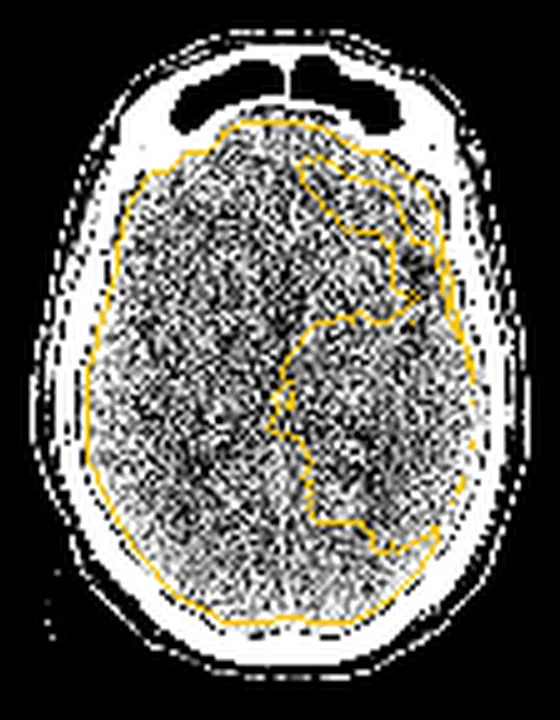
Aynı bölgenin, kaynak veri setinde patoloji içermediği belirtilen bir BT incelemesini kesit kesit kaydırın; organ sınırlarını açarak anatomiyi hasta görüntüsüyle karşılaştırın. Küçük rastlantısal bulgular tümüyle dışlanmamıştır.
Kaynak: TotalSegmentator v2.01 veri seti (Wasserthal ve ark., Radiology: Artificial Intelligence 2023), CC BY 4.0, vaka s0849; organ sınırları veri setinin otomatik segmentasyonundan, birleştirilerek sadeleştirildi. Görüntüler 2,5 mm kesit aralığına yeniden örneklendi.
Ayırıcı tanı
- İntrakraniyal arter diseksiyonu
- uzun konik daralma, lümen düzensizliği veya yalancı lümen görülebilir; damar duvarı hematomu için MRI yardımcıdır.
- Moyamoya hastalığı
- terminal ICA ve proksimal dallarda ilerleyici daralma ile bazal kollateral damar ağı eşlik eder.
- Vaskülit
- birden fazla arterde segmental daralma ve düzensizlik olabilir; yalnız CTA lümen görünümü kesin etiyoloji vermez.
- Vazospazm
- özellikle subaraknoid kanama veya akut nörolojik olay çevresinde segmental daralma; zamana bağlı gerileme klinik bağlamı destekler.
- İntrakraniyal emboli
- ani lümen kesilmesi ve distal dal dolum kusuru ön plandadır; sabit duvar plağı/kalsifikasyonu olmayabilir.
Tuzaklar
- Kalsifikasyon blooming etkisiyle damar lümenini daraltabilir; kaynak kesitleri kemik penceresi ve farklı düzlem rekonstrüksiyonlarıyla birlikte kontrol edin.
- Hareket ve kontrast bolusu zamanlaması distal dolumu azaltıp yalancı stenoz veya tıkanma görünümü yaratabilir; komşu arterlerin opasifikasyonunu kıyaslayın.
- Stenoz yüzdesi için kullanılan ölçüm yöntemini tutarlı uygulayın; farklı ölçüm şemaları farklı sonuçlar verebilir.
- CTA'daki fokal daralma aterosklerozu tek başına kanıtlamaz; diseksiyon, vaskülit, vazospazm ve Moyamoya klinik ve damar duvarı bulgularıyla ayrılır.
Kendini dene
CTA'da intrakraniyal stenoz derecesini ölçmek için DSA ile en yüksek uyumu gösteren yarı otomatik protokol hangisidir?
Cevabı göster
(1 − stenozun en küçük çapı / referansın ortalama çapı) × 100. Kaynakta DSA ile en yüksek uyum gösteren CTA protokolü, stenozun en küçük çapının referansın ortalama çapına oranı kullanılarak (1 − oran) × 100 biçiminde tanımlanmıştır.
CTA'da kavernöz sinüs yakınındaki intrakraniyal ICA için bildirilen görüntüleme sınırlılığı hangisidir?
Cevabı göster
Artefakt nedeniyle damar yapay olarak daralmış veya görünmez görünebilir. Kaynak, duyarlılık artefaktlarının kavernöz sinüs yakınında intrakraniyal ICA'yı yapay olarak daralmış veya görünmez gösterebileceğini bildirir.
Kaynaklar
Bu sayfadaki 40 cümle ve 2 quiz sorusu aşağıdaki kaynaklardan alıntılarla otomatik olarak karşılaştırıldı; 6 cümle bu karşılaştırmada düzeltildi (2026-10-09).
- Endovascular Treatment Strategies for Acute Intracranial Atherosclerotic Stenosis–Related Large Vessel Occlusionpmc.ncbi.nlm.nih.gov
- Endovascular Therapy for Intracranial Atherosclerotic Stenosis: Evidence and Controversiespmc.ncbi.nlm.nih.gov
- Integrating hemodynamic analysis with traditional imaging in intracranial atherosclerotic stenosis: current status and future perspectivespmc.ncbi.nlm.nih.gov
- Endovascular therapy vs. conventional medical treatment for symptomatic intracranial atherosclerotic stenosis: an updated meta-analysis of randomized controlled trialspmc.ncbi.nlm.nih.gov
- Elucidating the benefit of drug-eluting stent for symptomatic intracranial atherosclerotic stenosis: Meta-analysis of randomized controlled trialspmc.ncbi.nlm.nih.gov
- The Latest Information on Intracranial Atherosclerosis: Diagnosis and Treatment.pubmed.ncbi.nlm.nih.gov
- Treatment and imaging of intracranial atherosclerotic stenosis: current perspectives and future directions.pubmed.ncbi.nlm.nih.gov
- Optical Coherence Tomography in Cerebrovascular Disease: Open up New Horizons.pubmed.ncbi.nlm.nih.gov
- Stent-Retriever Thrombectomy and Rescue Treatment of M1 Occlusions Due to Underlying Intracranial Atherosclerotic Stenosis: Cohort Analysis and Review of the Literature.pubmed.ncbi.nlm.nih.gov
- Long-Term Evolutionary Change in the Lumen of Intracranial Atherosclerotic Stenosis Following Angioplasty and Stenting.pubmed.ncbi.nlm.nih.gov
- Clinical implications of haemodynamics in symptomatic intracranial atherosclerotic stenosis by computational fluid dynamics modelling: a systematic review.pubmed.ncbi.nlm.nih.gov
- Integrating hemodynamic analysis with traditional imaging in intracranial atherosclerotic stenosis: current status and future perspectives.pubmed.ncbi.nlm.nih.gov
- A systematic review of application of frequency-domain optical coherence tomography in cerebral large artery atherosclerosis.pubmed.ncbi.nlm.nih.gov
- Intracranial atherosclerotic stenosis: risk factors, diagnosis, and treatment.pubmed.ncbi.nlm.nih.gov
- Intracranial atherosclerotic disease mechanistic subtypes drive hypoperfusion patterns.pubmed.ncbi.nlm.nih.gov
- Residual Inflammatory Risk and Intracranial Atherosclerosis Plaque Vulnerability: Insights From High-Resolution Magnetic Resonance Imaging.pubmed.ncbi.nlm.nih.gov
- Risk Factors for Acute Ischemic Stroke Caused by Anterior Large Vessel Occlusion.pubmed.ncbi.nlm.nih.gov
- Neovascularization in Human Intracranial Atherosclerotic In-Stent Restenosis.pubmed.ncbi.nlm.nih.gov
- Complication of Stenting in Intracranial Arterial Stenosis.pubmed.ncbi.nlm.nih.gov
- A systematic review of intracranial atherosclerosis epidemiology-the largest contributor to the global ischemic stroke burden?pubmed.ncbi.nlm.nih.gov
- Intracranial Arterial Calcification and Its Impact on Subsequent Ischemic Cerebral Events: A Retrospective Cohort Study.pubmed.ncbi.nlm.nih.gov
- Dual versus mono antiplatelet therapy within 72 hours after onset for mild ischaemic stroke or transient ischaemic attack: meta-analysis of randomised controlled trials.pubmed.ncbi.nlm.nih.gov
- Congenital internal carotid artery hypoplasia in a symptomatic young adult: A case report.pubmed.ncbi.nlm.nih.gov
- Five-Year Delayed Rupture of a Suspected Infectious Intracranial Aneurysm: A Case Report.pubmed.ncbi.nlm.nih.gov
- A coexisting pseudoaneurysm hidden behind a saccular distal middle cerebral artery aneurysm in long-standing postirradiation vasculopathy: illustrative case.pubmed.ncbi.nlm.nih.gov
- Multimodal imaging in neuro-ophthalmology.pubmed.ncbi.nlm.nih.gov
- Clinical implications of vessel wall imaging-State-of-the-art review.pubmed.ncbi.nlm.nih.gov
- Artificial Intelligence for Computer-Aided Detection in Endovascular Interventions: Clinical Applications, Validation, and Translational Perspectives.pubmed.ncbi.nlm.nih.gov
- Computed Tomography Angiography Recognition of a Hemorrhagic Moyamoya Pattern May Alter Acute Blood Pressure Management.pubmed.ncbi.nlm.nih.gov
- Adult-onset moyamoya disease presenting with recurrent ischemic stroke: The role of CT Perfusion and digital subtraction angiography in bypass planning.pubmed.ncbi.nlm.nih.gov
- Clinical challenges and individualized management of hypertensive emergency complicated by acute ischemic stroke: A case report and literature review.pubmed.ncbi.nlm.nih.gov
- Impact of Intracranial Arterial Calcification on Outcomes of Mechanical Thrombectomy in Acute Ischemic Stroke: A Systematic Review and Meta-Analysis.pubmed.ncbi.nlm.nih.gov
- A Case of Bilateral Globus Pallidus Infarction Onset With Mental Disorder.pubmed.ncbi.nlm.nih.gov
- Effects of Computed Tomography Hyperdense Middle Cerebral Artery Sign on the Efficacy of Endovascular Therapy in Acute Ischemic Stroke With Large Infarcts: A Subgroup Analysis of the ANGEL-ASPECT Trial.pubmed.ncbi.nlm.nih.gov
- Machine learning in neuroradiology: Recent developments and applications.pubmed.ncbi.nlm.nih.gov
- Tissue clock-guided prediction and intervention of futile recanalization: towards precision therapy in mechanical thrombectomy.pubmed.ncbi.nlm.nih.gov
- Perioperative Point-of-Care Ultrasound Utilization in Abdominal Organ Transplantation. Part II: Postoperative Care.pubmed.ncbi.nlm.nih.gov
- Retrieval of an atherosclerotic plaque-vessel wall complex during mechanical thrombectomy: illustrative case.pubmed.ncbi.nlm.nih.gov
- Hybrid revascularization for acute common carotid artery occlusion after transcarotid transcatheter aortic valve implantation: illustrative case.pubmed.ncbi.nlm.nih.gov
- Aortic coarctation revealed by subarachnoid hemorrhage in a 17-year-old patient: A rare association with multiple basilar trunk aneurysms.pubmed.ncbi.nlm.nih.gov
- Multimodal imaging in conductive hearing loss: Optimising CT, MRI and CTA for accurate diagnosis and management.pubmed.ncbi.nlm.nih.gov
- Imaging of cerebrovascular complications from blunt skull base trauma.pubmed.ncbi.nlm.nih.gov
- Carotid plaque vulnerability and circle of Willis anatomy predict ipsilateral brain infarcts and long-term mortality in carotid endarterectomy patients.pubmed.ncbi.nlm.nih.gov
- Short-term outcomes of intracranial angioplasty with the Neuroform EZ stent in patients with hypoperfusion-related symptomatic intracranial atherosclerotic stenosis.pubmed.ncbi.nlm.nih.gov
- Prevalence, diagnosis and management of intracranial atherosclerosis in White populations: a narrative review.pubmed.ncbi.nlm.nih.gov
- Vertebrobasilar artery geometry and quantitative high-resolution vessel wall MRI for stratifying vulnerable plaques in young and middle-aged adults with stroke risk factors.pubmed.ncbi.nlm.nih.gov
- Panvascular Changes Under Intensive Medical Therapy in Intracranial Atherosclerotic Steno-Occlusive Disease.pubmed.ncbi.nlm.nih.gov
- Papilledema is not the point: intracranial pressure dysregulation as a common pathway in migraine and idiopathic intracranial hypertension.pubmed.ncbi.nlm.nih.gov
- Ischemic stroke as an initial manifestation of Loeys-Dietz syndrome type 3 caused by the recurrent p.Arg287Trp variant in <i>SMAD3</i>: a case report with familial context.pubmed.ncbi.nlm.nih.gov
- Severe stenosis of major intracranial arteries: an important risk factor for infarction complications after combined revascularization in adult patients with ischemic moyamoya disease.pubmed.ncbi.nlm.nih.gov
- Direct balloon dilatation angioplasty for intracranial atherosclerotic stenosis-related acute ischemic stroke: a single-arm retrospective study of technical feasibility and safety.pubmed.ncbi.nlm.nih.gov
- Structural Restoration of an Unruptured Vertebral Artery Dissection Following Autologous Mesenchymal Stem Cell Therapy: A Case Report.pubmed.ncbi.nlm.nih.gov
- Off-pump coronary artery bypass grafting in a patient with moyamoya syndrome: A case report and literature review.pubmed.ncbi.nlm.nih.gov
- Chinese expert consensus on the treatment and diagnosis of moyamoya disease and moyamoya syndrome (2024).pubmed.ncbi.nlm.nih.gov
- Moyamoya disease masquerading as CNS vasculitis: Diagnostic challenges and imaging insights.pubmed.ncbi.nlm.nih.gov
- Takayasu Arteritis in a 15-Year-Old Male Presenting with Constitutional Symptoms and Upper Limb Pulse Deficit: A Case Report a Resource-Limited Settings.pubmed.ncbi.nlm.nih.gov
- Case Report: Ischemic stroke revealing carotid fibromuscular dysplasia with multiple metabolic disturbances in a 42-year-old man.pubmed.ncbi.nlm.nih.gov
- Aneurysmal subarachnoid hemorrhage: predictive score for angiographic vasospasm.pubmed.ncbi.nlm.nih.gov
- Cerebral Vasospasm After Traumatic Subarachnoid Hemorrhage: A Narrative Review.pubmed.ncbi.nlm.nih.gov
- Rare circle of Willis variants associated with multiple intracranial aneurysms and subarachnoid hemorrhage: A case report.pubmed.ncbi.nlm.nih.gov
- Mechanical Thrombectomy for Acute Vertebrobasilar Occlusion Associated with Mycoplasma pneumoniae Pneumonia in a Child: A Case Report.pubmed.ncbi.nlm.nih.gov
- Case Report: Intra-procedural fatal cerebral lipiodol embolism without angiographically visible shunting during right inferior phrenic artery TACE.pubmed.ncbi.nlm.nih.gov
- Ophthalmologic assessment and intracranial pressure in children: diagnostic methods, clinical correlations, and future directions.pubmed.ncbi.nlm.nih.gov
- Photon-Counting CT: Technology, Current and Potential Future Clinical Applications, and Overview of Approved Systems and Those in Various Stages of Research and Development.pubmed.ncbi.nlm.nih.gov
- Two Cases of Innominate Artery Stenting via Alternative Upper-Extremity Arterial Access Using Tailored Distal Protection Strategies.pubmed.ncbi.nlm.nih.gov
- Balloon-mounting stent for intracranial arterial stenosis: A comprehensive and comparative systematic review and meta-analysis.pubmed.ncbi.nlm.nih.gov
- 2021 Taiwan Stroke Society Guidelines of blood pressure control for ischemic stroke prevention.pubmed.ncbi.nlm.nih.gov
- Integrated head and neck imaging of symptomatic patients with stroke using simultaneous non-contrast cardiovascular magnetic resonance angiography and intraplaque hemorrhage imaging as compared with digital subtraction angiography.pubmed.ncbi.nlm.nih.gov
- The WEVO Technique: A Novel Dual-Device Endovascular Strategy for Non-Geometric Wide Neck Bifurcation Aneurysms.pubmed.ncbi.nlm.nih.gov
- A surgical technique for lesions involving the ultra-high-riding carotid sheath based on the anatomical principles of the prevertebral fascia: cadaveric dissection and case illustrations.pubmed.ncbi.nlm.nih.gov
- Human cerebral arterial morphogenesis explored by 3D microscopic imaging of cleared embryos: Bridging the gap between embryonic and adult anatomy.pubmed.ncbi.nlm.nih.gov
- Exploratory analysis of morphology-hemodynamics-perfusion relationships after intracranial stenting in severe intracranial atherosclerotic stenosis.pubmed.ncbi.nlm.nih.gov
- Stenotic Lesions of the Intracranial Arteries in Relation to the Average Level and Variability of the Home Blood Pressure: A Cross-Sectional Study.pubmed.ncbi.nlm.nih.gov
- Interpretable machine learning for predicting early neurological deterioration in symptomatic intracranial atherosclerotic stenosis.pubmed.ncbi.nlm.nih.gov
- Association between basilar artery stenosis features, vertebral artery stenosis and perforator stroke after stenting.pubmed.ncbi.nlm.nih.gov
- Exchange-free single-pass gateway balloon-assisted neuroform atlas stenting for symptomatic high-grade intracranial atherosclerotic stenosis: clinical and angiographic outcomes.pubmed.ncbi.nlm.nih.gov
- A novel cerebrovascular drug-coated balloon catheter for treating symptomatic intracranial atherosclerotic stenosis lesions: Study protocol for a prospective, multicenter, single-arm, target-value clinical trial.pubmed.ncbi.nlm.nih.gov
- A retrospective study of individualized endovascular treatment for symptomatic intracranial atherosclerotic stenosis in patients with ischemic stroke/transient ischemic attack.pubmed.ncbi.nlm.nih.gov
- Effect of a Drug-Eluting Stent vs. Bare Metal Stent for the Treatment of Symptomatic Intracranial and Vertebral Artery Stenosis.pubmed.ncbi.nlm.nih.gov
- Magnetic Resonance Imaging and Clinical Factors Associated With Ischemic Stroke in Patients Suspected of Cervical Artery Dissection.pubmed.ncbi.nlm.nih.gov
- Inflammatory-hemodynamic interplay in severe intracranial stenosis: hs-CRP, vertebral artery flow, and posterior circulation infarction.pubmed.ncbi.nlm.nih.gov
- Non-invasive quantification of pressure drops in stenotic intracranial vessels: using deep learning-enhanced 4D flow MRI to characterize the regional haemodynamics of the pulsing brain.pubmed.ncbi.nlm.nih.gov
- Semi-automatic computed tomography angiography quantification assessment is an alternative method to digital subtraction angiography in intracranial stenosis: a multicenter study.pubmed.ncbi.nlm.nih.gov
- Sustained LDL-C target attainment and recurrent ischemic cerebrovascular events after ischemic stroke: effect modification by large-artery disease.pubmed.ncbi.nlm.nih.gov
- Safety assessment of symptomatic intracranial atherosclerotic stenosis: a comparison between sole balloon angioplasty and medical treatment.pubmed.ncbi.nlm.nih.gov
- Surgical treatment for cognitive impairment caused by internal jugular vein stenosis: a clinical study of atlas transverse process resection.pubmed.ncbi.nlm.nih.gov
- CT and MRI imaging of mass in the carotid space: a pictorial review.pubmed.ncbi.nlm.nih.gov
- Multifaceted Imaging of Renal Lesions With an Emphasis on Cross-Sectional Imaging.pubmed.ncbi.nlm.nih.gov
- Sphenoid sinus anatomical variants and pathologies: pictorial essay.pubmed.ncbi.nlm.nih.gov
- Case Report: Renal artery aneurysm rupture, axillary artery pseudoaneurysm, and catastrophic intraperitoneal hemorrhage caused by fibromuscular dysplasia with the involvement of multiple arterial beds.pubmed.ncbi.nlm.nih.gov
- Advances in cerebrovascular reserve function assessment for moyamoya disease.pubmed.ncbi.nlm.nih.gov
- Rare intradural lumbar disc herniation at L1/2: a case report.pubmed.ncbi.nlm.nih.gov
- Multimodality Imaging Approach in Diagnosis and Follow-Up of Aortic Coarctation in Adulthood.pubmed.ncbi.nlm.nih.gov
- State-of-the-art CT and MR imaging and assessment of atherosclerotic carotid artery disease: the reporting-a consensus document by the European Society of Cardiovascular Radiology (ESCR).pubmed.ncbi.nlm.nih.gov
- Computed Tomographic Evaluation of Congenital Left Ventricular Outflow Obstruction.pubmed.ncbi.nlm.nih.gov
- Machine learning-based prediction of cerebral artery territorial infarcts in acute ischemic stroke using non-contrast CT.pubmed.ncbi.nlm.nih.gov
- Territorial CT Perfusion Burden and Outcome After Aneurysmal Subarachnoid Haemorrhage: A Retrospective Single-Centre Cohort Study.pubmed.ncbi.nlm.nih.gov
- Moyamoya Vasculopathy and Atypical Moyamoya-like Patterns: Insights into Diagnosis and Therapeutic Implications.pubmed.ncbi.nlm.nih.gov
- Final Results of Cilostazol-Aspirin Therapy against Recurrent Stroke with Intracranial Artery Stenosis (CATHARSIS).pubmed.ncbi.nlm.nih.gov
- Quantification and assessment of extracranial and intracranial occlusive disease.pubmed.ncbi.nlm.nih.gov
- Getting to the heart of Carotid and Vertebral imaging in acute ischemic stroke: an all-encompassing cross-sectional comparative analysis of Colour Doppler Ultrasound, CT Angiography, and MR Angiography.pubmed.ncbi.nlm.nih.gov
- Extra-Carotid Arterial Webs: A Single-Center Series and Systematic Review.pubmed.ncbi.nlm.nih.gov
- Operative Procedures for Ultrasound Assessment of Extracranial Artery Disease: A Narrative Review by the Italian Society for Vascular Investigation (SIDV).pubmed.ncbi.nlm.nih.gov
- Endovascular Therapy for Intracranial Atherosclerotic Stenosis: Evidence and Controversies.pubmed.ncbi.nlm.nih.gov
- Spectral computed tomography characterization of coronary atherosclerotic plaque: principles, imaging biomarkers, and clinical significance.pubmed.ncbi.nlm.nih.gov
- Computed Tomography Coronary Angiography as a Gatekeeper for Invasive Coronary Assessment Before Transcatheter Aortic Valve Implantation.pubmed.ncbi.nlm.nih.gov
- Mechanisms of aortic dissection: From pathological changes to experimental and <i>in silico</i> models.pubmed.ncbi.nlm.nih.gov
- 2022 ACC/AHA guideline for the diagnosis and management of aortic disease: A report of the American Heart Association/American College of Cardiology Joint Committee on Clinical Practice Guidelines.pubmed.ncbi.nlm.nih.gov
- 2022 ACC/AHA Guideline for the Diagnosis and Management of Aortic Disease: A Report of the American Heart Association/American College of Cardiology Joint Committee on Clinical Practice Guidelines.pubmed.ncbi.nlm.nih.gov
- Sequential Helical-Axial-Helical Triple-Rule-Out CT Angiography: Technical Feasibility and Territory-Specific Image Quality in the Emergency Department.pubmed.ncbi.nlm.nih.gov
- Ferumoxytol-enhanced cardiovascular magnetic resonance imaging: applications and technical advances.pubmed.ncbi.nlm.nih.gov
- Clinical Imaging and Innovations in Aortic Aneurysm and Dissection.pubmed.ncbi.nlm.nih.gov
- Prevalence, prognosis, and treatment of atherosclerotic intracranial stenosis in Caucasians.pubmed.ncbi.nlm.nih.gov
- Endovascular Treatment Strategies for Acute Intracranial Atherosclerotic Stenosis-Related Large Vessel Occlusion.pubmed.ncbi.nlm.nih.gov
- Mechanical stress from the hyoid bone as a potential etiology of bony-related ischemic stroke secondary to carotid artery dissection: A case report and systematic review.pubmed.ncbi.nlm.nih.gov
Eğitim amaçlıdır; hasta başında tanı ve tedavi kararının yerine geçmez, tıbbi tavsiye değildir. Metinler yapay zekâ desteğiyle yazıldı ve otomatik bir adımda kaynak alıntılarıyla karşılaştırıldı; bu bir tıbbi doğrulama değildir ve içerik uzman radyolog incelemesinden geçmedi. Klinik kararlarda güncel kılavuzları, kurum protokollerini ve radyoloji raporunu esas alın.