Özet
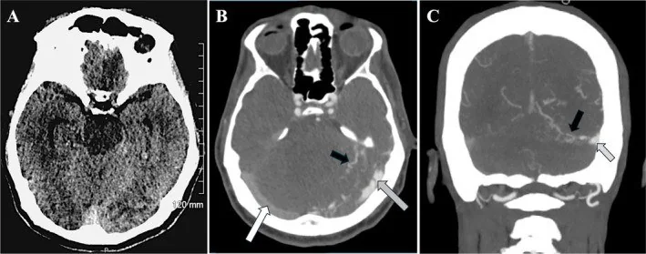
Kortikal venöz reflülü dural AV fistülde arteriyel kan sinüs veya dura içindeki fistül noktasından kortikal venlere ters ya da doğrudan akar ve venöz hipertansiyon oluşturur. Kontrastsız BT venöz konjesyonu, hemorajik infarktı ve olası kanamayı; BTA ise genişlemiş kortikal venleri ve sinüs çevresindeki anormal damarları gösterebilir. Reflünün varlığı ve yönü fistülün riskini belirlediğinden, statik BT bulguları şüpheyi yükseltir fakat akım sınıflamasının yerini tutmaz. Bu paterni benign sinüs drenajı sanmak venöz rüptür veya ilerleyen parankim hasarını gözden kaçırabilir.
Faz ve pencere
- Kontrastsız
- Venöz drenaj alanında vazojenik ödem, hemorajik infarkt ve çeşitli dağılımlarda (parankimal, subaraknoid, subdural veya ventriküler) kanama izlenebilir. Kanama parankimal, sulkal subaraknoid, subdural veya ventriküler dağılımda olabilir.
- BTA tanısal
- Dural sinüs çevresindeki besleyici meningeal damarlarla aynı incelemelerde belirgin genişlemiş serpijinöz kortikal venler, venöz ektazi/variks ve sinüs çıkışında daralma ya da oklüzyon araştırılır. Tek zamanlı BTA venöz geri akımın yönünü doğrudan ispatlamaz; dinamik anjiyografi veya kateter DSA gerekir.
Önerilen pencereler: Beyin (G 80 / M 40), Anjiyo (G 600 / M 150), Kemik (G 1800 / M 400).
BT bulguları
- Kortikal venöz reflü paterni — normal antegrad drenajdan farklı olarak arteriyel şanttan kortikal venlere yönelen anormal dolum; statik BTA'da şüpheli geniş venlerle tanınır, yön dinamik olarak doğrulanır.
- Genişlemiş kortikal venler — korteks boyunca uzanan, olağandan kalın ve kıvrımlı yüzey damarları.
- Venöz ektazi — reflü alanında fokal sakküler veya fusiform genişlemiş drenaj veni.
- Sinüs dışına uzanan drenaj — şant damarlarının dural sinüsten kortikal yüzey venlerine veya doğrudan kortikal vene devam etmesi.
- Venöz çıkış darlığı/tıkanması — fistülün boşaldığı sinüs segmentinde daralma veya kontrast dolum kusuru ve kollateral genişlemesi.
- Venöz konjesyon ödemi — venöz hipertansiyona bağlı olarak venöz drenaj sahasında gelişen vazojenik ödem.
- Hemorajik venöz infarkt — venöz drenaj alanına yakın bölgede gelişen parankimal kanama ve ödem.
- Kronik parankim etkisi — uzun süreli venöz hipertansiyonda progresif nörolojik defisit, bilişsel bozulma ve paradoksal ödem alanları görülebilir.
Ölçütler ve sınıflamalar
- Borden sınıflaması
- Kortikal venöz drenaj bulunan dural fistüller sinüse boşalıp kortikal vene geri akım varsa tip II, doğrudan kortikal vene boşalıyorsa tip III'tür; kortikal venöz drenaj olmayan tip I bu konuya girmez.
- Cognard sınıflaması
- Sinüs drenajıyla kortikal ven reflüsü tip IIb veya sinüs reflüsü de varsa IIa+b; doğrudan kortikal venöz drenaj ektazisiz tip III, ektazili tip IV; perimedüller uzanım tip V olarak adlandırılır.
Normalde
Normal kortikal venler ince kalibrasyonlu olup kanı yüzeyden dural sinüslere doğru toplar; korteks boyunca yaygın serpijinöz genişleme, venöz ektazi veya drenaj alanına uyan konjesyon görülmez. Aynı lobun karşı hemisfer yüzey venlerini ve ilgili transvers/sagittal sinüs çıkışını karşılaştırın; büyüklük asimetrisi tek başına reflü yönünü kanıtlamaz.
Normal BT ile kıyasla
Aynı bölgenin, kaynak veri setinde patoloji içermediği belirtilen bir BT incelemesini kesit kesit kaydırın; organ sınırlarını açarak anatomiyi hasta görüntüsüyle karşılaştırın. Küçük rastlantısal bulgular tümüyle dışlanmamıştır.
Kaynak: TotalSegmentator v2.01 veri seti (Wasserthal ve ark., Radiology: Artificial Intelligence 2023), CC BY 4.0, vaka s0103; organ sınırları veri setinin otomatik segmentasyonundan, birleştirilerek sadeleştirildi. Görüntüler 2,5 mm kesit aralığına yeniden örneklendi.
Ayırıcı tanı
- Kortikal/venöz tromboz
- dural sinüs trombozu, DAVF gelişiminin önemli bir tetikleyicisidir; akut tromboz venöz basıncı artırarak agresif fistül fenotipine yol açabilir.
- Venöz sinüs trombozu
- sinüs içinde kontrast dolum kusuru ve kollateral venlerin belirginleşmesi ile seyreder; statik görüntülemede venöz yapıların arteriyalize olup olmadığının kesin ayrımı zordur.
- Serebral AVM
- kortikal parankim içinde nidus ve pial besleyici arterler vardır; dural fistülde nidus yoktur ve şant sinüs/dura ile bağlantılıdır.
- Gelişimsel venöz anomali (DVA)
- normal venöz drenajı sağlayan ışınsal medüller venlerdir; DVA'nın kendisi venöz hipertansiyon veya kanama yapmaz, ancak altta yatan DAVF nedeniyle fonksiyonel obstrüksiyona uğrayabilir.
- Arteriyel oklüzyona bağlı kollateraller
- kronik arteriyel tıkanıklıklarda pial yüzey damarları artabilir; ancak DAVF'deki gibi dural sinüs çevresinde şant, arteriyalize venöz yapılar ve erken venöz dolum birlikte bulunur.
Tuzaklar
- Statik tek faz görüntüleme sırasında genişlemiş venlerin varlığından venöz drenajın yönünü kesin olarak çıkarmayın; zaman çözümlü dinamik görüntüleme gereklidir.
- Venöz infarkt bulgularını sadece klasik arteriyel vasküler alanlara göre değerlendirmeyin; fistül noktasına yakınlık ve venöz drenaj ilişkisi önemlidir.
- Venöz variks ile sakküler anevrizmayı karıştırmayın; variks genellikle fistül drenajıyla bağlantılıdır.
- Negatif rutin BTA, küçük dural fistülü veya reflüyü dışlamaz; klinik/görüntü şüphesi sürerse DSA ile venöz faz dahil drenaj haritası gerekir.
Kendini dene
Dural sinüs komşuluğunda fistül şüphesi ve aynı hemisferde ödemli geniş kortikal venler var. En önemli akım sorusu nedir?
Cevabı göster
Kortikal reflü var mı?. Kortikal venöz reflü, venöz hipertansiyon ve kanama riskini artıran drenaj özelliğidir. Dural fistülde parankimal nidus çapı temel tanımlayıcı değildir; anevrizma boynu veya arter oklüzyonu bu bulguların ana sorusu olmaz.
Sinüse boşalan fistülden kortikal vene retrograd drenaj gösterilmiş. Cognard tipi hangisidir?
Cevabı göster
Tip IIb. Sinüs drenajıyla kortikal ven reflüsü tip IIb'dir; venöz ektazi olmadan doğrudan kortikal drenaj tip III, ektazili doğrudan drenaj tip IV'tür. Tip I antegrad sinüs drenajını, tip V perimedüller drenajı ifade eder.
Kaynaklar
Bu sayfadaki 45 cümle ve 2 quiz sorusu aşağıdaki kaynaklardan alıntılarla tek tek karşılaştırıldı; 11 cümle bu karşılaştırmada düzeltildi (2026-10-08).
- Updates in the management of cranial dural arteriovenous fistulapmc.ncbi.nlm.nih.gov
- Intracranial Hemorrhage from Dural Arteriovenous Fistulas: What Can We Find with CT Angiography?pmc.ncbi.nlm.nih.gov
- [Dural Arteriovenous Fistula].pubmed.ncbi.nlm.nih.gov
- Diagnosis and management of intracranial dural arteriovenous fistulas.pubmed.ncbi.nlm.nih.gov
- Iatrogenic dural arteriovenous fistula after radical neck dissection for metastatic malignant disease: A case report.pubmed.ncbi.nlm.nih.gov
- Diagnostic accuracy of CTA and MRI/MRA in the evaluation of the cortical venous reflux in the intracranial dural arteriovenous fistula DAVF.pubmed.ncbi.nlm.nih.gov
- CT and MR imaging of non-cavernous cranial dural arteriovenous fistulas: Findings associated with cortical venous reflux.pubmed.ncbi.nlm.nih.gov
- A case of dural arteriovenous fistula draining to the diploic vein presenting with intracerebral hemorrhage.pubmed.ncbi.nlm.nih.gov
- Transarterial Embolization of Dural Arteriovenous Fistula in Superior Sagittal Sinus Under Bilateral External Carotid Artery Flow Control: Technical Note.pubmed.ncbi.nlm.nih.gov
- Spontaneous middle meningeal arteriovenous fistula without cortical venous reflux presenting with acute subdural hematoma: illustrative case.pubmed.ncbi.nlm.nih.gov
- Bilateral Cavernous Sinus Dural Arteriovenous Fistula Presenting With Subarachnoid Hemorrhage.pubmed.ncbi.nlm.nih.gov
- Clinical characteristics, imaging, treatment, and prognosis of dural arteriovenous fistula presented with Parkinsonism- a systematic review.pubmed.ncbi.nlm.nih.gov
- Radiographic Evaluation and Clinical Implications of Venous Connections Between Dural Arteriovenous Fistula of the Cavernous Sinus and Cerebellum and the Pontomedullary Venous System.pubmed.ncbi.nlm.nih.gov
- A Case of Spontaneous Acute Subdural Hemorrhage Caused by a Dural Arteriovenous Fistula on the Convexity without Cortical Venous Reflux.pubmed.ncbi.nlm.nih.gov
- Single-Session Curative Transarterial Onyx Embolization of a Convexity Dural AVF with a Concomitant Pial AVF Sharing the Vein of Labbé: A Case Report and Literature Review.pubmed.ncbi.nlm.nih.gov
- Stroke-Mimic: An Intracranial Dural Arteriovenous Fistula Revealed by New-Onset Seizures and Post-Ictal Deficit.pubmed.ncbi.nlm.nih.gov
- Flow control-assisted transarterial embolization of a high-flow superior sagittal sinus dural arteriovenous fistula: An illustrative case.pubmed.ncbi.nlm.nih.gov
- Spontaneous Superior Ophthalmic Vein Thrombosis Resulted in Posterior Fossa Venous Congestion in a Case of Cavernous Sinus Dural Arteriovenous Fistula: Successful Treatment with Direct Interruption Surgery.pubmed.ncbi.nlm.nih.gov
- Bilateral Post-Traumatic Carotid-Cavernous Fistula: A Case Report.pubmed.ncbi.nlm.nih.gov
- Post-traumatic Carotid-Cavernous Fistula Following Penetrating Orbital Injury: A Case Report and Literature Review.pubmed.ncbi.nlm.nih.gov
- Spinal dural arteriovenous fistula presenting with lower extremity weakness and intracranial hemorrhage: A case report.pubmed.ncbi.nlm.nih.gov
- PHACE syndrome: a systematic literature review and illustrative case report of a patient with severe cerebrovascular and neurodevelopmental sequelae.pubmed.ncbi.nlm.nih.gov
- Anatomy of the Middle Meningeal Artery: Implications for Middle Meningeal Artery Embolization in Chronic Subdural Hematoma.pubmed.ncbi.nlm.nih.gov
- International Society of Paediatric Surgical Oncology (IPSO) Surgical Practice Guidelines.pubmed.ncbi.nlm.nih.gov
- Cerebral venous disorders: clinical presentation, diagnostic strategy, and contemporary management.pubmed.ncbi.nlm.nih.gov
- Spontaneous closure of a superior sagittal sinus dural arteriovenous fistula with an extensive angioarchitectural network: A case report and systematic review of the literature.pubmed.ncbi.nlm.nih.gov
- Watch brain circulation in unexplained progressive myelopathy: a review of Cognard type V arterio-venous fistulas.pubmed.ncbi.nlm.nih.gov
- Central nervous system vascular malformations: A clinical review.pubmed.ncbi.nlm.nih.gov
- Intracranial non-galenic pial arteriovenous fistula: A review of the literature.pubmed.ncbi.nlm.nih.gov
- Pediatric aneurysms and vein of Galen malformations.pubmed.ncbi.nlm.nih.gov
- Intracranial Hemorrhage from Dural Arteriovenous Fistulas: What Can We Find with CT Angiography?pubmed.ncbi.nlm.nih.gov
- Intracranial dural arteriovenous fistulas: classification, imaging findings, and treatment.pubmed.ncbi.nlm.nih.gov
- Endovascular Management of Falcine Dural Arteriovenous Fistula-A Case Report and Review of Literature.pubmed.ncbi.nlm.nih.gov
- Grouting technique-combining transvenous Onyx and coils for the embolization of complex non-cavernous intracranial dural arteriovenous fistulas.pubmed.ncbi.nlm.nih.gov
- A Case of Direct Carotid-Cavernous Fistulae Successfully Treated by Bidirectional Double Catheter Technique: A Technical Note.pubmed.ncbi.nlm.nih.gov
- Endovascular Embolization in Neurovascular Disease: Material Science, Multimodal Management, and Future Horizons.pubmed.ncbi.nlm.nih.gov
- Evolving Management Paradigms in Dural Arteriovenous Fistulas: From Classification to Personalized Endovascular Therapy.pubmed.ncbi.nlm.nih.gov
- Endovascular embolization of dural arteriovenous fistulas: an 8-year single-centre experience and comparison with contemporary literature.pubmed.ncbi.nlm.nih.gov
- Congenital Intracranial Vascular Malformations in Children : Radiological Overview.pubmed.ncbi.nlm.nih.gov
- The 'corkscrew' sign: an indirect MRI hint for intracranial venous hypertension.pubmed.ncbi.nlm.nih.gov
- Pial arteriovenous fistula with a large intraparenchymal hemorrhage in a 9-year-old child: a case report and case-based mini review.pubmed.ncbi.nlm.nih.gov
- Preoperative embolization and craniotomy for the treatment of a tentorial dural arteriovenous fistula.pubmed.ncbi.nlm.nih.gov
- A case of spontaneous direct vertebral artery - External vertebral venous plexus fistula in the upper cervical portion.pubmed.ncbi.nlm.nih.gov
- Thoracolumbar spinal dural arteriovenous fistulae present with longer arteriovenous transit compared to cranial and cervical dural fistulae.pubmed.ncbi.nlm.nih.gov
- Rare sphenoid dural arteriovenous fistula treated with microsurgery: illustrative case.pubmed.ncbi.nlm.nih.gov
- Delayed Intracerebral Hemorrhage Following Transarterial Embolization for a Tentorial Dural Arteriovenous Fistula: Postoperative Venous Outflow Impairment Detected by Susceptibility-Weighted Imaging.pubmed.ncbi.nlm.nih.gov
- Morphological variations of the dural venous sinuses: a cadaveric study.pubmed.ncbi.nlm.nih.gov
- The endovascular treatment strategies of cerebrovascular injuries in traumatic brain injury.pubmed.ncbi.nlm.nih.gov
- Cerebral proliferative angiopathy in pediatric patients: case-based review with an illustrative case.pubmed.ncbi.nlm.nih.gov
- Pathophysiology, diagnosis and management of cerebral venous thrombosis: A comprehensive review.pubmed.ncbi.nlm.nih.gov
- The Venular Side of Cerebral Amyloid Angiopathy: Proof of Concept of a Neglected Issue.pubmed.ncbi.nlm.nih.gov
- Cerebral Superficial Siderosis : Etiology, Neuroradiological Features and Clinical Findings.pubmed.ncbi.nlm.nih.gov
- Systemic and CNS manifestations of inherited cerebrovascular malformations.pubmed.ncbi.nlm.nih.gov
- Focal venous hypertension as a pathophysiologic mechanism for tissue hypertrophy, port-wine stains, the Sturge-Weber syndrome, and related disorders: proof of concept with novel hypothesis for underlying etiological cause (an American Ophthalmological Society thesis).pubmed.ncbi.nlm.nih.gov
- Intracranial venous thrombosis imaging by anatomical location: an educational review with mimics and diagnostic pitfalls.pubmed.ncbi.nlm.nih.gov
- Clinical management of isolated transverse-sigmoid sinus dural arteriovenous fistulas: a three-case illustrative series and narrative review.pubmed.ncbi.nlm.nih.gov
- Non-restrictive cerebral venous dysgenesis in an 11-year-old girl: a case report.pubmed.ncbi.nlm.nih.gov
- Dilated MMA sign in cDAVF and other arterial feeders on 3D TOF MRA.pubmed.ncbi.nlm.nih.gov
- Long-term safety and effectiveness of the "OptEase" vena cava filter.pubmed.ncbi.nlm.nih.gov
- Co-occurring scalp arteriovenous malformation and dural arteriovenous fistula with diploic vein involvement: illustrative case.pubmed.ncbi.nlm.nih.gov
- Two decades of cranial dural arteriovenous fistula care: individualized, multidisciplinary treatment selection and outcomes from a single center.pubmed.ncbi.nlm.nih.gov
- Microsurgical management of cranial dural arteriovenous fistulas: Treating the "Sheep in Wolf's clothing"? - Three decades of experience and lessons learned.pubmed.ncbi.nlm.nih.gov
- Dural Arteriovenous Fistula with Bilateral Agenesia of Intracranial Venous Sinuses: A Case Report.pubmed.ncbi.nlm.nih.gov
- A rare case of multiple high-grade dural arteriovenous fistulas manifesting as progressive confusion and abnormal behavior.pubmed.ncbi.nlm.nih.gov
- Subarachnoid haemorrhage associated with septic cavernous sinus thrombosis secondary to odontogenic maxillary sinusitis.pubmed.ncbi.nlm.nih.gov
- Bilateral Low-Flow Type-D Dural Carotid-Cavernous Fistula: Diagnosis and Treatment with 3D Time-of-Flight Magnetic Resonance Angiography.pubmed.ncbi.nlm.nih.gov
- Spinal Dorsal Intradural Arteriovenous Fistulas: Natural History, Imaging, and Management.pubmed.ncbi.nlm.nih.gov
- [Vascular mass lesions of the spinal cord : Cavernous malformations, spinal arteriovenous malformations and haemangioblastomas].pubmed.ncbi.nlm.nih.gov
- Differentiating high-flow vascular lesions from clinical and hemodynamic mimics in the head and neck: a pattern-based multimodality imaging approach.pubmed.ncbi.nlm.nih.gov
- Microsurgical obliteration of a superior petrosal dural arteriovenous fistula presenting with a symptomatic brainstem cavernous malformation.pubmed.ncbi.nlm.nih.gov
- Cerebrovascular Malformations Associated With Hereditary Hemorrhagic Telangiectasia and HHT-Like Syndromes: A Comparative Overview.pubmed.ncbi.nlm.nih.gov
- Doppler Assessment of the Fetal Brain Circulation.pubmed.ncbi.nlm.nih.gov
- Neuroimaging for neurovascular complications of traumatic brain injury.pubmed.ncbi.nlm.nih.gov
- A Systematic Review and Case Illustrations of Misdiagnosing Intracranial Aneurysms.pubmed.ncbi.nlm.nih.gov
- Multimodal imaging in neuro-ophthalmology.pubmed.ncbi.nlm.nih.gov
- Radiological investigations in non-aneurysmal subarachnoid haemorrhage: A 5-year review.pubmed.ncbi.nlm.nih.gov
- Incidental vascular findings on brain magnetic resonance angiography.pubmed.ncbi.nlm.nih.gov
- Imaging Evaluation of Acute Traumatic Brain Injury.pubmed.ncbi.nlm.nih.gov
- Petrous apex's dural arteriovenous fistula (DAVF) mimicking a pineal region tumour: a rare cause of Hakim triad.pubmed.ncbi.nlm.nih.gov
- Adjunctive venous sinus stenting in transvenous embolization of vein of Galen malformations.pubmed.ncbi.nlm.nih.gov
- Rapidly Evolving Acute Brainstem Syndrome With Respiratory Insufficiency due to Intracranial Dural Arteriovenous Fistula.pubmed.ncbi.nlm.nih.gov
- Delayed-onset cavernous sinus dural arteriovenous fistula after nasoseptal flap reconstruction: illustrative case.pubmed.ncbi.nlm.nih.gov
- A new subtype of intracranial dural AVF according to the patterns of venous drainage.pubmed.ncbi.nlm.nih.gov
- Radiation Dose and Fluoroscopy Time of Endovascular Treatment in Patients with Intracranial Lateral Dural Arteriovenous Fistulae.pubmed.ncbi.nlm.nih.gov
- Prolonged venous transit on perfusion imaging predicts discharge Cog-4 scores in anterior-circulation large-vessel occlusion stroke.pubmed.ncbi.nlm.nih.gov
- Reversible Thalamic Dementia Caused by Venous Hypertension of Bilateral Thalami Resulting from Multiple Intracranial Dural Arteriovenous Fistulas Successfully Obliterated by Endovascular Treatment using Liquid Embolic Materials: A Case Report and Literature Review.pubmed.ncbi.nlm.nih.gov
- Spontaneous Resolution of a Bilateral Barrow Type D Indirect Carotid-Cavernous Fistula: A Rare Case Report and Literature Review.pubmed.ncbi.nlm.nih.gov
- Spinal Intradural Arteriovenous Shunt: Angiographic Characteristics and Treatment Outcomes.pubmed.ncbi.nlm.nih.gov
- Clinical and Angiographic Imaging Features of Isolated Cortical Venous Thrombosis.pubmed.ncbi.nlm.nih.gov
- Magnetic resonance imaging of the orbit, Part 2: Characterization of orbital pathologies.pubmed.ncbi.nlm.nih.gov
- Pulsatile Tinnitus: A Comprehensive Clinical Approach to Diagnosis and Management.pubmed.ncbi.nlm.nih.gov
- Non-Sinus Type Dural Arteriovenous Fistula: Others.pubmed.ncbi.nlm.nih.gov
- Progressive intracranial venous reflux with cognitive decline in a hemodialysis patient: illustrative case.pubmed.ncbi.nlm.nih.gov
- Dominant Orbitofrontal Pial Supply in Anterior Cranial Fossa Dural Arteriovenous Fistula: Angiographic Differentiation from Mixed Pial-Dural Arteriovenous Malformation and Anatomy-Based Treatment Selection.pubmed.ncbi.nlm.nih.gov
- Coil-Assisted Onyx Embolization of an Ophthalmic Artery-Supplied Anterior Cranial Fossa Dural Arteriovenous Fistula Mimicking an Intracranial Aneurysm.pubmed.ncbi.nlm.nih.gov
- Low-flow vascular malformations without arteriovenous shunting of the central nervous system: a pictorial review.pubmed.ncbi.nlm.nih.gov
- Frontal Base Mixed Pial-Dural Arteriovenous Malformations: A Distinct Entity Requiring Differentiated Treatment From Anterior Cranial Fossa Dural Arteriovenous Fistulas.pubmed.ncbi.nlm.nih.gov
- The Role of Angiographic Imaging in the Treatment of Spinal Vascular Malformations.pubmed.ncbi.nlm.nih.gov
- Pathophysiology of increased cerebrospinal fluid pressure associated to brain arteriovenous malformations: The hydraulic hypothesis.pubmed.ncbi.nlm.nih.gov
- Paediatric cranial ultrasound: abnormalities of the brain in term neonates and young infants.pubmed.ncbi.nlm.nih.gov
- Spontaneous bacteremia and spontaneous bacterial peritonitis share similar prognosis in patients with cirrhosis: a cohort study.pubmed.ncbi.nlm.nih.gov
- Remodeling of the Neurovascular Unit Following Cerebral Ischemia and Hemorrhage.pubmed.ncbi.nlm.nih.gov
- Diagnostic value of alternative techniques to gadolinium-based contrast agents in MR neuroimaging-a comprehensive overview.pubmed.ncbi.nlm.nih.gov
- Rescue clazosentan for symptomatic cerebral vasospasm following subarachnoid hemorrhage due to anterior cranial fossa dural arteriovenous fistula: A case report with serial angiographic evaluation.pubmed.ncbi.nlm.nih.gov
Eğitim amaçlıdır; hasta başında tanı ve tedavi kararının yerine geçmez. İçerik yapay zekâ destekli bir süreçle güncel literatüre göre cümle cümle kaynaklarla karşılaştırıldı, ancak uzman radyolog incelemesinden geçmedi. Klinik kararlarda güncel kılavuzları, kurum protokollerini ve radyoloji raporunu esas alın.