Özet
Karotis-kavernöz fistül (KKF), karotis arter sistemi ile kavernöz sinüs arasında anormal bağlantıdır; travma sonrası gelişebileceği gibi dural dallar üzerinden düşük akımlı da olabilir. BT'de proptozis ve oftalmik venöz sistem genişlemesi görülebilir; BTA veya dijital subtraksiyon anjiyografide doğrudan fistül, enjeksiyonun arteriyel fazında hemen opasifiye olur ve kavernöz sinüsten oftalmik venlere retrograd akım izlenebilir. Kavernöz sinüs genişlemesi tek başına özgül değildir; retrograd venöz akım, orbital konjesyon ve kortikal venöz reflü bulguları birlikte aranmalı, kortikal reflüye eşlik eden ödem veya kanama olasılığı dikkate alınmalıdır. Fistülün atlanması görme kaybı, göz içi basınç artışı veya intrakraniyal venöz hipertansiyon gibi ciddi sonuçları geciktirebilir; kesin sınıflama gerektiğinde kateter anjiyografi kullanılır.
Faz ve pencere
- Kontrastsız
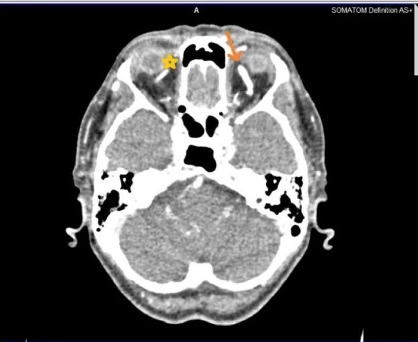
- İpsilateral üst oftalmik ven geniş ve kıvrımlı, glob öne itilmiş ve ekstraoküler kaslar kalınlaşmış olabilir; kavernöz sinüs asimetrisi seçilebilir. Bu bulgular şüphe uyandırır, fistül bağlantısını tek başına kanıtlamaz.
- BTA tanısal
- Uygun zamanlanmış arteriyel CTA'da kavernöz sinüs içinde erken arteriyel kontrastlanma, asimetrik genişleme ve üst oftalmik venin erken opasifikasyonu aranır; genişlemiş kıvrımlı oftalmik venler ve orbital konjesyon eşlik edebilir. Dural, küçük besleyiciler veya fistül ağzı CTA'da gösterilemeyebilir.
Önerilen pencereler: Anjiyo (G 600 / M 150), Yumuşak doku (G 400 / M 40), Kemik (G 1800 / M 400).
BT bulguları
- Erken kavernöz sinüs opasifikasyonu — arteriyel CTA'da aynı taraftaki sinüsün beklenen venöz dolumdan önce kontrastlanması fistül için doğrudan ipucudur.
- Üst oftalmik ven genişlemesi ve erken dolumu — KKF'de duyarlı bulgudur; karşı tarafla çap ve opasifikasyon zamanını kıyaslayın.
- Kavernöz sinüs asimetrisi veya genişlemesi KKF'de görülebilir; ancak tek başına özgül değildir.
- Proptozis — globun etkilenen tarafta öne yer değiştirmesi; karşı tarafla ve orbital kemik sınırlarıyla kıyaslanır.
- Ekstraoküler kas konjesyonu — birden fazla rektus kasında kalınlaşma, özellikle orbital venöz basınç artışıyla birlikte görülebilir.
- Orbital yağ dokusu ödemi — preseptal veya postseptal retikülasyonla göz çevresi venöz konjesyonunu destekler.
- Kortikal venöz reflüde kortikal venler genişleyebilir; eşlik eden ödem veya kanama intrakraniyal venöz hipertansiyonla ilişkili ciddi bulgulardır.
Ölçütler ve sınıflamalar
- Barrow sınıflaması
- A: kavernöz ICA ile sinüs arasında doğrudan şant. B: ICA dural dallarından sinüse. C: ECA dural dallarından sinüse. D: hem ICA hem ECA dural dallarından sinüse. BT bulguları tek başına her olguda besleyici dalları sınıflamaya yetmez.
Normalde
BTA'da kavernöz sinüs asimetrik genişlemesi tek başına özgül değildir; arteriyel fazda kavernöz sinüs kontrastlanması KKF için daha duyarlı bir bulgudur. KKF'de orbitada proptozis, oftalmik venöz sistem genişlemesi ve özellikle ekstraoküler kaslarda yumuşak doku şişliği görülebilir.
Normal BT ile kıyasla
Aynı bölgenin, kaynak veri setinde patoloji içermediği belirtilen bir BT incelemesini kesit kesit kaydırın; organ sınırlarını açarak anatomiyi hasta görüntüsüyle karşılaştırın. Küçük rastlantısal bulgular tümüyle dışlanmamıştır.
Kaynak: TotalSegmentator v2.01 veri seti (Wasserthal ve ark., Radiology: Artificial Intelligence 2023), CC BY 4.0, vaka s0849; organ sınırları veri setinin otomatik segmentasyonundan, birleştirilerek sadeleştirildi. Görüntüler 2,5 mm kesit aralığına yeniden örneklendi.
Ayırıcı tanı
- Kavernöz sinüs trombozu
- sinüs içi dolum defekti ve komşu sinüslerde tromboz; erken arteriyel sinüs opasifikasyonu göstermez.
- Kavernöz ICA anevrizması, kavernöz segment ICA'dan kaynaklanan ekstradural bir anevrizmadır ve BT'de gösterilebilir.
- Orbital selülit
- sinüs bağlantısı olmadan orbital yağda inflamatuvar kirlenme, koleksiyon veya komşu sinüs enfeksiyonu görülebilir.
- Tiroid orbitopatisi
- özellikle alt ve iç rektus kaslarında kas karnı fusiform kalınlaşır, tendonlar görece korunur; üst oftalmik venin erken dolması beklenen bulgu değildir.
- Dural kavernöz sinüs fistülü dışı AVM
- beyin parankiminde nidus, arteriyel besleyiciler ve kortikal venöz drenaj daha belirgindir.
Tuzaklar
- Kavernöz sinüs asimetrisi KKF'ye özgü değildir; erken arteriyel kontrastı ve üst oftalmik ven dolumunu zamanlama ile birlikte doğrulayın.
- Kontrast bolusu optimal zamanlanmadığında kavernöz sinüs asimetrisi yanıltıcı değerlendirmeye yol açabilir; arteriyel faz bulgularını yorumlarken zamanlama kalitesini göz önünde bulundurun.
- Düşük akımlı dural fistülde ince besleyici dallar CTA'da görünmeyebilir; belirgin klinik şüphede negatif CTA fistülü bütünüyle dışlamaz.
- Kavernöz sinüs trombozunda genişleme ve orbital konjesyon KKF'yi taklit edebilir; venöz dolum defektini ve sinüs içi kontrast paternini özellikle kontrol edin.
Kendini dene
Travma sonrası proptozis var. BTA'da kavernöz sinüs arteriyel fazda doluyor ve üst oftalmik ven geniş. Tanı nedir?
Cevabı göster
Karotis-kavernöz fistül. Arteriyel fazda kavernöz sinüsün erken dolması ve geniş üst oftalmik ven KKF'yi güçlü biçimde destekler. Tromboz dolum defekti yapar; tiroid orbitopatisi kas paternine, selülit ise orbital inflamatuvar değişikliklere odaklanır.
Kavernöz sinüste asimetrik genişleme var; ancak kontrast bolusu optimal zamanlanmamış. Bu bulgu tek başına KKF tanısı için yeterli midir?
Cevabı göster
Hayır; asimetri tek başına özgül değildir. Hayır. Asimetrik kavernöz sinüs genişlemesinin özgüllüğü düşüktür ve KKF için yalancı pozitif tanılara yol açabilir. Yalancı pozitif değerlendirmelerde kontrast bolusunun optimal zamanlanmamış olması daha olasıdır.
İlgili konular
Kaynaklar
Bu sayfadaki 48 cümle ve 2 quiz sorusu aşağıdaki kaynaklardan alıntılarla otomatik olarak karşılaştırıldı; 9 cümle bu karşılaştırmada düzeltildi (2026-10-09).
- Spontaneous Resolution of an Aggressive Direct Carotid Cavernous Fistula Following Partial Transvenous Embolization Treatment: A Case Report and Review of Literaturespmc.ncbi.nlm.nih.gov
- Neuroimaging for neurovascular complications of traumatic brain injurypmc.ncbi.nlm.nih.gov
- Neuroendovascular Surgery Applications in Craniocervical Traumapmc.ncbi.nlm.nih.gov
- Management of neurovascular emergencies with ophthalmic manifestationspmc.ncbi.nlm.nih.gov
- Clinical efficacy of endovascular treatment approach in patients with carotid cavernous fistula: A systematic review and meta-analysis ☆pmc.ncbi.nlm.nih.gov
- Cavernous sinus lesions.pubmed.ncbi.nlm.nih.gov
- [Orbito-palpebral vascular pathology].pubmed.ncbi.nlm.nih.gov
- [Basilar skull fractures].pubmed.ncbi.nlm.nih.gov
- Ophthalmological emergencies.pubmed.ncbi.nlm.nih.gov
- Big red flags in neuro-ophthalmology.pubmed.ncbi.nlm.nih.gov
- Imaging of Orbital Trauma and Emergent Non-traumatic Conditions.pubmed.ncbi.nlm.nih.gov
- Imaging of Painful Ophthalmologic Disorders.pubmed.ncbi.nlm.nih.gov
- Carotid-cavernous fistula from the perspective of an ophthalmologist A Review.pubmed.ncbi.nlm.nih.gov
- Posttraumatic cerebrovascular injuries in children. A systematic review.pubmed.ncbi.nlm.nih.gov
- Treatment of direct carotid-cavernous fistulas with flow diversion - does it work?pubmed.ncbi.nlm.nih.gov
- Exophthalmos following mechanical thrombectomy for anterior circulation stroke: A retrospective study and review of literature.pubmed.ncbi.nlm.nih.gov
- Endovascular repair of direct carotid-cavernous fistula in Ehlers-Danlos type IV.pubmed.ncbi.nlm.nih.gov
- Spontaneous resolution of direct carotid-cavernous fistulas: case series and literature review.pubmed.ncbi.nlm.nih.gov
- Carotid-Cavernous Fistula After Transsphenoidal Surgery: A Rare but Challenging Complication.pubmed.ncbi.nlm.nih.gov
- Anatomy of the Middle Meningeal Artery: Implications for Middle Meningeal Artery Embolization in Chronic Subdural Hematoma.pubmed.ncbi.nlm.nih.gov
- Post-traumatic Carotid-Cavernous Fistula Following Penetrating Orbital Injury: A Case Report and Literature Review.pubmed.ncbi.nlm.nih.gov
- Rethinking the sphenoparietal sinus: implications for lesser sphenoid wing dural arteriovenous fistulas. Illustrative cases.pubmed.ncbi.nlm.nih.gov
- Carotid Cavernous Fistula: A Case Report of a Rare Diagnosis and Presentation.pubmed.ncbi.nlm.nih.gov
- Spontaneous Resolution of a Bilateral Barrow Type D Indirect Carotid-Cavernous Fistula: A Rare Case Report and Literature Review.pubmed.ncbi.nlm.nih.gov
- Co-occurring scalp arteriovenous malformation and dural arteriovenous fistula with diploic vein involvement: illustrative case.pubmed.ncbi.nlm.nih.gov
- Incidental vascular findings on brain magnetic resonance angiography.pubmed.ncbi.nlm.nih.gov
- Multimodal imaging in neuro-ophthalmology.pubmed.ncbi.nlm.nih.gov
- When headache meets visual disturbance: An evidence-based review of common and critical entities.pubmed.ncbi.nlm.nih.gov
- Neuro-Ophthalmic Manifestations of Carotid Cavernous Fistulas: A Systematic Review and Meta-Analysis.pubmed.ncbi.nlm.nih.gov
- Imaging for Pediatric Head and Neck Emergencies: Critical Insights.pubmed.ncbi.nlm.nih.gov
- Guidelines for standard operation of imaging modalities in orbital diseases (2024).pubmed.ncbi.nlm.nih.gov
- Dominant Orbitofrontal Pial Supply in Anterior Cranial Fossa Dural Arteriovenous Fistula: Angiographic Differentiation from Mixed Pial-Dural Arteriovenous Malformation and Anatomy-Based Treatment Selection.pubmed.ncbi.nlm.nih.gov
- Probable cavernous sinus-adjacent meningeal metastasis from esophageal squamous cell carcinoma: A case report.pubmed.ncbi.nlm.nih.gov
- Consensus disease definitions for ophthalmic immune-related adverse events of immune checkpoint inhibitors.pubmed.ncbi.nlm.nih.gov
- Imaging of cerebrovascular complications from blunt skull base trauma.pubmed.ncbi.nlm.nih.gov
- Transvenous Approaches to Embolization of Dural Arteriovenous Fistulae of the Cavernous Sinus.pubmed.ncbi.nlm.nih.gov
- Stroke-Mimic: An Intracranial Dural Arteriovenous Fistula Revealed by New-Onset Seizures and Post-Ictal Deficit.pubmed.ncbi.nlm.nih.gov
- Ocular Color Doppler Ultrasound (OCDUS) in Diagnosis and Monitoring of Ophthalmological, Cerebrovascular and Systemic Diseases: A Narrative Review.pubmed.ncbi.nlm.nih.gov
- Delayed Rupture Presenting as Direct Carotid-Cavernous Fistula after Flow Diversion for a Cavernous Internal Carotid Artery Aneurysm with Enlarging Blebs: A Case Report.pubmed.ncbi.nlm.nih.gov
- Septic cerebral venous sinus thrombosis from maxillary sinusitis with concomitant right-sided sigmoid sinus hypoplasia: A case report and review of the literature.pubmed.ncbi.nlm.nih.gov
- Interocular symmetry and asymmetry in ocular health and disease.pubmed.ncbi.nlm.nih.gov
- Dilated MMA sign in cDAVF and other arterial feeders on 3D TOF MRA.pubmed.ncbi.nlm.nih.gov
- Oculomotor Nerve Palsy-Etiologies, Symptoms and Diagnosis: A Systematic Review with Meta-Analysis.pubmed.ncbi.nlm.nih.gov
- Pulsatile Tinnitus: A Comprehensive Clinical Approach to Diagnosis and Management.pubmed.ncbi.nlm.nih.gov
- The cavernous sinus: An anatomic study with clinical implication.pubmed.ncbi.nlm.nih.gov
- Etiologies of Proptosis: A review.pubmed.ncbi.nlm.nih.gov
- MR imaging of cavernous sinus thrombosis.pubmed.ncbi.nlm.nih.gov
- Intracerebral hemorrhage and carotid-cavernous fistulas: a rare connection.pubmed.ncbi.nlm.nih.gov
- A Practical Approach to Intracranial Dural Arteriovenous Fistulas: Pathogenesis, Classification and Management.pubmed.ncbi.nlm.nih.gov
- Intracranial dural arteriovenous fistulas with pial arterial supply: A narrative review.pubmed.ncbi.nlm.nih.gov
- Morphological Analysis of the Cavernous Segment of the Internal Carotid Artery: A Retrospective, Single Center Study of Its Clinical Significance.pubmed.ncbi.nlm.nih.gov
- Origin Variants of the Ascending Pharyngeal Artery and Sequential External Carotid Branching Classification.pubmed.ncbi.nlm.nih.gov
- Magnetic resonance imaging of the orbit, Part 1: Basic principles and radiological approach.pubmed.ncbi.nlm.nih.gov
- Bilateral Post-Traumatic Carotid-Cavernous Fistula: A Case Report.pubmed.ncbi.nlm.nih.gov
- Utilization of the PK Papyrus Covered Stent for the Treatment of Direct Carotid Cavernous Fistulas: A Case Series.pubmed.ncbi.nlm.nih.gov
- Clinico-Radiological Presentation and Management of Carotid-Cavernous Fistulae: Real-Time Institutional Experience From Pakistan.pubmed.ncbi.nlm.nih.gov
- Endovascular management of carotid-cavernous fistulas: a 16-year retrospective analysis of multimodal treatment strategies and long-term clinical outcomes.pubmed.ncbi.nlm.nih.gov
- Syndrome of Inappropriate Antidiuresis and Symptomatic Hyponatremia After Onyx Embolization of a Carotid-Cavernous Fistula: A Case Report.pubmed.ncbi.nlm.nih.gov
- Pipeline flow diverter and transvenous coiling for the treatment of direct carotid cavernous sinus fistulae: a retrospective case series.pubmed.ncbi.nlm.nih.gov
- Transvenous embolization of a carotid-cavernous fistula via the inferior ophthalmic vein: illustrative case.pubmed.ncbi.nlm.nih.gov
- A case of anterior cranial fossa dural arteriovenous fistula concomitant with moyamoya disease.pubmed.ncbi.nlm.nih.gov
- Recurrent ophthalmic arteries supplying posterior meningiomas: embryologic background and 3D anatomical visualization.pubmed.ncbi.nlm.nih.gov
- Intracranial venous thrombosis imaging by anatomical location: an educational review with mimics and diagnostic pitfalls.pubmed.ncbi.nlm.nih.gov
- Endovascular Embolization in Neurovascular Disease: Material Science, Multimodal Management, and Future Horizons.pubmed.ncbi.nlm.nih.gov
- The persistent proatlantal artery: types and subtypes.pubmed.ncbi.nlm.nih.gov
- Application of imaging studies for orbital tumors: a practical review and representative cases.pubmed.ncbi.nlm.nih.gov
- A Five-Step Approach to Acute Vision Loss: A Practical Narrative Review for Clinicians.pubmed.ncbi.nlm.nih.gov
- Magnetic resonance imaging of the orbit, Part 2: Characterization of orbital pathologies.pubmed.ncbi.nlm.nih.gov
- Update on thyroid-associated Ophthalmopathy with a special emphasis on the ocular surface.pubmed.ncbi.nlm.nih.gov
- CT angiogram findings in carotid-cavernous fistulas: stratification of imaging features to help radiologists avoid misdiagnosis.pubmed.ncbi.nlm.nih.gov
- Imaging the brain: diagnosis aided by structural features on neuroimaging studies.pubmed.ncbi.nlm.nih.gov
- 4D-DSA for Assessment of the Angioarchitecture and Grading of Cranial Dural AVF.pubmed.ncbi.nlm.nih.gov
- Neuroimaging for neurovascular complications of traumatic brain injury.pubmed.ncbi.nlm.nih.gov
- Narrower jugular bulbs and their tributaries are associated with skull base venous hyperintensity on arterial spin-labeling MRI.pubmed.ncbi.nlm.nih.gov
- Orbital tuberculosis mimicking a vascular emergency: A case report of superior ophthalmic vein engorgement.pubmed.ncbi.nlm.nih.gov
- An Orbital Abscess Caused by Fusobacterium nucleatum Mimicking Tolosa-Hunt Syndrome.pubmed.ncbi.nlm.nih.gov
- Systolic propulsion of the eyeballs in severe tricuspid regurgitation: a case series and review of the literature.pubmed.ncbi.nlm.nih.gov
- Tolosa-Hunt Syndrome and IgG4 Diseases in Neuro-Ophthalmology.pubmed.ncbi.nlm.nih.gov
- Rhinosinusitis: Evidence and experience - 2024.pubmed.ncbi.nlm.nih.gov
- Asymmetric Graves' Orbitopathy.pubmed.ncbi.nlm.nih.gov
- Superior Ophthalmic Vein Thrombosis in Trinidad and Tobago: A Case Series.pubmed.ncbi.nlm.nih.gov
- A Retrospective Study of Imaging of Invasive Rhino-Orbital-Cerebral Mucormycosis in the COVID-19 Pandemic in a Tertiary Care Center.pubmed.ncbi.nlm.nih.gov
- Rare sphenoid dural arteriovenous fistula treated with microsurgery: illustrative case.pubmed.ncbi.nlm.nih.gov
- Computed tomography perfusion stroke mimics on RAPID commercial software: A case-based review.pubmed.ncbi.nlm.nih.gov
- Jugular Venous Reflux Can Mimic Posterior Fossa Dural Arteriovenous Fistulas on MRI-MRA.pubmed.ncbi.nlm.nih.gov
- Central hypothyroidism as a rare sequela of traumatic carotid cavernous fistula in a pediatric patient.pubmed.ncbi.nlm.nih.gov
- Technical Refinement of a Hybrid Surgical and Endovascular Approach for Cavernous Dural Arteriovenous Fistula with Leptomeningeal Venous Reflux: Direct Superficial Middle Cerebral Vein Exposure for Transvenous Embolization.pubmed.ncbi.nlm.nih.gov
- Multimodal endovascular management of traumatic carotid-cavernous fistulas: case series and lessons learned.pubmed.ncbi.nlm.nih.gov
- Intracranial dural arteriovenous fistulas with and without pial arterial supply: a case series of 227 patients.pubmed.ncbi.nlm.nih.gov
- Utility of High-Dose Steroids in Treatment-Resistant Carotid-Cavernous Fistulas: A Case Report.pubmed.ncbi.nlm.nih.gov
- Postoperative Pituitary MRI Findings in Acromegaly: A Pictorial Review.pubmed.ncbi.nlm.nih.gov
- Color-Mapping of 4D-CTA for the Detection of Cranial Arteriovenous Shunts.pubmed.ncbi.nlm.nih.gov
- Prolonged trigeminocardiac reflex following transvenous coil embolization for cavernous sinus dural arteriovenous fistula with brainstem edema: illustrative case.pubmed.ncbi.nlm.nih.gov
- The "Transdural" Paradox: The Case of a Paraclinoid Internal Carotid Artery Aneurysm With an Intradural Neck and Extradural Dome.pubmed.ncbi.nlm.nih.gov
- A rare case report of vascular Ehlers-Danlos syndrome as a pulsatile neck mass with an internal carotid artery aneurysm.pubmed.ncbi.nlm.nih.gov
Eğitim amaçlıdır; hasta başında tanı ve tedavi kararının yerine geçmez, tıbbi tavsiye değildir. Metinler yapay zekâ desteğiyle yazıldı ve otomatik bir adımda kaynak alıntılarıyla karşılaştırıldı; bu bir tıbbi doğrulama değildir ve içerik uzman radyolog incelemesinden geçmedi. Klinik kararlarda güncel kılavuzları, kurum protokollerini ve radyoloji raporunu esas alın.