Özet
Baş ağrısı, nöbet, papilödem veya fokal nörolojik bulgularla gelen hastada kortikal ven ya da derin ven trombozu, venöz dönüşü bozarak infarkt ve kanama oluşturabilir. Kontrastsız BT'de tromboze damarın hiperdens çizgisi bazen doğrudan görülür; BT venografi dolum defektini ve trombüsün venöz anatomideki yerini gösterir. Parankim hasarı arter sınırlarına uymayan ödem, hemorajik infarkt ve derin sistem tutulduğunda bilateral talamik konjesyon biçiminde ortaya çıkabilir. Normal görünen kontrastsız BT trombozu dışlamaz; tanının atlanması ödem, hidrosefali ve nörolojik kötüleşmeye yol açabilir.
Faz ve pencere
- Kontrastsız
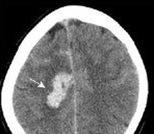
- Akut pıhtı kortikal sulkus boyunca uzanan hiperdens bir ven (kord işareti) veya düz sinüs/torculada yoğun dolu yapı olarak görülebilir. Venöz ödem hipodens ve damar alanı sınırlarını aşan dağılımdadır; kanama odakları eşlik edebilir. Doğrudan işaret yokluğu trombozu dışlamaz.
- Gecikmiş tanısal
- Venöz zamanlamalı BT venografide tromboze kortikal ven ya da internal serebral ven, Galen veni veya düz sinüs içinde kontrastla çevrili dolum defekti ve opaklaşmama aranır. Kollateral venlerin belirginleşmesi, derin sistemde iki taraflı venöz dönüş kaybı ve eşlik eden talamik/bazal gangliyon ödemi haritalanır.
Önerilen pencereler: Beyin (G 80 / M 40), Anjiyo (G 600 / M 150).
BT bulguları
- Kortikal kord işareti — sulkus içindeki kortikal ven kontrastsız BT'de çevre beyin dokusundan daha yüksek atenüasyonlu, çizgisel bir damar olarak izlenir.
- Venöz dolum defekti — BT venografide kortikal ven veya derin ven lümeninde kontrastla çevrili pıhtı ya da beklenen venöz opasifikasyonun kaybı görülür.
- Venöz infarkt dağılımı — hipodens ödem bir arterin sulama alanına uymaz, birden fazla arter alanını aşabilir ve korteksten subkortikal beyaz cevhere uzanır.
- Hemorajik venöz infarkt — düzensiz parankimal kanama, çevresindeki ödemle aynı venöz drenaj alanında yer alır; arteriyel infarkttaki tekdüze kama biçimini izlemek zorunda değildir.
- Derin venöz konjesyon — internal serebral venler, Galen veni veya düz sinüs trombozunda bilateral talamus ve bazal gangliyonlarda ödem ya da kanamalı hipodensite görülür.
- Kortikal ven kollateralleri — tıkalı ven çevresinde kıvrımlı, genişlemiş yüzeyel venler artmış venöz basınç ve alternatif drenajı düşündürür.
- Kitle etkisi ve hidrosefali — derin gri cevher ödemi üçüncü ventrikülü daraltabilir; daha ileri olguda ventrikül genişlemesi ve bazal sistern basısı eşlik edebilir.
Normalde
Normal kortikal venler kontrastsız incelemede çevre beyin dokusuna göre belirgin hiperdens bir kord oluşturmaz; venöz fazlı BT'de kortikal venler ve internal serebral venler kontrastı kesintisiz alır. İki talamusun atenüasyon ve hacmini, üçüncü ventrikül genişliğini ve sulkuslara eşlik eden venöz kanalların opasifikasyonunu karşılaştırın.
Normal BT ile kıyasla
Aynı bölgenin, kaynak veri setinde patoloji içermediği belirtilen bir BT incelemesini kesit kesit kaydırın; organ sınırlarını açarak anatomiyi hasta görüntüsüyle karşılaştırın. Küçük rastlantısal bulgular tümüyle dışlanmamıştır.
Kaynak: TotalSegmentator v2.01 veri seti (Wasserthal ve ark., Radiology: Artificial Intelligence 2023), CC BY 4.0, vaka s0103; organ sınırları veri setinin otomatik segmentasyonundan, birleştirilerek sadeleştirildi. Görüntüler 2,5 mm kesit aralığına yeniden örneklendi.
Ayırıcı tanı
- Arteriyel infarkt
- hipodensite çoğunlukla belirli bir arter dağılımını izler; venöz trombüs gibi drenaj dışı kanamalı ödem ve genişlemiş kortikal ven beklenmez.
- Hiperdens kortikal arter
- damar sulkus yerine tipik arter güzergâhındadır ve arteriyel fazlı BTA'da tıkalı arterle devam eder; venöz zamanlı incelemede kortikal venler doludur.
- Subaraknoid kanama
- hiperdensite sulkus veya sisternleri doldurabilir ve nadiren de olsa kortikal ven trombozunun yarattığı çizgisel hiperdensiteyi (kord işareti) taklit edebilir.
- Kontrast tutulumu veya yalancı hiperdens ven
- önceki kontrastlı inceleme sonrasında damarlar yaygın yoğun görünebilir; klinik zamanlama ve birden fazla venöz yapının benzer opasifikasyonu kontrol edilir.
- Derin gri cevher enfarktı
- bilateral talamik değişiklikler arteriyel perforan damar hastalıklarında da görülebilir; derin ven trombozunda iç serebral ven/Galen/düz sinüs dolum kusuru ve daha yaygın venöz ödem aranır.
Tuzaklar
- Kontrastsız BT'de yoğun kortikal ven görülmemesi trombozu dışlamaz; küçük ven trombüsü ve subakut pıhtı doğrudan seçilmeyebilir, venöz fazlı inceleme gerekir.
- Fizyolojik polisitemi gibi durumlar ven sinüslerde genel hiperatenüasyona yol açabilir; tek bir venin fokal yoğunluğunu diğer venler ve hematokrit bağlamında karşılaştırın.
- Asimetrik ya da hipoplastik venöz yapı trombüs gibi yorumlanabilir; kontrastlı incelemede damar duvarı boyunca kesintisiz lümen, karşı taraf ve kemik oluk kalibresiyle birlikte değerlendirilir.
- Derin venöz trombozda bilateral talamik ödemi posterior dolaşım arter infarktı olarak etiketlemeyin; düz sinüs ve internal serebral ven opasifikasyonunu özellikle arayın.
Kendini dene
Baş ağrısı ve bilinç bulanıklığında iki talamusta ödem, internal serebral venlerde dolum defekti var. En olası tanı nedir?
Cevabı göster
Derin serebral ven trombozu. Bilateral talamik venöz konjesyon ve internal serebral ven dolum defekti derin ven trombozunu gösterir. Arteriyel infarkt damar alanı dağılımına uyar; PRES genellikle posterior subkortikal ödemle belirgindir; gliom fokal kitle etkisi yapar.
Kortikal kanama ve ödem birden fazla arter alanını aşıyor; komşu sulkusta hiperdens çizgisel damar görülüyor. Hangi neden öne çıkar?
Cevabı göster
Kortikal ven trombozu. Damar alanını aşan hemorajik ödem ve sulkustaki hiperdens ven kortikal ven trombozunu destekler. MCA embolisi tipik arter dağılımını izler; hipertansif kanama derin çekirdektedir; anevrizmal kanama sisternlerde yoğunlaşır.
Kaynaklar
Bu sayfadaki 40 cümle ve 2 quiz sorusu aşağıdaki kaynaklardan alıntılarla tek tek karşılaştırıldı; 4 cümle bu karşılaştırmada düzeltildi (2026-10-08).
- Intracranial venous thrombosis imaging by anatomical location: an educational review with mimics and diagnostic pitfallspmc.ncbi.nlm.nih.gov
- Conduction Aphasia in a Case of Left Cortical Veins and Left Lateral Sinus Thrombosis Due to Multiple Risk Factors: A Case Report and Review of the Literaturepmc.ncbi.nlm.nih.gov
- Isolated Psychiatric Presentation of Cerebral Venous Thrombosis in a Patient Without Risk Factors: A Diagnostic Challengepmc.ncbi.nlm.nih.gov
- Posterior Circulation Stroke After Subclavian Artery Thrombosis Following Low-Voltage Electrical Injury in an Active Woman With Previously Unrecognized Prothrombotic Risk Factors: A Case Reportpmc.ncbi.nlm.nih.gov
- A pediatric case of varicella-associated cerebral venous sinus thrombosis with anti-GAD65 autoimmune encephalitis and multisystem complications – a case reportpmc.ncbi.nlm.nih.gov
- Adrenocortical hyperplasia: a review of clinical presentation and imaging.pubmed.ncbi.nlm.nih.gov
- Functional imaging of adrenal cortex.pubmed.ncbi.nlm.nih.gov
- Corticothalamic axon morphologies and network architecture.pubmed.ncbi.nlm.nih.gov
- Why acute unilateral vestibular cortex lesions mostly manifest without vertigo.pubmed.ncbi.nlm.nih.gov
- Aldosterone-Producing Cell Clusters in Normal and Pathological States.pubmed.ncbi.nlm.nih.gov
- Cortical hemichorea-hemiballism.pubmed.ncbi.nlm.nih.gov
- Cortical thickness analysis in operculo-insular epilepsy.pubmed.ncbi.nlm.nih.gov
- Atypical age-related changes in cortical thickness in autism spectrum disorder.pubmed.ncbi.nlm.nih.gov
- Cellular and Genetic Causes of Idiopathic Hyperaldosteronism.pubmed.ncbi.nlm.nih.gov
- Ectopic adrenal cortical adenoma in the gastric wall: case report.pubmed.ncbi.nlm.nih.gov
- Aberrant cortical neurodevelopment in major depressive disorder.pubmed.ncbi.nlm.nih.gov
- Localizing ECoG electrodes on the cortical anatomy without post-implantation imaging.pubmed.ncbi.nlm.nih.gov
- Layer 6 corticothalamic neurons activate a cortical output layer, layer 5a.pubmed.ncbi.nlm.nih.gov
- Probabilistic functional tractography of the human cortex revisited.pubmed.ncbi.nlm.nih.gov
- Efficacy and safety of anticoagulation for pediatric cerebral sinovenous thrombosis: systematic review and meta-analysis.pubmed.ncbi.nlm.nih.gov
- When headache meets visual disturbance: An evidence-based review of common and critical entities.pubmed.ncbi.nlm.nih.gov
- Cerebral venous sinus thrombosis in pregnancy and the postpartum period: epidemiology, management, and maternal mortality-a systematic review and meta-analysis.pubmed.ncbi.nlm.nih.gov
- Extensive cerebral venous sinus thrombosis with hemorrhagic venous infarction as the initial presentation of acute myeloid leukemia: A case report.pubmed.ncbi.nlm.nih.gov
- Overview of the utility of noncontrast CT brain in the diagnosis of cerebral vein thrombosis.pubmed.ncbi.nlm.nih.gov
- Congenital Intracranial Vascular Malformations in Children : Radiological Overview.pubmed.ncbi.nlm.nih.gov
- Venous-significant intracranial hemorrhage in neonatal HIE treated with therapeutic hypothermia: imaging phenotypes, diagnostic pitfalls, and prognostic implications.pubmed.ncbi.nlm.nih.gov
- Intracranial venous thrombosis imaging by anatomical location: an educational review with mimics and diagnostic pitfalls.pubmed.ncbi.nlm.nih.gov
- Role of diffusion weighted imaging, apparent diffusion coefficient and magnetic resonence perfusion among acute ischemic stroke patient.pubmed.ncbi.nlm.nih.gov
- Diagnostic neuroimaging in neuro-ophthalmic disorders.pubmed.ncbi.nlm.nih.gov
- Heart Disease and Stroke Statistics-2023 Update: A Report From the American Heart Association.pubmed.ncbi.nlm.nih.gov
- Neuroimaging for neurovascular complications of traumatic brain injury.pubmed.ncbi.nlm.nih.gov
- Blind spots in brain imaging: a pictorial essay.pubmed.ncbi.nlm.nih.gov
- Non-Thrombotic Filling Defects in Cerebral Veins and Sinuses: When Normal Structures Mimic a Disease.pubmed.ncbi.nlm.nih.gov
- Cerebral hemorrhage after left ventricular assist device implantation in a patient with dilated cardiomyopathy: a case report.pubmed.ncbi.nlm.nih.gov
- Brain intraparenchymal hemorrhage following urgent cesarean section: a case report.pubmed.ncbi.nlm.nih.gov
- Enterococcus faecalis Aortic Valve Endocarditis Masquerading As Urinary Tract Infection: Diagnostic Delay, Septic Embolic Strokes, and a Fatal Therapeutic Impasse.pubmed.ncbi.nlm.nih.gov
- Acute otitis media complicated by mastoiditis and otogenic transverse-sigmoid sinus thrombosis in an adult: a case report on infectious pathogenesis and multidisciplinary therapy.pubmed.ncbi.nlm.nih.gov
- Venous Thromboembolism in Neonates, Children, and Adolescents: A Comprehensive Narrative Review of Risk Factors, Diagnosis, Treatment, and Prevention.pubmed.ncbi.nlm.nih.gov
- Bilateral Thalamic Infarction Secondary to Deep Cerebral Venous Thrombosis: A Diagnostic Challenge Associated With Hormonal Therapy.pubmed.ncbi.nlm.nih.gov
- Vascular Cognitive Impairment and Dementia: Clinical Features, Neuropathology, and Biomarkers.pubmed.ncbi.nlm.nih.gov
- Clinical Presentation, Neuroimaging Findings, and Predictors of Brain Parenchymal Lesions in Cerebral Vein and Dural Sinus Thrombosis: A Retrospective Study.pubmed.ncbi.nlm.nih.gov
- Commentary: an eye on PET quantification.pubmed.ncbi.nlm.nih.gov
- Isolated Cortical Venous Thrombosis as a Mimic for Cortical Subarachnoid Hemorrhage.pubmed.ncbi.nlm.nih.gov
- Signs in Neuroradiology: A Pictorial Review.pubmed.ncbi.nlm.nih.gov
- Imaging Evaluation of Acute Traumatic Brain Injury.pubmed.ncbi.nlm.nih.gov
- Case Report: Mechanical thrombectomy of cerebral venous sinuses using the NeVa stent retriever.pubmed.ncbi.nlm.nih.gov
- Risk factors for cerebral venous sinus thrombosis in Xinjiang, China: a single-center case-control study with a focus on high-altitude exposure.pubmed.ncbi.nlm.nih.gov
- Differentiating effective salvage from ineffective delayed compensation in anterior circulation stroke: a dynamic quantitative collateral index.pubmed.ncbi.nlm.nih.gov
- Multidelay ASL of the pediatric brain.pubmed.ncbi.nlm.nih.gov
- Spinal vascular lesions: anatomy, imaging techniques and treatment.pubmed.ncbi.nlm.nih.gov
- Quantitative Cerebrovascular Analysis for Improved Prediction of Post-Stroke Complications.pubmed.ncbi.nlm.nih.gov
- Deep learning and high-resolution magnetic resonance vascular wall imaging: current challenges and future perspectives.pubmed.ncbi.nlm.nih.gov
- Refractory deep cerebral venous thrombosis managed by mechanical thrombectomy and balloon angioplasty: A case report.pubmed.ncbi.nlm.nih.gov
- Direct superior ophthalmic vein puncture for cavernous sinus dural arteriovenous fistula with dual orbital venous outflow: illustrative case.pubmed.ncbi.nlm.nih.gov
- Cross-sectional imaging in an inferior vena cava agenesis associated with antiphospholipid syndrome, lower limb, and portomesenteric deep vein thrombosis and renal insufficiency in a young patient: A rare presentation.pubmed.ncbi.nlm.nih.gov
- Giant Right Sphenoid Wing Meningioma as a Reversible Frontal Network Lesion: A Pseudo-bvFTD Case with Venous-Sparing Skull-Base Resection.pubmed.ncbi.nlm.nih.gov
- Fetal intracranial hemorrhage and infarct: Main sonographic and MRI characteristics: A review article.pubmed.ncbi.nlm.nih.gov
- Bilateral thalamic and brainstem anaplastic astrocytoma: A case report.pubmed.ncbi.nlm.nih.gov
- Preventable cerebral venous sinus thrombosis from diet- and alcohol-induced hyperhomocysteinemia: illustrative case.pubmed.ncbi.nlm.nih.gov
- Septic cerebral venous sinus thrombosis from maxillary sinusitis with concomitant right-sided sigmoid sinus hypoplasia: A case report and review of the literature.pubmed.ncbi.nlm.nih.gov
- Cerebral venous sinus thrombosis in a young female misdiagnosed as migraine ending in a permanent vegetative state: a case report and review of the literature.pubmed.ncbi.nlm.nih.gov
- Post-Traumatic Cerebral Infarction: A Narrative Review of Pathophysiology, Diagnosis, and Treatment.pubmed.ncbi.nlm.nih.gov
- Many faces of acute bowel ischemia: overview of radiologic staging.pubmed.ncbi.nlm.nih.gov
- Retrospective Observational Study of Brain MRI Findings in Patients with Acute SARS-CoV-2 Infection and Neurologic Manifestations.pubmed.ncbi.nlm.nih.gov
- Occipital Pial AVM Rupture in a Young Adult: Dual Intranidal Aneurysms, Solitary Parasagittal SSS Drainage, and Hematoma-Corridor Microsurgical Cure.pubmed.ncbi.nlm.nih.gov
- Spontaneous Non-Aneurysmal Convexity Subarachnoid Hemorrhage: A Scoping Review of Different Etiologies beyond Cerebral Amyloid Angiopathy.pubmed.ncbi.nlm.nih.gov
- TFE3-rearranged renal cell car cinoma with massive calcification: Imaging-pathologic correlation.pubmed.ncbi.nlm.nih.gov
- Photon-counting detector CT in oncology: a new era of cancer imaging.pubmed.ncbi.nlm.nih.gov
- Challenging Resection of Bilateral Parasagittal and Falcine Meningioma Involving Both Anterior Third and Middle Third of the Superior Sagittal Sinus: A Case Report and Literature Review.pubmed.ncbi.nlm.nih.gov
- When the Brainstem Is the Target: Cryptogenic Paramedian Pontine Infarction Associated With Patent Foramen Ovale.pubmed.ncbi.nlm.nih.gov
- Artery of Percheron Stroke: A Case Report With a Diagnostic Challenge.pubmed.ncbi.nlm.nih.gov
- Stepwise clinical and diagnostic strategy for coma of unknown origin.pubmed.ncbi.nlm.nih.gov
- 2025 Heart Disease and Stroke Statistics: A Report of US and Global Data From the American Heart Association.pubmed.ncbi.nlm.nih.gov
- Imaging of Cerebral Venous Thrombosis.pubmed.ncbi.nlm.nih.gov
- Case Report: Structured MRI assessment of posterior thalamic infarction in a distribution compatible with posterior choroidal artery territory presenting as Déjerine-Roussy syndrome in an adolescent: differentiating arterial ischemia from venous thrombosis and thalamic neoplasm.pubmed.ncbi.nlm.nih.gov
- Post-stroke headache: a review of epidemiology, pathophysiology, and clinical management.pubmed.ncbi.nlm.nih.gov
- Tissue infarction secondary to brucellosis: a systematic review of case reports and case series.pubmed.ncbi.nlm.nih.gov
- Diagnosis and Management of Pineal Germinoma: From Eye to Brain.pubmed.ncbi.nlm.nih.gov
- Mixed Germ Cell Tumor with a Yolk Sac Tumor Component in the Medulla Oblongata of a 50-year-old Patient: A Case Report and Literature Review.pubmed.ncbi.nlm.nih.gov
- Comprehensive review of the cerebral aqueduct: anatomy, embryology, imaging, genetics, molecular, and pathology.pubmed.ncbi.nlm.nih.gov
- The clinical and molecular landscape of thalamic glioma.pubmed.ncbi.nlm.nih.gov
- Association between improvement of clinical signs and decrease of ventricular volume after ventriculoperitoneal shunting in dogs with internal hydrocephalus.pubmed.ncbi.nlm.nih.gov
- Traumatic bilateral deep cerebral venous thrombosis: illustrative case.pubmed.ncbi.nlm.nih.gov
- Perfusion imaging abnormalities in acute cerebral venous sinus thrombosis: A case series.pubmed.ncbi.nlm.nih.gov
- When Cancer Clots: An Extensive Radiologic Analysis of Cancer-Associated Thromboembolism.pubmed.ncbi.nlm.nih.gov
- Reversible cerebral vasoconstriction syndrome: A narrative review.pubmed.ncbi.nlm.nih.gov
- Endoscopic Variceal Therapy With Multisystemic Glue Embolism: A Case Report and Narrative Review of a Rare but Severe Complication.pubmed.ncbi.nlm.nih.gov
- Extensive Cerebral Venous Sinus Thrombosis With Obstructive Hydrocephalus in a Young Female With Underlying Essential Thrombocythemia: A Case Report.pubmed.ncbi.nlm.nih.gov
- Bilateral thalamic infarction secondary to internal cerebral vein thrombosis in a postpartum patient: a rare case report.pubmed.ncbi.nlm.nih.gov
- Ruptured intraventricular aneurysm in the setting of diffuse cerebral venous congenital abnormalities in a pediatric patient.pubmed.ncbi.nlm.nih.gov
- Bilateral Vein of Trolard Thrombosis Presenting with Seizure and Minimal Deficits: A Rare Case Report.pubmed.ncbi.nlm.nih.gov
- Tentorial Dural Arteriovenous Fistulas as a Cause of Thalamic Edema: 2 Cases of an Important Differential Diagnosis to Consider.pubmed.ncbi.nlm.nih.gov
- Heterogeneity in Regional Damage Detected by Neuroimaging and Neuropathological Studies in Older Adults With COVID-19: A Cognitive-Neuroscience Systematic Review to Inform the Long-Term Impact of the Virus on Neurocognitive Trajectories.pubmed.ncbi.nlm.nih.gov
- Long-term safety and effectiveness of the "OptEase" vena cava filter.pubmed.ncbi.nlm.nih.gov
- Cerebral venous disorders: clinical presentation, diagnostic strategy, and contemporary management.pubmed.ncbi.nlm.nih.gov
Eğitim amaçlıdır; hasta başında tanı ve tedavi kararının yerine geçmez. İçerik yapay zekâ destekli bir süreçle güncel literatüre göre cümle cümle kaynaklarla karşılaştırıldı, ancak uzman radyolog incelemesinden geçmedi. Klinik kararlarda güncel kılavuzları, kurum protokollerini ve radyoloji raporunu esas alın.