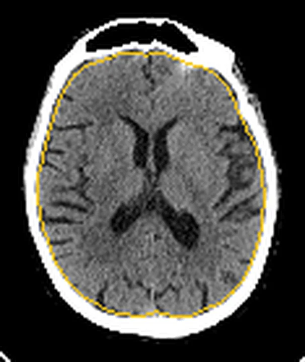
,_posterior_atrophy_(PA)_and_frontal_cortical_atrophy_(fGCA).png)
Özet
Bölgesel atrofi, hacim kaybının tüm beyne eşit dağılmadığı ve belirli lob ya da anatomik yapılarda baskınlaştığı görüntüleme örüntüsüdür. Bilişsel veya davranışsal yakınması olan hastalarda kontrastsız BT büyük ölçekli frontal, temporal ya da posterior baskınlığı gösterebilir; hafif hipokampal değişiklikleri değerlendirmede MR daha duyarlıdır. Asimetri, temporal boynuz ve hipokampal kontur ile eski doku kaybı birlikte incelenmelidir. Fokal ensefalomalazi bölgesel atrofiyi taklit edebileceğinden yanlış patern atfı klinik-radyolojik eşleşmeyi bozabilir.
Faz ve pencere
- Kontrastsız tanısal
- Frontal veya temporal konveksite sulkuslarında, Sylvian fissürde ya da parietooksipital sulkuslarda lobar ağırlıklı genişleme; hipokampal seviyede temporal boynuz ve koroid fissür çevresinde BOS aralığı artışı görülebilir. BT'de geçerli HU eşiği yoktur; dansite değil anatomik dağılım değerlendirilir.
Önerilen pencereler: Beyin (G 80 / M 40), Subdural (G 200 / M 75).
BT bulguları
- Frontal baskın sulkal açılma — frontal konveksite sulkusları karşılaştırılabilir parietal sulkuslardan daha geniştir.
- Anterior temporal hacim kaybı — temporal kutup çevresinde sulkuslar açılır ve temporal boynuz hacmi artabilir.
- Medial temporal hacim kaybı — hipokampal formasyon küçülür, koroid fissür ve temporal boynuz geniş görünür.
- Posterior baskınlık — parietal ve oksipital konveksitedeki sulkuslar frontal bölgeye göre daha belirgin açılır.
- Lobar asimetri — bir hemisferde giral hacim azalması ve sulkal genişleme karşı tarafa göre öne çıkar.
- Ventrikül konturunda eşlik eden değişim — temporal boynuz veya frontal boynuz genişlemesi, çevre parankim hacim kaybıyla birlikte okunur.
- Fokal kaviter doku kaybı — ensefalomalazide BOS yoğunluğunda kama ya da düzensiz kavite ve komşu gliozis/volüm kaybı görülebilir.
Normalde
Normal frontal ve parietal konveksite sulkusları benzer derinlikte, temporal boynuzlar ise hipokampal baş ve gövdeye komşu dar BOS çizgileri şeklindedir. Kıyaslamada frontal kutup, anterior temporal lob, hipokampal oluk ve parietooksipital konveksiteyi aynı seviyelerde sağ-sol ve ön-arka yönde karşılaştırın.
Normal BT ile kıyasla
Aynı bölgenin, kaynak veri setinde patoloji içermediği belirtilen bir BT incelemesini kesit kesit kaydırın; organ sınırlarını açarak anatomiyi hasta görüntüsüyle karşılaştırın. Küçük rastlantısal bulgular tümüyle dışlanmamıştır.
Kaynak: TotalSegmentator v2.01 veri seti (Wasserthal ve ark., Radiology: Artificial Intelligence 2023), CC BY 4.0, vaka s0103; organ sınırları veri setinin otomatik segmentasyonundan, birleştirilerek sadeleştirildi. Görüntüler 2,5 mm kesit aralığına yeniden örneklendi.
Ayırıcı tanı
- Kronik arteriyel enfarkt
- kama biçimli doku kaybı belirli damar alanına uyar ve çevresinde fokal sulkal genişleme bulunur.
- Eski travmatik kontüzyon
- çoğunlukla inferior frontal veya anterior temporal yüzeyde düzensiz ensefalomalazi odakları görülür.
- Normal basınçlı hidrosefali
- ventrikül genişlemesi lobar kortikal atrofiyle uyumsuz olabilir; konveksite sulkusları dar kalabilir.
- Asimetrik kortikal gelişim veya eski cerrahi
- hacim farklılığı olabilir; kemik defekti, cerrahi öykü veya uzun dönem stabilite yol gösterir.
Tuzaklar
- Tek taraflı sulkal genişlemeyi dejeneratif lobar patern saymadan önce komşu dilimlerde damar alanına uyan kaviter doku kaybı arayın.
- Aksiyel BT'de hipokampal uzun eksen eğik kesilir; medial temporal atrofiye dair ince derecelendirme için koronal MR tercih edilir.
- Frontal, temporal veya posterior baskınlık tek başına belirli demans etiyolojisini kanıtlamaz; görüntü örüntüsü klinik fenotiple eşleştirilir.
Kendini dene
Davranış değişikliği olan hastada anterior temporal sulkuslar asimetrik geniş; fokal kavite yok. Bulguyu en iyi ne adlandırır?
Cevabı göster
Anterior temporal baskın atrofi. Lobar ağırlıklı sulkal açılma ve kaviter damar alanı defekti olmaması anterior temporal atrofi örüntüsünü destekler. Kronik enfarkt kaviter olabilir; hidrosefali ventriküler paternle, akut ödem ise sulkus silinmesiyle seyreder.
Sol inferior frontal lobda BOS yoğunluklu kama biçimli boşluk ve çevresinde hacim kaybı var. En olası bulgu hangisidir?
Cevabı göster
Kronik enfarkt sonrası ensefalomalazi. Kama biçimli parankim kavitesi ve komşu hacim kaybı eski vasküler hasara uyar. Lobar atrofi daha yaygın sulkal açılma yapar; akut ödem doku hacmini artırıp sulkusları siler, ekstraaksiyel koleksiyon parankim dışında yer alır.
İlgili konular
Kaynaklar
Bu sayfadaki 36 cümle ve 2 quiz sorusu aşağıdaki kaynaklardan alıntılarla otomatik olarak karşılaştırıldı (2026-10-08).
- Retinal and Choroidal Vascular Metrics Predict Cerebral Atrophy and Cognitive Impairment in Noninfarcted Large Artery Stenosispmc.ncbi.nlm.nih.gov
- Structural Lung Disease in Children and Adolescents With Severe Neurological Disorderspmc.ncbi.nlm.nih.gov
- Postmortem brain MRI reveals differential associations of subcortical and limbic volumes with cortical thinning and neurodegenerative pathologiespmc.ncbi.nlm.nih.gov
- Recent Advances in Functional Liver Volumetry: Emphasis on 99mTc‐GSA SPECT/CT Fusion Imagingpmc.ncbi.nlm.nih.gov
- Posterior-to-anterior periventricular white matter hyperintensity area ratio: a supportive MRI feature for differentiating cerebral amyloid angiopathy in lobar intracerebral hemorrhagepmc.ncbi.nlm.nih.gov
- Clinical features of TBK1 carriers compared with C9orf72, GRN and non-mutation carriers in a Belgian cohort.pubmed.ncbi.nlm.nih.gov
- Morphologic changes in hepatitis virus-related liver cirrhosis: Relationship to hemodynamics of portal vein on dynamic contrast-enhanced CT.pubmed.ncbi.nlm.nih.gov
- Effect of diagnostic criteria on prevalence of frontotemporal dementia in the elderly.pubmed.ncbi.nlm.nih.gov
- Schistosoma Mansoni and Haematobium : Radiological Diagnostic Clues and Pathophysiology.pubmed.ncbi.nlm.nih.gov
- Imaging studies of kleptomania in a middle-aged woman with obsessive-compulsive disorder: a case report.pubmed.ncbi.nlm.nih.gov
- Lobar and segmental liver atrophy associated with hilar cholangiocarcinoma and the impact of hilar biliary anatomical variants: a pictorial essay.pubmed.ncbi.nlm.nih.gov
- Quantifying brain atrophy in Frontotemporal Dementia: a head-to-head comparison of neuroimaging techniques.pubmed.ncbi.nlm.nih.gov
- Enhancing brain age estimation with structural MRI and synthesized cerebral blood volume maps.pubmed.ncbi.nlm.nih.gov
- Posterior-to-anterior periventricular white matter hyperintensity area ratio: a supportive MRI feature for differentiating cerebral amyloid angiopathy in lobar intracerebral hemorrhage.pubmed.ncbi.nlm.nih.gov
- DTI-ALPS as a Biomarker of Small Vessel Disease Progression and Amyloid-β in Normal Aging: A 3-Year Longitudinal Study.pubmed.ncbi.nlm.nih.gov
- Retinal and Choroidal Vascular Metrics Predict Cerebral Atrophy and Cognitive Impairment in Noninfarcted Large Artery Stenosis.pubmed.ncbi.nlm.nih.gov
- Cognitive and Neuroimaging Divergence Between Juvenile and Adult FUS Amyotrophic Lateral Sclerosis.pubmed.ncbi.nlm.nih.gov
- Postmortem brain MRI reveals differential associations of subcortical and limbic volumes with cortical thinning and neurodegenerative pathologies.pubmed.ncbi.nlm.nih.gov
- Advances in research on dental occlusion and brain function: neuroimaging correlates and molecular mechanisms.pubmed.ncbi.nlm.nih.gov
- Phenotypic diversity of frontotemporal lobar degeneration in two novel GRN variants from Colombia.pubmed.ncbi.nlm.nih.gov
- The Relations Between Recognition Disorders of Familiar People and Other Unique Entities in Patients with Semantic and Behavioral Variants of Right Frontotemporal Degeneration: A Review of Single-Case Studies.pubmed.ncbi.nlm.nih.gov
- Real-world comparison of brain [<sup>18</sup>F]FDG-PET imaging with CSF Alzheimer's disease biomarkers in a tertiary memory clinic setting.pubmed.ncbi.nlm.nih.gov
- Prosopagnosia: face blindness and its association with neurological disorders.pubmed.ncbi.nlm.nih.gov
- Donanemab: Appropriate use recommendations.pubmed.ncbi.nlm.nih.gov
- Radiological assessment of dementia: the Italian inter-society consensus for a practical and clinically oriented guide to image acquisition, evaluation, and reporting.pubmed.ncbi.nlm.nih.gov
- Neurological manifestations and neuroimaging findings in patients with SARS-CoV2-a systematic review.pubmed.ncbi.nlm.nih.gov
- Alternating Hemispheric Episodes in Bilateral Rasmussen Encephalitis: The Crucial Role of Sequential Multimodal Neuroimaging.pubmed.ncbi.nlm.nih.gov
- Continuum of Irreversible and Potentially Reversible Traumatic Brain Injury Sequelae: Axonal Injury and Hydrocephalus.pubmed.ncbi.nlm.nih.gov
- Structural MRI phenotyping in Alzheimer's disease: Comparison of visual rating scales, volumetry, and cortical thickness in a Serbian single-centre cohort.pubmed.ncbi.nlm.nih.gov
- A comparative assessment of age- and gender-related variations in brain morphometry using linear and volumetric MRI analysis.pubmed.ncbi.nlm.nih.gov
- The Hemispheric Imbalance: A Double Encounter with Dyke-Davidoff-Masson Syndrome.pubmed.ncbi.nlm.nih.gov
- Computed tomography-based nnU-Net for region-specific brain structural changes across the alzheimer's continuum and frontotemporal dementia subtypes.pubmed.ncbi.nlm.nih.gov
- Case Report: Seizure freedom after resection of a temporal choroidal fissure cyst in drug-resistant epilepsy.pubmed.ncbi.nlm.nih.gov
- Two-year outcomes following modified transsylvian peri-insular hemispherotomy.pubmed.ncbi.nlm.nih.gov
- A systematic approach to detection of epileptogenic foci on imaging: protocols, entities, search pattern, and reporting tools.pubmed.ncbi.nlm.nih.gov
- Brain Folding Trajectories in Amyotrophic Lateral Sclerosis.pubmed.ncbi.nlm.nih.gov
- Sulcal depth-based individualized structural covariance networks reveal multi-level brain reorganization associated with cognitive performance in COPD.pubmed.ncbi.nlm.nih.gov
- Magnetic Resonance Imaging-Based Cortical and Subcortical Volumetric Changes in Alzheimer's Disease: Association with Clinical Severity.pubmed.ncbi.nlm.nih.gov
- Regional Brain Volume Variation Across Adulthood: A Cross-Sectional MRI Analysis of Age, Sex, and Hemispheric Asymmetry.pubmed.ncbi.nlm.nih.gov
- Syndrome-specific vulnerability of neuronal and glial 4-repeat frontotemporal lobar degeneration-tau.pubmed.ncbi.nlm.nih.gov
- Redefining Idiopathic Normal Pressure Hydrocephalus Using AI-Driven Brain Volumetry.pubmed.ncbi.nlm.nih.gov
- Cerebrospinal fluid dysregulation and post-traumatic hydrocephalus after paediatric traumatic brain injury.pubmed.ncbi.nlm.nih.gov
- Navigating Diagnostic Ambiguity in Palliative Pediatric Neuro-Oncology: A Conceptual Framework for Suspected Shunt Malfunction and Refractory Hyponatremia.pubmed.ncbi.nlm.nih.gov
- Foetal disruptive brain injuries: Diagnosing the underlying pathogenetic mechanisms with cranial ultrasonography.pubmed.ncbi.nlm.nih.gov
- Fetal intracranial hemorrhage and infarct: Main sonographic and MRI characteristics: A review article.pubmed.ncbi.nlm.nih.gov
- Paediatric cranial ultrasound: assessment of the preterm brain.pubmed.ncbi.nlm.nih.gov
- Computational Neuroanatomy of Language: A Compartmental Framework From Cranial Landmarks to Subcortical Networks.pubmed.ncbi.nlm.nih.gov
- From non-human to human primates: a translational approach to enhancing resection, safety, and indications in glioma surgery while preserving sensorimotor abilities.pubmed.ncbi.nlm.nih.gov
- Prenatal assessment of brain malformations on neuroimaging: an expert panel review.pubmed.ncbi.nlm.nih.gov
- Anterior Superior Longitudinal Fasciculus Microstructure Correlates With Bimanual Visuomotor Performance in Healthy Younger but Not Older Adults.pubmed.ncbi.nlm.nih.gov
- Correlation between brain symmetry index and motor function in Parkinson's disease: a cross-sectional study.pubmed.ncbi.nlm.nih.gov
- Mapping Neuroanatomical Heterogeneity of Brain Aging Within a Clinically Defined Aging Reference Cohort.pubmed.ncbi.nlm.nih.gov
- Case Report: Structured MRI assessment of posterior thalamic infarction in a distribution compatible with posterior choroidal artery territory presenting as Déjerine-Roussy syndrome in an adolescent: differentiating arterial ischemia from venous thrombosis and thalamic neoplasm.pubmed.ncbi.nlm.nih.gov
- When Stroke Leads to Hydrocephalus: A Case of Unilateral Ventricular Enlargement.pubmed.ncbi.nlm.nih.gov
- Encephalomalacia from Physical Trauma in an Adult: A Case Report and Review of Literature.pubmed.ncbi.nlm.nih.gov
- Post-Traumatic Middle Cerebral Artery Pseudoaneurysm Following Depressed Skull Fracture: Case Report and PRISMA-Guided Literature Review.pubmed.ncbi.nlm.nih.gov
- Case Report: Prism for PTSD in severe traumatic brain injury with psychiatric comorbidities: two cases.pubmed.ncbi.nlm.nih.gov
- The Unspoken Danger of the Mount Fuji Sign Leading to Sudden Death.pubmed.ncbi.nlm.nih.gov
- Degenerative pathologies on cortical biopsy, dopaminergic depletion, and shunt efficacy in iNPH.pubmed.ncbi.nlm.nih.gov
- Structural neuroimaging markers of normal pressure hydrocephalus versus Alzheimer's dementia and Parkinson's disease, and hydrocephalus versus atrophy in chronic TBI-a narrative review.pubmed.ncbi.nlm.nih.gov
- Radiological predictors of shunt response in the diagnosis and treatment of idiopathic normal pressure hydrocephalus: a systematic review and meta-analysis.pubmed.ncbi.nlm.nih.gov
- Deep grey nuclei automated assessment in acute-subacute phase of middle cerebral artery cortical stroke helps predict the 3-month outcome.pubmed.ncbi.nlm.nih.gov
- Exploring the Role of TSPO-PET Imaging Among MRI-Negative Patients with Temporal Lobe Epilepsy: From the Perspective of Heterogeneity.pubmed.ncbi.nlm.nih.gov
- Paediatric cranial ultrasound: abnormalities of the brain in term neonates and young infants.pubmed.ncbi.nlm.nih.gov
- Emerging Neural Recording and Neurostimulation Technologies Based on Brain-Computer Interface: A Promising Approach for Neuropsychiatric Disorders.pubmed.ncbi.nlm.nih.gov
- Radiologic Classification of Hippocampal Sclerosis in Epilepsy.pubmed.ncbi.nlm.nih.gov
- High-resolution Structural Magnetic Resonance Imaging and Quantitative Susceptibility Mapping.pubmed.ncbi.nlm.nih.gov
- Qualitative and quantitative hippocampal MRI assessments in intractable epilepsy.pubmed.ncbi.nlm.nih.gov
- Autopsy Confirmed Normal Pressure Hydrocephalus (NPH) Presenting With Corticobasal Syndrome.pubmed.ncbi.nlm.nih.gov
- Biomarkers of post-stroke cognitive impairment-a systematic literature review.pubmed.ncbi.nlm.nih.gov
- Targeting protein aggregate co-pathologies in neurodegeneration: a viable therapeutic strategy?pubmed.ncbi.nlm.nih.gov
- Evolutionary implications of <i>NOTCH2NLC</i> mutations: brain structural changes in neuronal intranuclear inclusion disease revealed by comprehensive morphometry.pubmed.ncbi.nlm.nih.gov
- Longitudinal patterns of regional cerebral blood flow during anti-amyloid therapy in mild cognitive impairment: a pilot ASL MRI case series.pubmed.ncbi.nlm.nih.gov
- Comparative Analysis of Four Commercially Available Artificial Intelligence Software for Brain Volumetry.pubmed.ncbi.nlm.nih.gov
- Subcortical morphological alterations and volume loss in infantile hydrocephalus: A surface- and volume-based analysis.pubmed.ncbi.nlm.nih.gov
- Alterations in glymphatic system and brain morphology in patients with temporal lobe epilepsy.pubmed.ncbi.nlm.nih.gov
- Enhanced Detection of Age-Related and Cognitive Declines Using Automated Hippocampal-To-Ventricle Ratio in Alzheimer's Patients.pubmed.ncbi.nlm.nih.gov
- Assessment of Cerebral Perfusion Asymmetry in Chronic Migraine Patients Using Intravoxel Incoherent Motion (IVIM)-Derived Pseudodiffusion Coefficient and Perfusion Fraction.pubmed.ncbi.nlm.nih.gov
- Quantitative Navigated Transcranial Magnetic Stimulation Motor Mapping and nTMS-Guided Corticospinal Tractography in Healthy Individuals and Patients with Motor-Eloquent Brain Tumors.pubmed.ncbi.nlm.nih.gov
- Atlas of cortical sulcal imprints on Pan endocasts.pubmed.ncbi.nlm.nih.gov
- Motifs of brain cortical folding from birth to adulthood.pubmed.ncbi.nlm.nih.gov
- Volume-inflation registration (INFREG) for morphometric analysis of human focal cortical dysplasia type II.pubmed.ncbi.nlm.nih.gov
- Behavior determines times to care home admission and death in semantic dementia: Insights from a cohort of 90 cases.pubmed.ncbi.nlm.nih.gov
- Early-onset frontotemporal dementia misdiagnosed as psychosis: a case from rural Nepal.pubmed.ncbi.nlm.nih.gov
- A 57-Year-Old Male With Behavioral Variant Frontotemporal Dementia and MATR3 and NOS3 Mutations.pubmed.ncbi.nlm.nih.gov
- Neuroimaging standards for research into small vessel disease and its contribution to ageing and neurodegeneration.pubmed.ncbi.nlm.nih.gov
- COVID-19 and Eye: A Review of Ophthalmic Manifestations of COVID-19.pubmed.ncbi.nlm.nih.gov
- Long-term safety and effectiveness of the "OptEase" vena cava filter.pubmed.ncbi.nlm.nih.gov
- Comprehensive review of pulmonary embolism imaging: past, present and future innovations in computed tomography (CT) and other diagnostic techniques.pubmed.ncbi.nlm.nih.gov
- Abdominal and gastrointestinal manifestations in COVID-19 patients: Is imaging useful?pubmed.ncbi.nlm.nih.gov
- Traumatic Brain Injury and Chronic Traumatic Encephalopathy: Not Only Trigger for Neurodegeneration but Also for Cerebral Amyloid Angiopathy?pubmed.ncbi.nlm.nih.gov
- Cerebral small vessel disease: Recent advances and future directions.pubmed.ncbi.nlm.nih.gov
- Pathophysiology and probable etiology of cerebral small vessel disease in vascular dementia and Alzheimer's disease.pubmed.ncbi.nlm.nih.gov
- CSF1R mutations in an Italian population of early-onset dementia: a case series.pubmed.ncbi.nlm.nih.gov
- A multimodal clinical diagnostic approach using MRI and <sup>18</sup>F-FDG-PET for antemortem diagnosis of TDP-43 in cases with low-intermediate Alzheimer's disease neuropathologic changes and primary age-related tauopathy.pubmed.ncbi.nlm.nih.gov
- Spatial covariance of mGluR5 density and structural degeneration in behavioral variant frontotemporal degeneration.pubmed.ncbi.nlm.nih.gov
- Rostral associations of MRI atrophy of the amygdala and entorhinal cortex across the AD spectrum.pubmed.ncbi.nlm.nih.gov
- Soft tissue changes in Dementia: A comprehensive systematic review.pubmed.ncbi.nlm.nih.gov
- FuzzyFed-CNN: secure and explainable multimodal federated learning for early Alzheimer's diagnosis.pubmed.ncbi.nlm.nih.gov
- Neural Correlates of Huntington's Disease Based on Electroencephalography (EEG): A Mechanistic Review and Discussion of Excitation and Inhibition (E/I) Imbalance.pubmed.ncbi.nlm.nih.gov
- A review of brain regions and associated post-concussion symptoms.pubmed.ncbi.nlm.nih.gov
- MRI-Guided Electrode Implantation for Chronic Intracerebral Recordings in a Rat Model of Post-Traumatic Epilepsy-Challenges and Gains.pubmed.ncbi.nlm.nih.gov
- Neurophysiological evidence for crossmodal (face-name) person-identity representation in the human left ventral temporal cortex.pubmed.ncbi.nlm.nih.gov
- Pathological insights into cerebral amyloid angiopathy underlying intracerebral haemorrhage: population-based autopsy study.pubmed.ncbi.nlm.nih.gov
- PET Imaging in Alzheimer Disease in the Era of Antiamyloid Therapy in the United States: Clinical Utility, Quantification, and Policy Landscape.pubmed.ncbi.nlm.nih.gov
- Structure-aware medical image fusion via mean curvature enhancement in the contourlet domain.pubmed.ncbi.nlm.nih.gov
- Deep learning interpretability in neuroimaging: A comprehensive survey and methodological recommendations.pubmed.ncbi.nlm.nih.gov
- Early-stage differentiation between Alzheimer's disease and frontotemporal lobe degeneration: Clinical, neuropsychology, and neuroimaging features.pubmed.ncbi.nlm.nih.gov
Eğitim amaçlıdır; hasta başında tanı ve tedavi kararının yerine geçmez, tıbbi tavsiye değildir. Metinler yapay zekâ desteğiyle yazıldı ve otomatik bir adımda kaynak alıntılarıyla karşılaştırıldı; bu bir tıbbi doğrulama değildir ve içerik uzman radyolog incelemesinden geçmedi. Klinik kararlarda güncel kılavuzları, kurum protokollerini ve radyoloji raporunu esas alın.