Özet
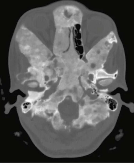
Kraniofasiyal fibröz displazi, normal kemik iliği ve lameller kemiğin fibröz doku ile örgü kemiğe dönüştüğü benign fibro-osseöz bir lezyondur; kalvaryum ve kafa tabanında tek kemik ya da birden çok kemiği tutabilir. Kontrastsız ince kesit BT, karakteristik buzlu cam matriksi, kemik ekspansiyonunu ve kanal-foramen ilişkisini en iyi gösteren yöntemdir. Lezyon matriksi yaşla ve kemik içindeki bileşenlerle değişken görünebildiğinden dağılım ve kortikal remodeling de tanımlanmalıdır. Optik kanal veya kraniyal sinir geçişlerinin daralmasını fark etmemek anatomik riskin değerlendirilmesini eksik bırakabilir; yalnız kanal daralması ise klinik sinir işlev kaybını tek başına göstermez.
Faz ve pencere
- Kontrastsız tanısal
- Kemik algoritmasında medüller boşluğu silen buzlu cam dansiteli ya da değişken mineralize matriks, ekspansiyon, kortikal incelme ve düzgün remodeling değerlendirilir; tutulum optik kanal, orbital apeks, kafa tabanı forameni ve klivusla ilişkili olarak haritalanır. Kontrastlı faz lezyon matriksini tanımlamak için gerekli değildir.
Önerilen pencereler: Kemik (G 1800 / M 400).
BT bulguları
- Medüller diploik alanı dolduran buzlu cam benzeri homojen veya hafif heterojen mineralize matriks
- Diploik mesafede ekspansiyon, dış tabulada düzgün kabalaşma ve iç-dış korteksin yeniden şekillenmesi
- Lezyon kenarında çoğu kez keskin tümör kapsülü yerine çevre kemiğe geçişli, daralan sınır
- Kalvaryum, sfenoid, frontal kemik, maksilla veya diğer kraniofasiyal kemiklerde tek odaklı ya da çok odaklı dağılım
- Optik kanal, superior orbital fissür, foramen rotundum/ovale ve diğer kafa tabanı geçişlerinde kemik daralması veya kanal çevresinde sarılma
- Matriksin bazı alanlarda daha sklerotik, litik ya da kistik görünmesi; klasik buzlu cam görünümünün tüm kesitlerde aynı yoğunlukta olmayabilmesi
- Agresif kortikal yıkım, yumuşak doku kitlesi veya yeni belirgin ağrı öyküsü varsa alışılmış düzgün remodeling paterninden sapmanın not edilmesi
Normalde
Normal kalvaryumda diploik aralık trabeküler kemik içerir; iç ve dış tabula ince, düzenli kortikal sınırlardır ve kafa tabanı kanalları çevre kemikle açık konturunu korur. Etkilenen kemiği karşı tarafla ve komşu kemiklerle kıyaslayarak diploik ekspansiyon, buzlu cam matriks, kortikal remodeling ve optik kanal ya da kraniyal sinir forameni çapındaki asimetriyi ara.
Normal BT ile kıyasla
Aynı bölgenin, kaynak veri setinde patoloji içermediği belirtilen bir BT incelemesini kesit kesit kaydırın; organ sınırlarını açarak anatomiyi hasta görüntüsüyle karşılaştırın. Küçük rastlantısal bulgular tümüyle dışlanmamıştır.
Kaynak: TotalSegmentator v2.01 veri seti (Wasserthal ve ark., Radiology: Artificial Intelligence 2023), CC BY 4.0, vaka s0103; organ sınırları veri setinin otomatik segmentasyonundan, birleştirilerek sadeleştirildi. Görüntüler 2,5 mm kesit aralığına yeniden örneklendi.
Ayırıcı tanı
- Ossifiye fibrom
- özellikle çene ve yüz kemiklerinde iyi sınırlı, çevre kemikten ayrılan ekspansil kitle görünümü fibröz displazinin geçişli sınırından ayrılır.
- Fibröz displazinin radyografik görünümleri literatürde “pagetoid” olarak da tanımlanmıştır.
- İntraosseöz meningiom
- genellikle fokal hiperostoz ve komşu dural/yumuşak doku ilişkisi oluşturur; medüller buzlu cam matriks beklenen özellik değildir.
- Metastaz veya miyelom
- litik, sklerotik ya da çok odaklı kemik destrüksiyonu yapabilir; düzgün ekspansil remodeling ve buzlu cam matriks yerine kortikal yıkım görülebilir.
- Kraniofasiyal osteom
- yoğun kemik dansiteli, sınırlı dışa büyüyen lezyondur; diploik boşluğu yaygın buzlu cam dokuyla değiştiren patern göstermez.
Tuzaklar
- Buzlu cam matriks her olguda homojen değildir; heterojen veya kistik alanlar tanıyı dışlamaz, ancak dağılımı ve korteksin davranışını yeniden gözden geçirmeyi gerektirir.
- Kanalın BT'de dar görünmesi optik nöropati ya da kraniyal nöropati demek değildir; anatomik daralmayı tarif edin, işlev kaybını görüntüden varsaymayın.
- Lezyon sınırının keskin olmaması malignite anlamına gelmez; fibröz displazide kemikten lezyona geçiş sıklıkla yumuşaktır, destrüktif kortikal kırılma ve yumuşak doku kitlesi ayrı uyarı bulgularıdır.
Kendini dene
Genç erişkinde sfenoid kemikte geçişli sınırlı, ekspansil buzlu cam matriks ve düzgün kortikal remodeling var. En olası tanı nedir?
Cevabı göster
Fibröz displazi. Geçişli sınırla birlikte buzlu cam matriks ve düzgün kemik remodeling fibröz displaziyi destekler. Ossifiye fibrom daha iyi sınırlı ekspansil kitle; intraosseöz meningiom fokal hiperostoz; metastaz ise sıklıkla farklı litik/sklerotik ve yıkıcı özellikler gösterir.
Kafa tabanı fibröz displazisinde optik kanal BT'de dar; hangi çıkarım doğrudur?
Cevabı göster
Anatomik daralma gösterilmiştir. BT kemik kanalının anatomik daralmasını gösterebilir, ancak görme işlevini veya nöropatiyi tek başına belirlemez. Kontrastsız kemik matriksi tanısal değerlendirmeyi sağlar; daralma malign dönüşüm kanıtı değildir.
Kaynaklar
Bu sayfadaki 35 cümle ve 2 quiz sorusu aşağıdaki kaynaklardan alıntılarla otomatik olarak karşılaştırıldı (2026-10-09).
- Clinical guidelines for the management of craniofacial fibrous dysplasiapmc.ncbi.nlm.nih.gov
- Pediatric Fibrous Dysplasia of the Skull Base: Update on Management and Treatmentpmc.ncbi.nlm.nih.gov
- Imaging of Skull Base Tumorspmc.ncbi.nlm.nih.gov
- Surgical treatment of craniofacial fibrous dysplasia in adults.pubmed.ncbi.nlm.nih.gov
- Craniofacial Fibrous Dysplasia Involvements of Mccune-Albright Syndrome: A Review with an Additional Case.pubmed.ncbi.nlm.nih.gov
- Clinical guidelines for the management of craniofacial fibrous dysplasia.pubmed.ncbi.nlm.nih.gov
- Radiation-induced dedifferentiated chondrosarcoma with orbital invasion.pubmed.ncbi.nlm.nih.gov
- Computed Tomography in Craniofacial Fibrous Dysplasia: A Case Series with Review of Literature and Classification Update.pubmed.ncbi.nlm.nih.gov
- Craniofacial fibrous dysplasia: A review of current literature.pubmed.ncbi.nlm.nih.gov
- Craniofacial Fibrous Dysplasia Involving the Orbit: A Case Report and Literature Review.pubmed.ncbi.nlm.nih.gov
- Reduction of giant parietooccipital fibrous dysplasia using dynamic mirror image guidance: a case report and review of the literature.pubmed.ncbi.nlm.nih.gov
- [Craniofacial fibrous dysplasia].pubmed.ncbi.nlm.nih.gov
- Craniofacial fibrous dysplasia.pubmed.ncbi.nlm.nih.gov
- Computed tomography imaging findings of craniofacial fibrous dysplasia.pubmed.ncbi.nlm.nih.gov
- The indication and application of computer-assisted navigation in oral and maxillofacial surgery-Shanghai's experience based on 104 cases.pubmed.ncbi.nlm.nih.gov
- Radiological Imaging Findings of Craniofacial Fibrous Dysplasia.pubmed.ncbi.nlm.nih.gov
- An Unusual Skull With Bilateral Multiple Bony Lesions on the Parietal and Frontal Bones of the Superior Surface of the Calvaria: A Case Report.pubmed.ncbi.nlm.nih.gov
- Monostotic fibrous dysplasia of jaw bones: a case series.pubmed.ncbi.nlm.nih.gov
- Review of imaging modalities and radiological findings of calvarial lesions.pubmed.ncbi.nlm.nih.gov
- Imaging of Skull Base Tumors.pubmed.ncbi.nlm.nih.gov
- Intraosseous Venous Malformations of the Head and Neck.pubmed.ncbi.nlm.nih.gov
- Orbital apex disorders: Imaging findings and management.pubmed.ncbi.nlm.nih.gov
- Multidisciplinary approach and clinical management of McCune-Albright syndrome.pubmed.ncbi.nlm.nih.gov
- Fifty Shades of PSMA-Avid Rib Lesions: A Comprehensive Review.pubmed.ncbi.nlm.nih.gov
- The pathogenesis of cerebral small vessel disease and vascular cognitive impairment.pubmed.ncbi.nlm.nih.gov
- Orbital tumours and tumour-like lesions: exploring the armamentarium of multiparametric imaging.pubmed.ncbi.nlm.nih.gov
- International Society of Paediatric Surgical Oncology (IPSO) Surgical Practice Guidelines.pubmed.ncbi.nlm.nih.gov
- Commentary: an eye on PET quantification.pubmed.ncbi.nlm.nih.gov
- Long-term safety and effectiveness of the "OptEase" vena cava filter.pubmed.ncbi.nlm.nih.gov
- Decoding calvarial lesions: Contemporary neurosurgical management integrating radiological and histopathological features.pubmed.ncbi.nlm.nih.gov
- Low-grade Central Osteosarcoma with High-grade Transformation in the Distal Tibia: A Rare Case Report.pubmed.ncbi.nlm.nih.gov
- Magnetic Resonance Imaging Features of Hepatic Hydatid Disease: A Pictorial Review with Emphasis on Atypical Presentations and Differential Diagnosis.pubmed.ncbi.nlm.nih.gov
- Osteoblastic and hyperostotic craniofacial lesion detected by 99mTc-labeled methylene diphosphonate bone scintigraphy and single-photon emission computed tomography/computed tomography: a pictorial essay.pubmed.ncbi.nlm.nih.gov
- TGF-β Family Signaling in Connective Tissue and Skeletal Diseases.pubmed.ncbi.nlm.nih.gov
- Diseases of the tongue: a review of etiologies and treatments.pubmed.ncbi.nlm.nih.gov
- Spectrum of Biliary and Nonbiliary Neoplasms Growing and Spreading Within the Lumen of the Bile Ducts.pubmed.ncbi.nlm.nih.gov
- Spectrum of Biliary Lesions/Neoplasms in Hepatic Parenchyma with Reference to a Precursor of Small Duct-Type Intrahepatic Cholangiocarcinoma: Comprehensive Categorization into Three Groups.pubmed.ncbi.nlm.nih.gov
- Calvarial lesions: overview of imaging features and neurosurgical management.pubmed.ncbi.nlm.nih.gov
- MR imaging of orbital inflammatory pseudotumors with extraorbital extension.pubmed.ncbi.nlm.nih.gov
- Revisiting Fat Content in Bone Lesions: Paradigms in Bone Lesion Detection.pubmed.ncbi.nlm.nih.gov
- Osteoarthritis: Epidemiology, Diagnosis, and Treatment.pubmed.ncbi.nlm.nih.gov
- The solitary skull mass: An atypical presentation of anaplastic large cell lymphoma.pubmed.ncbi.nlm.nih.gov
- When plasmacytoma defies expectations: An unusual maxillary presentation with mixed imaging features in a young patient.pubmed.ncbi.nlm.nih.gov
- Posttraumatic intradiploic cyst-like lesion of the frontal bone without cortical disruption: A case report.pubmed.ncbi.nlm.nih.gov
- Pott's Puffy Tumor: A Case Report of a Rare Complication of Frontal Sinusitis.pubmed.ncbi.nlm.nih.gov
- How Clinical and Radiological Findings in Chronic Mandibular Osteomyelitis Do Not Always Correlate: Diagnostic Dilemmas in Dental-Related Bone Inflammations.pubmed.ncbi.nlm.nih.gov
- Aneurysmal bone cyst associated with a dentigerous cyst in the mandible: A diagnostic challenge-case report.pubmed.ncbi.nlm.nih.gov
- Navigating Fibrous Dysplasia of the Middle Turbinate: A Decade-Long Journey and Comprehensive Management Strategies.pubmed.ncbi.nlm.nih.gov
- Why was the concordance rate of imaging and clinical diagnosis in cemento-osseous dysplasia low? A retrospective study of 55 cases.pubmed.ncbi.nlm.nih.gov
- ANO5 p.I616F variant drives florid cemento-osseous dysplasia by disrupting transmembrane domain structure.pubmed.ncbi.nlm.nih.gov
- Multimodal Imaging of Osteosarcoma: From First Diagnosis to Radiomics.pubmed.ncbi.nlm.nih.gov
- Perineurioma-like EMA-positive calvarial neoplasms: clinicopathological study of eight cases.pubmed.ncbi.nlm.nih.gov
- Malignant Transformation of Musculoskeletal Lesions with Imaging-Pathology Correlation-Part 1: Bone Lesions.pubmed.ncbi.nlm.nih.gov
- Unveiling Primary Bone Tumors of the Spine: A Review of Essential Imaging Clues.pubmed.ncbi.nlm.nih.gov
- Clival and paraclival pathologies: imaging features and differential diagnosis.pubmed.ncbi.nlm.nih.gov
- A practical overview of CT and MRI features of developmental, inflammatory, and neoplastic lesions of the sphenoid body and clivus.pubmed.ncbi.nlm.nih.gov
- Cavernous venous malformation of the frontal sinus: CT and MRI imaging findings.pubmed.ncbi.nlm.nih.gov
- Benign and malignant lesions of the mandible and maxilla: imaging findings and systematic approach.pubmed.ncbi.nlm.nih.gov
- A pictorial essay of thoracic wall diseases: multiple pathologies in the same anatomical site.pubmed.ncbi.nlm.nih.gov
- Clinical summary of fibroblast activation protein inhibitor-based radiopharmaceuticals: cancer and beyond.pubmed.ncbi.nlm.nih.gov
- Salvaging Vision: A Study of Non-Traumatic Optic Neuropathies.pubmed.ncbi.nlm.nih.gov
- Surgery versus watchful waiting in patients with craniofacial fibrous dysplasia--a meta-analysis.pubmed.ncbi.nlm.nih.gov
- Osteomyelitis of the Jaw Bones and Its Mimics: Resolving the Diagnostic Enigma.pubmed.ncbi.nlm.nih.gov
- Ossifying fibroma of the maxilla: A case report with literature review.pubmed.ncbi.nlm.nih.gov
- Radiological review of skull lesions.pubmed.ncbi.nlm.nih.gov
- Skull Base Tumors and Tumor-Like Lesions: A Pictorial Review.pubmed.ncbi.nlm.nih.gov
- Jansen metaphyseal chondrodysplasia: analysis of craniofacial manifestations.pubmed.ncbi.nlm.nih.gov
- Three-Dimensional Constructive Interference in Steady State (3D CISS) Imaging and Clinical Applications in Brain Pathology.pubmed.ncbi.nlm.nih.gov
- Sellar and parasellar lesions: multidisciplinary management.pubmed.ncbi.nlm.nih.gov
- Diagnostic neuroimaging in neuro-ophthalmic disorders.pubmed.ncbi.nlm.nih.gov
- Radiological and Surgical Findings of Patients with Intradiploic Mass Lesion: Case Series and Systematic Review.pubmed.ncbi.nlm.nih.gov
- Update of Key Clinical, Histological and Molecular Features of Malignant Bone Tumours Arising in the Craniofacial Skeleton.pubmed.ncbi.nlm.nih.gov
- Task force Guideline of Brazilian Society of Otology ‒ hearing loss in children - Part I ‒ Evaluation.pubmed.ncbi.nlm.nih.gov
- Comprehensive review on endonasal endoscopic sinus surgery.pubmed.ncbi.nlm.nih.gov
- Polyostotic fibrous dysplasia: Unveiling the complexity of a rare bone disorder through a case report.pubmed.ncbi.nlm.nih.gov
- Unusual Manifestations of Sinonasal Osteomas: A Narrative Review.pubmed.ncbi.nlm.nih.gov
- What's that smell? A pictorial review of the olfactory pathways and imaging assessment of the myriad pathologies that can affect them.pubmed.ncbi.nlm.nih.gov
- Fibrous dysplasia: an overview of disease process, indications for surgical management, and a case report.pubmed.ncbi.nlm.nih.gov
- Lesion Expansion in Craniofacial Fibrous Dysplasia: Natural History and Indicators of Progression in the Frontal, Zygomatic, and Sphenoid Bones.pubmed.ncbi.nlm.nih.gov
- Intracranial aneurysmal bone cysts in association with fibrous dysplasia: Case report and scoping review of case reports.pubmed.ncbi.nlm.nih.gov
- Fibrous Dysplasia Presenting as an Exophytic Gingival Mass: A Rare Clinical Presentation.pubmed.ncbi.nlm.nih.gov
- Conventional radiography for the assessment of focal bone lesions of the appendicular skeleton: fundamental concepts in the modern imaging era.pubmed.ncbi.nlm.nih.gov
- Society of Skeletal Radiology- white paper. Guidelines for the diagnostic management of incidental solitary bone lesions on CT and MRI in adults: bone reporting and data system (Bone-RADS).pubmed.ncbi.nlm.nih.gov
- Massive Mandibular Fibrous Dysplasia Mimicking Calcifying Epithelial Odontogenic Tumor Radiologically: A Case Report.pubmed.ncbi.nlm.nih.gov
- Orbital pseudotumor and skull base infiltration as the sole manifestation of granulomatosis with polyangiitis.pubmed.ncbi.nlm.nih.gov
- Clival fibrous dysplasia in which short interval disease progression posed a diagnostic challenge in a skeletally mature patient: a case report.pubmed.ncbi.nlm.nih.gov
- Fibrous dysplasia: Rare sphenoidal and parietal involvement.pubmed.ncbi.nlm.nih.gov
- Minimally invasive fixation and management of sacral fibrous dysplasia with impending pathological fracture: illustrative case.pubmed.ncbi.nlm.nih.gov
Eğitim amaçlıdır; hasta başında tanı ve tedavi kararının yerine geçmez, tıbbi tavsiye değildir. Metinler yapay zekâ desteğiyle yazıldı ve otomatik bir adımda kaynak alıntılarıyla karşılaştırıldı; bu bir tıbbi doğrulama değildir ve içerik uzman radyolog incelemesinden geçmedi. Klinik kararlarda güncel kılavuzları, kurum protokollerini ve radyoloji raporunu esas alın.