Özet
Kraniyosinostoz, bir veya daha fazla sütürün erken füzyonudur; pozisyonel kafa şekil bozukluğu ve yaşa bağlı fizyolojik sütür kapanması benzer kontur veya sütür görünümü oluşturabilir. Bebeklerde değerlendirme klinik kafa şekli muayenesiyle başlar; tanı belirsizse seçilmiş olgularda BT sütür füzyonunu doğrulamaya ve eşlik eden kafatası şekil değişikliklerini değerlendirmeye yardımcı olabilir. Bebeklerde klinik muayene ve gerektiğinde radyasyonsuz ultrasonografi ilk değerlendirmeyi destekler; BT özellikle karmaşık kafa şekil bozukluklarında planlama için, kemik ayrıntısı ve 3B rekonstrüksiyon amacıyla kullanılabilir. Pozisyonel plagiosefali lambdoid sinostozdan daha sık görüldüğü için bu iki durum ayırt edilmelidir; fizyolojik metopik ridgingin sinostoz sayılması gereksiz cerrahiye yol açabilir.
Faz ve pencere
- Kontrastsız tanısal
- İnce kesit BT'de şüpheli sütür boyunca aralığın devamlılığı, serrasyon kaybı, perisutural skleroz veya kemik köprüleşmesi ile kafatası konturu değerlendirilir.
- Kemik algoritması tanısal
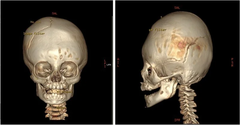
- Aksiyel ve ortogonal rekonstrüksiyonlarla bütün sütür seyri incelenir; 3B yüzey görüntüsü kafa şekli, frontal bossing, oksipital düzleşme ve mastoid kontur farkını gösterir.
Önerilen pencereler: Kemik (G 1800 / M 400), Beyin (G 80 / M 40).
BT bulguları
- Sütür aralığı — Açık sütür çizgisi boyunca aralık ve serrasyonlar sürer; füzyonda aralık kaybolabilir, serrasyon azalabilir, kenarlar doğrusal hale gelebilir veya kemik köprüleşmesi görülebilir.
- Perisutural kemik — skleroz ve kemik köprü, yalnız tek seviyede kaybolmuş sütür görünümünden daha anlamlıdır.
- Pozisyonel plagiosefali — lambdoid sütür açık kalır; ipsilateral oksipital düzleşmeye ipsilateral frontal bossing eşlik edebilir.
- Lambdoid sinostoz şekli — etkilenen sütür füzyonuna ipsilateral oksipital düzleşme ve mastoid bölge çıkıntısı eşlik edebilir.
- Metopik varyant — metopik sütürün fizyolojik kapanması tek başına kraniyosinostoz tanısı koydurmaz; trigonosefali ve orbital bulgular da değerlendirilmelidir.
- Birden çok sütür — karşı sütürlerin açıklığı ve kafatasının kompansatuvar genişleme yönü, tek sütür bulgusunun anlamını belirlemeye yardım eder.
Normalde
Normal bebekte açık sütür, kemik görüntülerinde radyolüsent, serrasyonlu ve doğrusal olmayan bir çizgi olarak izlenir; yenidoğan sütürlerinin görünümü yaşa göre değişken olabilir. Şüpheli tarafta aynı sütürün karşı tarafını, diğer kalvaryal sütürleri ve frontal/oksipital kemik konturunu kıyaslayın; yalnız sütürün kapanmasına değil, çevresindeki skleroz ve kafa şekline bakın.
Normal BT ile kıyasla
Aynı bölgenin, kaynak veri setinde patoloji içermediği belirtilen bir BT incelemesini kesit kesit kaydırın; organ sınırlarını açarak anatomiyi hasta görüntüsüyle karşılaştırın. Küçük rastlantısal bulgular tümüyle dışlanmamıştır.
Kaynak: TotalSegmentator v2.01 veri seti (Wasserthal ve ark., Radiology: Artificial Intelligence 2023), CC BY 4.0, vaka s0103; organ sınırları veri setinin otomatik segmentasyonundan, birleştirilerek sadeleştirildi. Görüntüler 2,5 mm kesit aralığına yeniden örneklendi.
Ayırıcı tanı
- Pozisyonel plagiosefali
- sütür açıktır; asimetri oksipital düzleşme ve aynı tarafta frontal bossing ile birlikte görülebilir.
- Lambdoid kraniyosinostoz
- lambdoid sütürde füzyon bulguları ve buna eşlik eden mastoid/oksipital şekil değişikliği gösterir.
- Fizyolojik metopik kapanma
- metopik sütür kapanabilir; eşlik eden trigonosefali veya orbital daralma yoksa tek başına sinostoz tanısı koydurmaz.
- Kafa deformasyonuna yol açan konjenital pozisyon
- sütürleri açık bırakabilir; görüntüde füzyon yerine bası/deformasyon konturu öne çıkar.
Tuzaklar
- Metopik sütürün fizyolojik kapanmasını yalnız sütür çizgisinin görünmemesine dayanarak metopik sinostoz saymayın; trigonosefali ve orbital bulguları da değerlendirin.
- Pozisyonel plagiosefalide posterior asimetriyi lambdoid füzyon kanıtı saymayın; sütürün bütün uzunluğunu multiplanar kemik görüntülerde doğrulayın.
- Bir tek kesitte görülen kısa açıklık kaybı parsiyel hacim etkisi olabilir; sütürü aksiyel, koronal ve sagittal rekonstrüksiyonlarda izleyin.
- Kafatası şeklindeki asimetriyi tek başına BT'de sinostoz olarak etiketlemeyin; ilgili sütürde kemik köprüleşmesi ve eşleşen şekil paternini arayın.
Kendini dene
Bebekte posterior asimetri var; lambdoid sütür açık, ipsilateral oksiput düz ve ipsilateral frontal bossing mevcut. En olası tanı nedir?
Cevabı göster
Pozisyonel plagiosefali. Pozisyonel plagiosefalide lambdoid sütür patenttir; oksipital düzleşmeye ipsilateral frontal bossing eşlik edebilir. Lambdoid sinostozda lambdoid sütür füzyonu beklenir.
Metopik sütür BT'de kapanmış; alın üçgen biçiminde (trigonosefali) ve orbitler birbirine yakın görünüyor. Hangi tanı daha olasıdır?
Cevabı göster
Metopik kraniyosinostoz. Trigonosefali ve azalmış interorbital mesafe metopik sinostozla ilişkili bulgulardır; fizyolojik metopik kapanma tek başına tanı koydurmaz. Tek bir BT bulgusu tanısal olmadığından bulgular birlikte değerlendirilmelidir.
İlgili konular
Kaynaklar
Bu sayfadaki 35 cümle ve 2 quiz sorusu aşağıdaki kaynaklardan alıntılarla otomatik olarak karşılaştırıldı; 10 cümle bu karşılaştırmada düzeltildi (2026-10-09).
- Craniosynostosis : Updates in Radiologic Diagnosispmc.ncbi.nlm.nih.gov
- Practical Computed Tomography Scan Findings for Distinguishing Metopic Craniosynostosis from Metopic Ridgingpmc.ncbi.nlm.nih.gov
- The Use of Machine Learning to Enable Objective Assessment of Surgical Outcomes in Sagittal Craniosynostosispmc.ncbi.nlm.nih.gov
- Long-term morphometric and functional outcomes of frontofacial advancement in syndromic craniosynostosispmc.ncbi.nlm.nih.gov
- Geometric Fidelity of Magnetic Resonance Imaging and Computed Tomography-Derived Virtual 3D Models of Porcine Cadaver Mandibles: Conventional Versus Artificial Intelligence-Based Segmentationpmc.ncbi.nlm.nih.gov
- Automated Sagittal Craniosynostosis Classification from CT Images Using Transfer Learning.pubmed.ncbi.nlm.nih.gov
- Morphological Basis for Airway Surgical Intervention in Apert Syndrome.pubmed.ncbi.nlm.nih.gov
- Orbital volume, ophthalmic sequelae and severity in unilateral coronal synostosis.pubmed.ncbi.nlm.nih.gov
- Craniometric and Volumetric Analysis of Posterior Vault Distraction Osteogenesis: 10 Year Update.pubmed.ncbi.nlm.nih.gov
- Cranial distraction osteogenesis for syndromic craniosynostosis: long-term follow-up and effect on postoperative cranial growth.pubmed.ncbi.nlm.nih.gov
- Posterior Vault Distraction Osteogenesis: Rates of Ossification in Bone Gaps After Consolidation.pubmed.ncbi.nlm.nih.gov
- Growth patterns of the airway in Crouzon syndrome patients with different types of cranial vault suture synostosis.pubmed.ncbi.nlm.nih.gov
- Craniosynostosis: imaging review and primer on computed tomography.pubmed.ncbi.nlm.nih.gov
- Evaluation of fronto-orbital reconstruction surgery for the treatment of metopic synostosis in Chinese population.pubmed.ncbi.nlm.nih.gov
- Racial disparity in orbital morphology and spatial relations in unoperated Crouzon patients.pubmed.ncbi.nlm.nih.gov
- Digital Analysis of Cranial Sutures Computed Tomography Data in Adolescents.pubmed.ncbi.nlm.nih.gov
- Defining and Correcting Asymmetry in Isolated Unilateral Frontosphenoidal Synostosis: Differences in Orbital Shape, Facial Scoliosis, and Skullbase Twist Compared to Unilateral Coronal Synostosis.pubmed.ncbi.nlm.nih.gov
- Optic canal characteristics in pediatric syndromic craniosynostosis.pubmed.ncbi.nlm.nih.gov
- Premature Fusion of the Posterior Intraoccipital Synchondrosis, A Minor Suture Mimic of Lambdoid Craniosynostosis.pubmed.ncbi.nlm.nih.gov
- Allometry of human calvaria bones during development from birth to 8 years of age shows a nonlinear growth pattern.pubmed.ncbi.nlm.nih.gov
- Clinical application of "black bone" imaging in paediatric craniofacial disorders.pubmed.ncbi.nlm.nih.gov
- New developments in the biology of fibroblast growth factors.pubmed.ncbi.nlm.nih.gov
- Congenital Vault Depression in an Infant Requiring Manual Pelvis Disengagement During Delivery.pubmed.ncbi.nlm.nih.gov
- Functional adaptation of the infant craniofacial system to mechanical loadings arising from masticatory forces.pubmed.ncbi.nlm.nih.gov
- A Unique Case of Pure Bilateral Lambdoid Craniosynostosis and Progression to Mercedes-Benz Syndrome Following Spring Cranioplasty.pubmed.ncbi.nlm.nih.gov
- Contralateral Coronal-Lambdoid Craniosynostosis Treated with Distraction Osteogenesis and Spring-Mediated Cranioplasty.pubmed.ncbi.nlm.nih.gov
- Predictive Value of Sonographic Parameters on the Effects of Cranial Molding Helmet Therapy in Infants with Positional Plagiocephaly.pubmed.ncbi.nlm.nih.gov
- Early Outcomes of Endoscopic Spring-Assisted Repair of Metopic Craniosynostosis.pubmed.ncbi.nlm.nih.gov
- Association between craniosynostosis and phospholipid metabolism: Insights from single-cell and transcriptomic analysis.pubmed.ncbi.nlm.nih.gov
- Exploring Different Management Modalities of Nonsyndromic Craniosynostosis.pubmed.ncbi.nlm.nih.gov
- Non-syndromic craniosynostosis: contemporary concepts in biology, diagnosis, and distraction-based operative strategy.pubmed.ncbi.nlm.nih.gov
- Genetic Bone Diseases: A Scoping Review of Pathology, Symptoms, Diagnosis, Treatment, and New Horizons.pubmed.ncbi.nlm.nih.gov
- When Virchow's law fails: Crouzon syndrome as a test case for quantitative suture closure-endocranial shape relationships.pubmed.ncbi.nlm.nih.gov
- Developments in diagnostic and surgical techniques in children with sagittal suture craniosynostosis: a systematic review spanning the last 30 years.pubmed.ncbi.nlm.nih.gov
- The Role of Virtual Surgical Planning in Surgery for Complex Craniosynostosis.pubmed.ncbi.nlm.nih.gov
- Microcephaly, Agenesis of the Corpus Callosum, and Suspected Blake Pouch Cyst Presenting With Failure to Thrive in an Infant.pubmed.ncbi.nlm.nih.gov
- Isolated frontosphenoidal craniosynostosis with pathogenic <i>FGFR3</i> variant: a case report and genetic insights.pubmed.ncbi.nlm.nih.gov
- Imaging Modalities in Craniosynostosis: A Systematic Review and Proposal of the ARCANA Protocol for Multimodal Radiation-Free Assessment.pubmed.ncbi.nlm.nih.gov
- A tissue engineering approach to regenerate the cranial suture skeletal stem cell niche with a multicompartment biomaterial scaffold.pubmed.ncbi.nlm.nih.gov
- Craniosynostosis: Imaging and Surgical Implications.pubmed.ncbi.nlm.nih.gov
- Long-term morphometric and functional outcomes of frontofacial advancement in syndromic craniosynostosis.pubmed.ncbi.nlm.nih.gov
- Concurrent posterior vault expansion and extradural Chiari decompression in syndromic and non-syndromic craniosynostosis: a case series.pubmed.ncbi.nlm.nih.gov
- Craniosynostosis - Recognition, clinical characteristics, and treatment.pubmed.ncbi.nlm.nih.gov
- Craniosynostosis : Updates in Radiologic Diagnosis.pubmed.ncbi.nlm.nih.gov
- Craniosynostosis in Siblings, an Extremely Rare Occurrence: A Case Report.pubmed.ncbi.nlm.nih.gov
- Complications in craniosynostosis surgery in patients with rickets: illustrative case and systematic review of literature.pubmed.ncbi.nlm.nih.gov
- Neuro-Ophthalmological Manifestations of Craniosynostosis: Current Perspectives.pubmed.ncbi.nlm.nih.gov
- Trends and insights in non-syndromic craniosynostosis research globally: a bibliometric analysis of the Web of Science database (2005-2024).pubmed.ncbi.nlm.nih.gov
- Preservation versus Structural Cranioplasty: A New Concept for Craniosynostosis Treatment.pubmed.ncbi.nlm.nih.gov
- Geometric Morphometric Study on Distinguishing Metopic Craniosynostosis from Metopic Ridging.pubmed.ncbi.nlm.nih.gov
- Molecular Mechanisms Involved in Craniosynostosis.pubmed.ncbi.nlm.nih.gov
- Clinical and molecular characterization of TCF12 variants in an Asian pediatric cohort with craniosynostosis.pubmed.ncbi.nlm.nih.gov
- Diagnostic performance of digital markings on head computed tomography for evaluating intracranial hypertension in neonates and infants.pubmed.ncbi.nlm.nih.gov
- Geometric Fidelity of Magnetic Resonance Imaging and Computed Tomography-Derived Virtual 3D Models of Porcine Cadaver Mandibles: Conventional Versus Artificial Intelligence-Based Segmentation.pubmed.ncbi.nlm.nih.gov
- Cranial anatomy, palaeoneurology, palaeobiology and stratigraphic age of the large-bodied ornithopod, <i>Muttaburrasaurus langdoni</i> Bartholomai and Molnar, 1981, from the mid-Cretaceous of Australia.pubmed.ncbi.nlm.nih.gov
- Rare Diseases of the Orbit.pubmed.ncbi.nlm.nih.gov
Eğitim amaçlıdır; hasta başında tanı ve tedavi kararının yerine geçmez, tıbbi tavsiye değildir. Metinler yapay zekâ desteğiyle yazıldı ve otomatik bir adımda kaynak alıntılarıyla karşılaştırıldı; bu bir tıbbi doğrulama değildir ve içerik uzman radyolog incelemesinden geçmedi. Klinik kararlarda güncel kılavuzları, kurum protokollerini ve radyoloji raporunu esas alın.