Özet
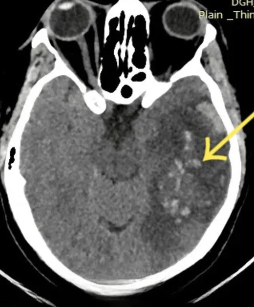
Baş ağrısı, nöbet veya fokal defisitle gelen ve venöz sinüs trombozu riski taşıyan hastada, arteriyel enfarktı taklit eden hemorajik ödem venöz çıkış tıkanıklığının işareti olabilir. Kontrastsız BT kanama ve ödemi hızla gösterir; venöz fazda BT venografi sinüs ve büyük venlerde trombüsü ya da dolum defektini araştırır. Lezyonlar arteriyel sulama alanlarına uymaz, parasagittal veya bilateral olabilir ve venöz hipertansiyona bağlı kanama içerebilir. Yalnızca parankim kanamasına odaklanmak altta yatan trombozu ve ilerleyici venöz konjesyonu gözden kaçırabilir.
Faz ve pencere
- Kontrastsız tanısal
- Ödemli parankimde noktasal veya birleşen hiperdens kan odakları ve hematomalar görülebilir; diffüz ödem nedeniyle sulkuslar daralabilir; tromboze sinüs bazen spontan hiperdens olarak izlenebilir. Arteriyel sınırlara uymayan parasagittal, kortikal veya bilateral dağılım venöz kökeni düşündürür.
- Venöz tanısal
- Venöz zamanlamalı BT venografide superior sagittal, transvers-sigmoid sinüsler ve derin venöz sistem kontrastla opaklaşır; trombüs ilgili segmentte dolum defekti veya opaklaşmama olarak aranır. Damar açıklığı BT venografi veya MR venografi gibi venöz görüntülemeyle değerlendirilir.
Önerilen pencereler: Beyin (G 80 / M 40), Anjiyo (G 600 / M 150).
BT bulguları
- Arteriyel olmayan dağılım — ödem ve kanama birden fazla arter alanını geçebilir.
- Parasagittal hemorajik odak — falx komşuluğundaki frontal veya parietal korteks-altı dokuda ödemle çevrili kan.
- Bilateral talamik ödem/kanama — derin venöz sistem veya straight sinus tıkanıklığını akla getirir.
- Kortikal ven hiperdensliği — kontrastsız incelemede tromboze kortikal ven boyunca kord benzeri atenüasyon artışı.
- Sinüs dolum defekti — venöz zamanlamalı görüntülerde kontrastla çevrili trombüs veya segmental opaklaşmama.
- Venöz konjesyon etkisi — sulkusların daralması, lokal kitle etkisi ve eşlik eden subaraknoid ya da intraventriküler kanama.
Normalde
Normal parasagittal düzeyde iki hemisferin korteksi ve beyaz cevheri benzer dansitededir; falx boyunca simetrik sulkuslar seçilir ve venöz sinüs lümeni kesintisiz izlenir. Şüpheli tarafta ödemin sulkusları kapatıp kapatmadığını, peteşiyel kanın ödem alanı içine dağılımını ve karşı hemisferde eş lezyon bulunup bulunmadığını karşılaştırın.
Normal BT ile kıyasla
Aynı bölgenin, kaynak veri setinde patoloji içermediği belirtilen bir BT incelemesini kesit kesit kaydırın; organ sınırlarını açarak anatomiyi hasta görüntüsüyle karşılaştırın. Küçük rastlantısal bulgular tümüyle dışlanmamıştır.
Kaynak: TotalSegmentator v2.01 veri seti (Wasserthal ve ark., Radiology: Artificial Intelligence 2023), CC BY 4.0, vaka s0103; organ sınırları veri setinin otomatik segmentasyonundan, birleştirilerek sadeleştirildi. Görüntüler 2,5 mm kesit aralığına yeniden örneklendi.
Ayırıcı tanı
- Arteriyel hemorajik dönüşüm
- tipik bir arter sulama alanını izler ve ilgili arter tıkanıklığı/iskemisi eşlik edebilir.
- Hipertansif hematomlar genellikle derin yapıları etkilerken, venöz infarktlar arteriyel sulama sınırlarını aşarak daha yaygın ve asimetrik bir dağılım gösterir.
- Kanamalı metastaz
- kontrastlanan kitle lezyonu ve intralezyonel kanama ile görülebilir.
- Travmatik kontüzyon
- sıklıkla kortikal yüzeyde ve darbe karşıtı bölgelerde kümelenir; travma bulguları eşlik eder.
- Ensefalit
- temporal/limbik ödem ve bazen kanama yapabilir; sinüs dolum defekti beklenmez.
Tuzaklar
- Birden fazla arter alanını geçen kanamalı ödemi yalnızca embolik arter enfarktı diye sınıflamayın; venöz sinüsleri ve derin venleri ayrıca inceleyin.
- Kontrastsız BT'de hiperdens görünen sinüsü tek başına trombüs saymayın; yüksek hematokrit ve akım artefaktını düşünerek venöz dolum defekti arayın.
- Normal kontrastsız BT venöz trombozu dışlamaz; erken dönemde parankim bulgusu silik olabilir ve doğrudan damar görüntülemesi gerekir.
Kendini dene
Baş ağrısı ve nöbeti olan hastada bilateral talamik ödem ile küçük kanama odakları var. En olası damar sorunu nedir?
Cevabı göster
Straight sinus trombozu. Bilateral talamik konjesyon ve hemorajik ödem derin venöz drenaj tıkanıklığını, özellikle straight sinus düzeyini düşündürür. Derin gri cevher ve talamik lezyonlar, arteriyel sulama sınırlarını aşan venöz infarktların tipik yerleşimidir; bu bulgu seti için venöz tromboz önceliklidir.
Parasagittal kanamalı ödem iki arter alanını aşıyor. Hangi inceleme trombüsü doğrudan gösterebilir?
Cevabı göster
BT venografi. Venöz zamanlamalı BT venografi sinüs ve büyük venlerdeki opaklaşma kusurunu gösterir. Kontrastsız BT bulguları dolaylı olabilir; şüpheli olgularda trombüsü dolum defekti olarak değerlendirmek için BT venografi veya MR venografi yapılır.
Kaynaklar
Bu sayfadaki 37 cümle ve 2 quiz sorusu aşağıdaki kaynaklardan alıntılarla tek tek karşılaştırıldı; 6 cümle bu karşılaştırmada düzeltildi (2026-10-08).
- Imaging of Cerebral Venous Thrombosispmc.ncbi.nlm.nih.gov
- Toward a Broader Classification of Deep Venous Cerebral Anatomy in Extremely Preterm Infants: A Literature-Based Approach and Its Relevance to Intraventricular Hemorrhage.pubmed.ncbi.nlm.nih.gov
- Contralateral extensive cerebral hemorrhagic venous infarction caused by retrograde venous reflux into the opposite basal vein of Rosenthal in posttraumatic carotid-cavernous fistula: A case report and literature review.pubmed.ncbi.nlm.nih.gov
- Isolated cortical vein thrombosis: case series.pubmed.ncbi.nlm.nih.gov
- Cerebral venous thrombosis: A typical presentation of COVID-19 in the young.pubmed.ncbi.nlm.nih.gov
- Clinical utility of susceptibility-weighted imaging in vascular diseases of the brain.pubmed.ncbi.nlm.nih.gov
- Outflow-Restricted Developmental Venous Anomaly Masquerading as a Tumefactive Lesion on Imaging.pubmed.ncbi.nlm.nih.gov
- Anatomical Venous Variants in Children With Cerebral Sinovenous Thrombosis.pubmed.ncbi.nlm.nih.gov
- Safety assessment of anticoagulation therapy in patients with hemorrhagic cerebral venous thrombosis.pubmed.ncbi.nlm.nih.gov
- Neonatal deep white matter venous infarction and liquefaction: a pseudo-abscess lesion.pubmed.ncbi.nlm.nih.gov
- Simultaneous Occurrence of Cerebral Venous Sinus Thrombosis and Immune Thrombocytopenic Purpura in a Patient with a History of COVID-19 Infection.pubmed.ncbi.nlm.nih.gov
- Cerebral venous thrombosis: report of 2 cases of hemorrhagic venous infarction.pubmed.ncbi.nlm.nih.gov
- Recurrent Hemorrhagic Venous Infarctions Caused by Thrombosis of a Pontine Developmental Venous Anomaly and Protein S Mutation.pubmed.ncbi.nlm.nih.gov
- Imaging the Cerebral Veins in Pediatric Patients: Beyond Dural Venous Sinus Thrombosis.pubmed.ncbi.nlm.nih.gov
- Severe Headache, Seizures and Supraventricular Tachycardia in a 33-year-old Filipino Male with Confirmed COVID-19: A Case Report.pubmed.ncbi.nlm.nih.gov
- Intracranial venous thrombosis imaging by anatomical location: an educational review with mimics and diagnostic pitfalls.pubmed.ncbi.nlm.nih.gov
- A Prothrombotic Shift: Cerebral Venous Sinus Thrombosis in the Recovery Phase of Dengue Fever-A Case Report.pubmed.ncbi.nlm.nih.gov
- Cerebral venous sinus thrombosis in pregnancy and the postpartum period: epidemiology, management, and maternal mortality-a systematic review and meta-analysis.pubmed.ncbi.nlm.nih.gov
- Extensive cerebral venous sinus thrombosis with hemorrhagic venous infarction as the initial presentation of acute myeloid leukemia: A case report.pubmed.ncbi.nlm.nih.gov
- Tissue clock-guided prediction and intervention of futile recanalization: towards precision therapy in mechanical thrombectomy.pubmed.ncbi.nlm.nih.gov
- Management of neurovascular emergencies with ophthalmic manifestations.pubmed.ncbi.nlm.nih.gov
- Non-contrast CT findings suggestive of secondary intracerebral haemorrhage.pubmed.ncbi.nlm.nih.gov
- Paediatric cranial ultrasound: abnormalities of the brain in term neonates and young infants.pubmed.ncbi.nlm.nih.gov
- Radiological Findings of Retrograde Venous Cerebral Air Embolism Infarcts: A Case Report and Literature Review.pubmed.ncbi.nlm.nih.gov
- Perfusion imaging abnormalities in acute cerebral venous sinus thrombosis: A case series.pubmed.ncbi.nlm.nih.gov
- Cerebral venous disorders: clinical presentation, diagnostic strategy, and contemporary management.pubmed.ncbi.nlm.nih.gov
- Imaging in Acute Anterior Circulation Ischemic Stroke: Current and Future.pubmed.ncbi.nlm.nih.gov
- Venous-significant intracranial hemorrhage in neonatal HIE treated with therapeutic hypothermia: imaging phenotypes, diagnostic pitfalls, and prognostic implications.pubmed.ncbi.nlm.nih.gov
- Case Report: Mechanical thrombectomy of cerebral venous sinuses using the NeVa stent retriever.pubmed.ncbi.nlm.nih.gov
- Beyond Recanalization: A Mechanistic and Procedural Framework for Adjunct Pharmacologic Therapy During Mechanical Thrombectomy.pubmed.ncbi.nlm.nih.gov
- Multiparametric ultrasound in acute scrotum: a comprehensive review with special emphasis on contrast-enhanced ultrasound.pubmed.ncbi.nlm.nih.gov
- Emergency management of cerebral air gas embolism as a complication of CT-guided lung biopsy.pubmed.ncbi.nlm.nih.gov
- The Association Between Venous Sinus Opacification Time and Futile Recanalization in Patients with Large Vessel Occlusion Acute Ischemic Stroke.pubmed.ncbi.nlm.nih.gov
- Giant Right Sphenoid Wing Meningioma as a Reversible Frontal Network Lesion: A Pseudo-bvFTD Case with Venous-Sparing Skull-Base Resection.pubmed.ncbi.nlm.nih.gov
- Risk factors influencing cerebral venous infarction after meningioma resection.pubmed.ncbi.nlm.nih.gov
- Supplementary Motor Area Syndrome after Removal of an Unusual Extensive Parasagittal Meningioma: Analysis of Twelve Reported Cases.pubmed.ncbi.nlm.nih.gov
- Refractory deep cerebral venous thrombosis managed by mechanical thrombectomy and balloon angioplasty: A case report.pubmed.ncbi.nlm.nih.gov
- Bilateral Vein of Trolard Thrombosis Presenting with Seizure and Minimal Deficits: A Rare Case Report.pubmed.ncbi.nlm.nih.gov
- Admission hyperdense sinus sign predicts poorer outcomes in cerebral venous sinus thrombosis.pubmed.ncbi.nlm.nih.gov
- Acute otitis media complicated by mastoiditis and otogenic transverse-sigmoid sinus thrombosis in an adult: a case report on infectious pathogenesis and multidisciplinary therapy.pubmed.ncbi.nlm.nih.gov
- The Anticoagulation Dilemma: Concurrent Traumatic Brain Hemorrhage and Cerebral Venous Thrombosis in a Blast Injury Patient.pubmed.ncbi.nlm.nih.gov
- The laminar subarachnoid hemorrhage of infant venous stroke: Evidence of a 4<sup>th</sup> meningeal layer?pubmed.ncbi.nlm.nih.gov
- Subpial hemorrhages in neonates: imaging features, clinical factors and outcomes.pubmed.ncbi.nlm.nih.gov
- A lumbar arteriovenous fistula presenting with intraventricular hemorrhage and hydrocephalus.pubmed.ncbi.nlm.nih.gov
- A Case-Based Review of Cerebral Venous Infarcts With Perfusion Imaging and Comparison to Arterial Ischemic Stroke.pubmed.ncbi.nlm.nih.gov
- Parenchymal Insults in Abuse-A Potential Key to Diagnosis.pubmed.ncbi.nlm.nih.gov
- Single-Center Retrospective Case Series of 33 Patients with Ophthalmic-Cerebral Embolic Stroke Following Cosmetic Facial Injection, 2017-2025.pubmed.ncbi.nlm.nih.gov
- Role of diffusion weighted imaging, apparent diffusion coefficient and magnetic resonence perfusion among acute ischemic stroke patient.pubmed.ncbi.nlm.nih.gov
- Three-territory sign in infective endocarditis and concomitant stroke: a retrospective analysis on characteristics and associated factors.pubmed.ncbi.nlm.nih.gov
- Bilateral simultaneous thalamic hematomas - Unusual presentation of intracerebral hemorrhage: A case report.pubmed.ncbi.nlm.nih.gov
- Inflammation of central nervous system fiber tracts is active in patients over six months post-intracerebral hemorrhage.pubmed.ncbi.nlm.nih.gov
- Fetal intracranial hemorrhage and infarct: Main sonographic and MRI characteristics: A review article.pubmed.ncbi.nlm.nih.gov
- Late surgical site hemorrhage is a possible radiologic indicator of early tumor recurrence in grossly totally resected high-grade meningioma: A case report.pubmed.ncbi.nlm.nih.gov
- A rare case of primary cardiac sarcoma with both intracranial metastasis and embolic infarction in a pediatric patient.pubmed.ncbi.nlm.nih.gov
- Spontaneous intracerebral hemorrhage associated with lenvatinib in advanced hepatocellular carcinoma without conventional risk factors: a case report literature review.pubmed.ncbi.nlm.nih.gov
- Correlation of Contusion Morphology and Volume With Functional Recovery and in Hospital Mortality in Traumatic Brain Injury.pubmed.ncbi.nlm.nih.gov
- Cerebral contusions - Pathomechanism, predictive factors for progression and historical and current management.pubmed.ncbi.nlm.nih.gov
- The Dynamics of Neuroinflammation in Traumatic Brain Injury: Molecular Markers Useful for Establishing the Post-Traumatic Interval in Forensic Practice.pubmed.ncbi.nlm.nih.gov
- A Review of the Role of Neuroimaging in Neurotoxicity Monitoring in Children with Acute Lymphoblastic Leukemia.pubmed.ncbi.nlm.nih.gov
- Spectrum of neuroimaging mimics in children with COVID-19 infection.pubmed.ncbi.nlm.nih.gov
- When Cancer Clots: An Extensive Radiologic Analysis of Cancer-Associated Thromboembolism.pubmed.ncbi.nlm.nih.gov
- Radiologic evaluation of the kidney transplant donor and recipient.pubmed.ncbi.nlm.nih.gov
- Imaging Evaluation of Acute Traumatic Brain Injury.pubmed.ncbi.nlm.nih.gov
- Hyperthyroidism-Induced Dural Venous Sinus Thrombosis: A Case Report.pubmed.ncbi.nlm.nih.gov
- Isolated Cortical Vein Thrombosis in a Young Woman on Transdermal Ethinyl Estradiol: Case Report and Literature Review.pubmed.ncbi.nlm.nih.gov
- Posterior Reversible Encephalopathy Syndrome: A Case Report With Contemporary Literature Review and Neuropathological Evidence From Autopsy.pubmed.ncbi.nlm.nih.gov
- Infection and inflammation: radiological insights into patterns of pediatric immune-mediated CNS injury.pubmed.ncbi.nlm.nih.gov
- Prognostic value of modified magnetic resonance arterial and venous collateral scores in unilateral chronic middle cerebral artery occlusion.pubmed.ncbi.nlm.nih.gov
- Territorial CT Perfusion Burden and Outcome After Aneurysmal Subarachnoid Haemorrhage: A Retrospective Single-Centre Cohort Study.pubmed.ncbi.nlm.nih.gov
- Computational fluid dynamics assessment of altered hemodynamics in the Circle of Willis during acute ischemic stroke and the impact of cerebral collateral development.pubmed.ncbi.nlm.nih.gov
- Dysfunction of hippocampal cells and its role in cognitive impairment.pubmed.ncbi.nlm.nih.gov
- Childhood Cerebral Vasculitis : A Multidisciplinary Approach.pubmed.ncbi.nlm.nih.gov
- Diagnostic neuroimaging in neuro-ophthalmic disorders.pubmed.ncbi.nlm.nih.gov
- Acute Innominate Vein Dissection During Pacemaker Revision Procedure: A Case Report and Conservative Management.pubmed.ncbi.nlm.nih.gov
- Clinical benefits and current challenges of photon-counting detector CT in vascular imaging.pubmed.ncbi.nlm.nih.gov
- Understanding Photon-Counting CT: Physics, Detector Technology, and Image Reconstructions.pubmed.ncbi.nlm.nih.gov
- Multimodality imaging considerations for genicular artery embolization in knee osteoarthritis.pubmed.ncbi.nlm.nih.gov
- An Atypical Finding in a Patient With Acute Left Flank Pain.pubmed.ncbi.nlm.nih.gov
- Lemierre-like syndrome presenting with extensive intracranial and central venous thrombosis without pulmonary embolism: illustrative case.pubmed.ncbi.nlm.nih.gov
- Multidisciplinary Management of Emergency Neurosurgery for Intracerebral Hemorrhage During Pregnancy: A Case Report.pubmed.ncbi.nlm.nih.gov
- Tumor-like cortical venous thrombosis: Case report and literature review.pubmed.ncbi.nlm.nih.gov
- Solitary Brain Metastasis From Hepatocellular Carcinoma Masquerading as Acute Ischaemic Stroke: Radiological Diagnosis and Clinical Implications.pubmed.ncbi.nlm.nih.gov
- Unusual Venous-Origin Intraparenchymal Hematoma in the Context of a Post-traumatic Carotid-Cavernous Fistula.pubmed.ncbi.nlm.nih.gov
- Characteristics and Outcomes of Traumatic Acute Subdural Hematoma in Elderly Patients Receiving Antithrombotic Therapy: A Single-Center Retrospective Cohort Study.pubmed.ncbi.nlm.nih.gov
- When headache meets visual disturbance: An evidence-based review of common and critical entities.pubmed.ncbi.nlm.nih.gov
- Retrospective clinical study of endoscopic transfrontal approach vs. transSylvian-transinsular craniotomy for hypertensive intracerebral hemorrhage in basal ganglia: efficacy comparison and value of anatomical cognition of Sylvian fissure.pubmed.ncbi.nlm.nih.gov
- The silent risk: cardiometabolic burden and etiology of young stroke in a South Asian migrant cohort, United Arab Emirates.pubmed.ncbi.nlm.nih.gov
- Epidemiology and Outcome of Early Acute Kidney Injuries in Patients with Acute Intracerebral Hemorrhage Admitted to the Apex Neurosciences Center in Dubai.pubmed.ncbi.nlm.nih.gov
- Stroke secondary to cancer-associated coagulopathy: a systematic review.pubmed.ncbi.nlm.nih.gov
- Low-flow vascular malformations without arteriovenous shunting of the central nervous system: a pictorial review.pubmed.ncbi.nlm.nih.gov
- Open Surgical Management of Renal Cell Carcinoma with Infradiaphragmatic Venous Tumor Thrombus (Mayo Levels 0-III): The Epitome of Surgical Self-Reliance in Urology.pubmed.ncbi.nlm.nih.gov
- Clinical Management in Traumatic Brain Injury.pubmed.ncbi.nlm.nih.gov
- Bilateral basal ganglia hemorrhage: a systematic review of etiologies, management strategies, and clinical outcomes.pubmed.ncbi.nlm.nih.gov
- Recent Advances in Pathophysiology of Traumatic Brain Injury.pubmed.ncbi.nlm.nih.gov
- Clinical characteristics and prognostic analysis of patients with herpesvirus meningitis/encephalitis based on cerebrospinal fluid mNGS positivity.pubmed.ncbi.nlm.nih.gov
- Cerebrovascular Events in Pediatric Inflammatory Bowel Disease: A Review of Published Cases.pubmed.ncbi.nlm.nih.gov
- An Integrated Approach on the Diagnosis of Cerebral Veins and Dural Sinuses Thrombosis (a Narrative Review).pubmed.ncbi.nlm.nih.gov
- The pathogenesis of cerebral small vessel disease and vascular cognitive impairment.pubmed.ncbi.nlm.nih.gov
- Headache Attributed to Infection: A Clinical and Pathophysiological Overview.pubmed.ncbi.nlm.nih.gov
- Overview of the utility of noncontrast CT brain in the diagnosis of cerebral vein thrombosis.pubmed.ncbi.nlm.nih.gov
- Non-Thrombotic Filling Defects in Cerebral Veins and Sinuses: When Normal Structures Mimic a Disease.pubmed.ncbi.nlm.nih.gov
- The Imaging of Primary Fallopian Tube Carcinoma: A Literature Review.pubmed.ncbi.nlm.nih.gov
- Neuroradiology in women of childbearing age.pubmed.ncbi.nlm.nih.gov
Eğitim amaçlıdır; hasta başında tanı ve tedavi kararının yerine geçmez. İçerik yapay zekâ destekli bir süreçle güncel literatüre göre cümle cümle kaynaklarla karşılaştırıldı, ancak uzman radyolog incelemesinden geçmedi. Klinik kararlarda güncel kılavuzları, kurum protokollerini ve radyoloji raporunu esas alın.