
Özet
Akut fokal nörolojik defisitli hastada arter lümenindeki taze trombüs, kontrastsız BT'de çevre beyin dokusundan daha parlak görünerek küçük bir kan odağı gibi algılanabilir. Kontrastsız BT kanamayı dışlamanın yanında damar boyunca uzanan hiperdens pıhtı işaretini de gösterebilir; BTA lümen açıklığını ve tıkanıklığın yerini ortaya koyar. Hiperdens damar bulgusu, damarın beklenen anatomik seyri ve eşlik eden erken iskemik değişikliklerle birlikte değerlendirilmelidir. İşaretin kanama sanılması damar tıkanıklığının tanınmasını geciktirebilir; her hiperdens damar bulgusu da tek başına trombüsü kanıtlamaz.
Faz ve pencere
- Kontrastsız tanısal
- Taze pıhtı, özellikle orta serebral arterin M1 segmentinde damar lümeni boyunca çizgisel veya tübüler hiperatenuasyon olarak izlenir; komşu beyin dokusuna göre ve karşı taraftaki eş damar ile kıyaslanır. Kalsifikasyon damar duvarında daha keskin, kemik kadar yoğun odak şeklinde olabilir; bu incelemede iyot kontrastı gerekmez.
- BTA tanısal
- Şüpheli segmentte kontrastla dolmayan lümen, distal dolum azalması veya kesintisi CTA ile belirlenir; tıkanıklığın yeri ve eş damarın açıklığı doğrulanır.
Önerilen pencereler: İnme (G 40 / M 40), Beyin (G 80 / M 40), Anjiyo (G 600 / M 150).
BT bulguları
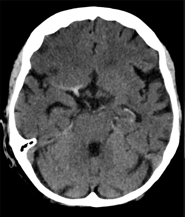
- Hiperdens MCA işareti — M1 boyunca, karşı taraftaki MCA'dan belirgin yoğun lineer lümen görünümü.
- MCA nokta işareti — Sylvian fissürde distal M2 dalının kesitsel hiperdens odağı.
- Vertebrobaziler hiperdensite — baziler arter veya vertebral arter güzergâhında uzanan yoğunluk; kafa tabanı artefaktıyla birlikte kontrol edilir.
- Damar seyriyle devamlılık — hiperdens yapının ardışık aksiyel kesitlerde aynı arterin anatomik hattında sürmesi.
- Karşı taraf asimetrisi — aynı düzeydeki eş damara kıyasla tek taraflı belirgin atenüasyon artışı.
- Erken iskemi eşliği — tıkalı damarın beslediği alanda gri-beyaz cevher ayrımında silinme veya sulkal daralma.
- BTA'da kontrastla opaklaşmayan damar segmenti, kontrastsız BT'deki hiperdens pıhtı düzeyiyle örtüşür.
Normalde
Normal kontrastsız BT'de bilateral arterler simetrik kalibre ve yoğunlukta izlenir; tek taraflı fokal hiperdensite veya kesinti patolojik kabul edilir. Özellikle Sylvian fissürdeki M1 ve M2 dalları aynı kesitte karşılaştırılmalı; hiperdensitenin damar duvarında kavisli bir kalsifikasyon mu, yoksa lümen boyunca uzanan trombüs mü olduğu kemik penceresinde ayrıştırılmalıdır.
Normal BT ile kıyasla
Aynı bölgenin, kaynak veri setinde patoloji içermediği belirtilen bir BT incelemesini kesit kesit kaydırın; organ sınırlarını açarak anatomiyi hasta görüntüsüyle karşılaştırın. Küçük rastlantısal bulgular tümüyle dışlanmamıştır.
Kaynak: TotalSegmentator v2.01 veri seti (Wasserthal ve ark., Radiology: Artificial Intelligence 2023), CC BY 4.0, vaka s0103; organ sınırları veri setinin otomatik segmentasyonundan, birleştirilerek sadeleştirildi. Görüntüler 2,5 mm kesit aralığına yeniden örneklendi.
Ayırıcı tanı
- İntraparenkimal kanama
- hiperdens odak damar hattını izlemek yerine beyin dokusu içinde yuvarlak veya düzensiz bir hematom oluşturur.
- Subaraknoid kanama
- kan bazal sisternleri veya sulkusları doldurur; tek bir arter lümeniyle sınırlı kalmaz.
- İntrakraniyal arter kalsifikasyonu
- kemik yoğunluğuna yaklaşan, damar duvarında kavisli veya noktasal odak; BTA'da lümen açıklığı korunabilir.
- Yüksek hematokrit
- birden çok arter ve venöz sinüste daha yaygın, görece simetrik yoğunluk artışı oluşturabilir.
- Kafa tabanı ışın sertleşmesi
- özellikle baziler arter çevresinde kemik komşuluğuna bağlı yapay hiperatenuasyon yaratabilir.
Tuzaklar
- Başın rotasyonu bilateral karşılaştırmayı zorlaştırabilir; damar güzergâhı ardışık kesitlerde izlenmeli ve çok düzlemli yeniden yapılandırmalarla doğrulanmalıdır.
- Baziler arterde kafa tabanı kemiklerinden kaynaklanan ışın sertleşmesi pıhtı izlenimi verebilir; yoğunluğu sagittal düzlemde ve komşu kemik artefaktıyla beraber inceleyin.
- Kalsifik plak lümen içi pıhtı sanılabilir; kemik penceresinde duvar boyunca kavisli, çok yoğun görünümü ve BTA lümenini karşılaştırın.
- Hiperdens damar işaretinin görülmemesi büyük damar tıkanıklığını dışlamaz; damar açıklığı kuşkusu sürüyorsa BTA bulgusunu esas alın.
Kendini dene
Acil BT'de sol M1 boyunca ince hiperdens çizgi var; BTA'da aynı yerde lümen dolmuyor. En olası açıklama nedir?
Cevabı göster
İntralüminal trombüs. Damar hattındaki hiperdensiteyle aynı yerde BTA dolum kesintisi bulunması oklüzif trombüsü destekler. Duvar kalsifikasyonu kemik gibi çok yoğun ve periferik görünür; subaraknoid kan sulkusu doldurur, kontüzyon ise parankim içindedir.
Baziler arter komşuluğundaki parlaklık aksiyel kesitte seçiliyor, sagittalde kemik artefaktıyla artıyor ve BTA lümeni açık. En uygun yorum hangisidir?
Cevabı göster
Işın sertleşmesi. Kafa tabanı kemiklerinin oluşturduğu ışın sertleşmesi baziler arterin yalancı hiperdens görünmesine yol açabilir; lümenin BTA'da açık olması trombüsü desteklemez. Pons kanaması parankim içi odak, sinüs trombozu ise venöz sinüs dağılımında bulgu oluşturur.
Kaynaklar
Bu sayfadaki 38 cümle ve 2 quiz sorusu aşağıdaki kaynaklardan alıntılarla tek tek karşılaştırıldı; 7 cümle bu karşılaştırmada düzeltildi (2026-10-09).
- Pitfall in necrotizing pancreatitis: Organized hematoma with arteriovenous fistula mimicking a splenic artery pseudoaneurysmpmc.ncbi.nlm.nih.gov
- Intracranial venous thrombosis imaging by anatomical location: an educational review with mimics and diagnostic pitfallspmc.ncbi.nlm.nih.gov
- The Great Pretender in the Sella: A Rare Doughnut-Shaped Partially Thrombosed Internal Carotid Artery Aneurysm Mimicking Pituitary Macroadenomapmc.ncbi.nlm.nih.gov
- Automated detection of hyperdense artery sign on non-contrast CT for rapid identification of large vessel occlusion: a multicenter validation studypmc.ncbi.nlm.nih.gov
- A Rare Case of Uterine Artery Pseudoaneurysm Causing Secondary Postpartum Hemorrhage Following Cesarean Section: A Diagnostic and Management Challenge in a Resource‐Limited Settingpmc.ncbi.nlm.nih.gov
- Noncontrast CT-based habitat analysis for differentiating hemorrhagic transformation from contrast extravasation after endovascular therapy of acute ischemic stroke.pubmed.ncbi.nlm.nih.gov
- Effects of Computed Tomography Hyperdense Middle Cerebral Artery Sign on the Efficacy of Endovascular Therapy in Acute Ischemic Stroke With Large Infarcts: A Subgroup Analysis of the ANGEL-ASPECT Trial.pubmed.ncbi.nlm.nih.gov
- Polycythemia, a radiologic mimic in plain CT head: A case report.pubmed.ncbi.nlm.nih.gov
- Punctate Hyperdensity on Noncontrast-enhanced Computed Tomography before Rupture of a Mycotic Cerebral Aneurysm in a Patient with Infective Endocarditis: A Case Report.pubmed.ncbi.nlm.nih.gov
- Non-contrast CT findings suggestive of secondary intracerebral haemorrhage.pubmed.ncbi.nlm.nih.gov
- Intracranial venous thrombosis imaging by anatomical location: an educational review with mimics and diagnostic pitfalls.pubmed.ncbi.nlm.nih.gov
- Stroke or Seizure? Diagnostic Role of Neuroimaging in Acute Neurologic Mimics.pubmed.ncbi.nlm.nih.gov
- Current state and advancements of imaging in acute ischemic stroke: a practical review.pubmed.ncbi.nlm.nih.gov
- Spontaneous calcified cerebral emboli: a comprehensive review and proposed diagnostic criteria.pubmed.ncbi.nlm.nih.gov
- Non-Thrombotic Filling Defects in Cerebral Veins and Sinuses: When Normal Structures Mimic a Disease.pubmed.ncbi.nlm.nih.gov
- Angioedema as anaphylactic reaction following tenecteplase administration in acute ischemic stroke: A case report.pubmed.ncbi.nlm.nih.gov
- Mechanical thrombectomy for tandem internal carotid artery-middle cerebral artery occlusion in a patient with a persistent hypoglossal artery: illustrative case.pubmed.ncbi.nlm.nih.gov
- Understanding Photon-Counting CT: Physics, Detector Technology, and Image Reconstructions.pubmed.ncbi.nlm.nih.gov
- Clinical benefits and current challenges of photon-counting detector CT in vascular imaging.pubmed.ncbi.nlm.nih.gov
- Photon-Counting CT: Technology, Current and Potential Future Clinical Applications, and Overview of Approved Systems and Those in Various Stages of Research and Development.pubmed.ncbi.nlm.nih.gov
- Using Multimodal Imaging to Correlate Link Between Intracranial Aneurysms and Acute Ischemic Stroke.pubmed.ncbi.nlm.nih.gov
- Refining reperfusion assessment with flat-panel detector CT: association with final infarct volume after endovascular thrombectomy.pubmed.ncbi.nlm.nih.gov
- Prediction of functional outcome after mechanical thrombectomy using radiomics and deep learning features derived from intra-thrombus and peri-thrombus regions.pubmed.ncbi.nlm.nih.gov
- Hyperdense middle cerebral artery sign is not associated with functional outcome following mechanical thrombectomy.pubmed.ncbi.nlm.nih.gov
- Prognostic value of the hyperdense middle cerebral artery sign in patients undergoing mechanical thrombectomy.pubmed.ncbi.nlm.nih.gov
- Utility of Non-Contrast CT and MRA Fusion Images for Depicting Occluded Vessels in Mechanical Thrombectomy.pubmed.ncbi.nlm.nih.gov
- Acute reperfusion therapies for acute ischemic stroke patients with unknown time of symptom onset or in extended time windows: an individualized approach.pubmed.ncbi.nlm.nih.gov
- Validation of hyperintense middle cerebral artery sign in acute ischemic stroke: Comparison between magnetic resonance imaging and angiography.pubmed.ncbi.nlm.nih.gov
- Multimodal CT in stroke imaging: new concepts.pubmed.ncbi.nlm.nih.gov
- Computed tomography perfusion-defined ischemic core predicts functional outcome after basilar artery thrombectomy.pubmed.ncbi.nlm.nih.gov
- Early Detection of Hyperdense Basilar Artery Signs Through Comparison With Previous Images.pubmed.ncbi.nlm.nih.gov
- Imaging of Cerebral Venous Thrombosis.pubmed.ncbi.nlm.nih.gov
- Imaging of cardiac valves by computed tomography.pubmed.ncbi.nlm.nih.gov
- Spectrum of Neuroimaging Findings in CNS Lymphoproliferative Disorders.pubmed.ncbi.nlm.nih.gov
- External Validation of Atherosclerotic Neuroimaging Biomarkers in Emergent Large-Vessel Occlusion.pubmed.ncbi.nlm.nih.gov
- Amyloid-related imaging abnormalities (ARIA) in anti-amyloid therapies for Alzheimer's disease: An update from the Alzheimer's Association ARIA workgroup.pubmed.ncbi.nlm.nih.gov
- Valvular and Outflow Tract Metastases From Germ Cell Tumors: Contrasting Left-Sided Embolism and Right-Sided Silence.pubmed.ncbi.nlm.nih.gov
- Post-cardiac arrest imaging: Unraveling etiology and predicting neurological outcome.pubmed.ncbi.nlm.nih.gov
- Endovascular Treatment Strategies for Acute Intracranial Atherosclerotic Stenosis-Related Large Vessel Occlusion.pubmed.ncbi.nlm.nih.gov
- The first CT-based classification system for pulmonary actinomycosis: correlating imaging patterns with therapeutic strategies and prognosis in a highly misdiagnosed disease.pubmed.ncbi.nlm.nih.gov
- Middle cerebral artery dissection: pathophysiology, diagnosis, and therapeutic options.pubmed.ncbi.nlm.nih.gov
- Brain Midline Shift Measurement and Its Automation: A Review of Techniques and Algorithms.pubmed.ncbi.nlm.nih.gov
- Fast nonlinear regression method for CT brain perfusion analysis.pubmed.ncbi.nlm.nih.gov
- A Review of the Effectiveness of Neuroimaging Modalities for the Detection of Traumatic Brain Injury.pubmed.ncbi.nlm.nih.gov
- Comparing Outcomes of Thrombectomy Versus Intravenous Thrombolysis Based on Middle Cerebral Artery M2 Occlusion Features.pubmed.ncbi.nlm.nih.gov
- Mechanical Thrombectomy Beyond 2b Reperfusion: Should We Pursue a Higher Reperfusion Grade after Achievement of 2b?pubmed.ncbi.nlm.nih.gov
- Regional leptomeningeal collateral score by computed tomographic angiography correlates with 3-month clinical outcome in acute ischemic stroke.pubmed.ncbi.nlm.nih.gov
- Reversible cerebral vasoconstriction syndrome: A narrative review.pubmed.ncbi.nlm.nih.gov
- Time-Based Decision Making for Reperfusion in Acute Ischemic Stroke.pubmed.ncbi.nlm.nih.gov
- Contrast Extravasation Post Thrombectomy in Patients With Acute Cerebral Stroke: A Review and Recommendations for Future Studies.pubmed.ncbi.nlm.nih.gov
- When headache meets visual disturbance: An evidence-based review of common and critical entities.pubmed.ncbi.nlm.nih.gov
- Update on hypoxic-ischemic brain injury: Prognosis and management.pubmed.ncbi.nlm.nih.gov
- Impact of Intracranial Arterial Calcification on Outcomes of Mechanical Thrombectomy in Acute Ischemic Stroke: A Systematic Review and Meta-Analysis.pubmed.ncbi.nlm.nih.gov
- Tmax volume can predict clinical type in patients with acute ischemic stroke.pubmed.ncbi.nlm.nih.gov
- Sporadic Hemangioblastoma of the Cavernous Sinus and Meckel's Cave.pubmed.ncbi.nlm.nih.gov
- Pediatric arterial ischemic stroke.pubmed.ncbi.nlm.nih.gov
- Spontaneous bacteremia and spontaneous bacterial peritonitis share similar prognosis in patients with cirrhosis: a cohort study.pubmed.ncbi.nlm.nih.gov
- Imaging Evaluation of Acute Traumatic Brain Injury.pubmed.ncbi.nlm.nih.gov
- The role of contrast-enhanced transcranial color-coded real-time sonography combined with the common carotid artery compression test in preoperative evaluation of anterior communicating artery patency in patients with insufficient temporal bone windows.pubmed.ncbi.nlm.nih.gov
- Perforator-Sparing Microsurgical Clipping of Tandem Dominant-Hemisphere Middle Cerebral Artery Aneurysms: Geometry-Guided Reconstruction of a Wide-Neck Bifurcation and Dorsal M1 Fusiform Lesion.pubmed.ncbi.nlm.nih.gov
- Emerging Techniques and Future Directions: Fast and Portable Magnetic Resonance Imaging.pubmed.ncbi.nlm.nih.gov
- A review of the pathogenic mechanism and clinical management progress of in-stent calcification restenosis.pubmed.ncbi.nlm.nih.gov
- Beyond Angiography: Cardiac CT for Planning Complex PCI in Calcified Coronary Lesions.pubmed.ncbi.nlm.nih.gov
- From intravascular imaging to adaptive vascular care: intelligent photonics and digital twins in panvascular disease.pubmed.ncbi.nlm.nih.gov
- Artificial Intelligence That Changes Clinical Neurology Practice: Translating Algorithms Into Actionable Care.pubmed.ncbi.nlm.nih.gov
- The ABCs of Subarachnoid Hemorrhage Blood Volume Measurement: A Simplified Quantitative Method Predicts Outcomes and Delayed Cerebral Ischemia.pubmed.ncbi.nlm.nih.gov
- The Hyperdense Middle Cerebral Artery Sign in Drip-and-Ship Models of Acute Stroke Management.pubmed.ncbi.nlm.nih.gov
- Bio-clinical evaluation of collateral score in acute middle cerebral artery occlusion.pubmed.ncbi.nlm.nih.gov
- NaF-PET Imaging for Detection of Early Arterial Microcalcification and Monitoring of Targeted Therapy: A Narrative Review.pubmed.ncbi.nlm.nih.gov
- Multimodal imaging in neuro-ophthalmology.pubmed.ncbi.nlm.nih.gov
- Multimodality imaging in the detection and management of coronary and peripheral arterial disease in patients with cancer receiving cardiotoxic antineoplastic treatments: A clinical consensus statement of the ESC Council of Cardio-Oncology and the European Association of Cardiovascular Imaging (EACVI) of the ESC.pubmed.ncbi.nlm.nih.gov
- Multicenter Validation of Artificial Intelligence Predicting Anterior Circulation Large Vessel Occlusion Using Noncontrast Head CT.pubmed.ncbi.nlm.nih.gov
- Hyperdense Aneurysm Visualized at the Occlusion Site During Thrombectomy: A Case Report.pubmed.ncbi.nlm.nih.gov
- Diagnostic value of the perfusion mismatch ratio in identifying intracranial atherosclerotic disease related occlusion.pubmed.ncbi.nlm.nih.gov
- Deep learning for intracranial hemorrhage detection and classification in brain CT scans: a systematic review and hybrid model approach.pubmed.ncbi.nlm.nih.gov
- Neuroimaging Patterns of Intracranial Infections: Meningitis, Cerebritis, and Their Complications.pubmed.ncbi.nlm.nih.gov
- Neurological manifestations and neuroimaging findings in patients with SARS-CoV2-a systematic review.pubmed.ncbi.nlm.nih.gov
- Low-flow vascular malformations without arteriovenous shunting of the central nervous system: a pictorial review.pubmed.ncbi.nlm.nih.gov
- Hyperintense acute reperfusion marker (HARM) and thrombus analysis in acute ischemic stroke.pubmed.ncbi.nlm.nih.gov
- Pitfall in necrotizing pancreatitis: Organized hematoma with arteriovenous fistula mimicking a splenic artery pseudoaneurysm.pubmed.ncbi.nlm.nih.gov
- The Great Pretender in the Sella: A Rare Doughnut-Shaped Partially Thrombosed Internal Carotid Artery Aneurysm Mimicking Pituitary Macroadenoma.pubmed.ncbi.nlm.nih.gov
- Segmentation of the Hyperdense Artery Sign on Noncontrast CT in Ischemic Stroke Using Artificial Intelligence.pubmed.ncbi.nlm.nih.gov
- Clinical image analysis to build patient-specific models of acute ischemic stroke patients.pubmed.ncbi.nlm.nih.gov
- Pre-treatment and post-treatment nasopharyngeal carcinoma imaging: imaging updates, pearls and pitfalls.pubmed.ncbi.nlm.nih.gov
- Life-threatening Carotid Complications Caused by Extraluminal Migration of Ingested Foreign Bodies; a Case Report and Narrative Review of Literature.pubmed.ncbi.nlm.nih.gov
- Predictors of Mortality in Posterior Circulation Ischemic Stroke.pubmed.ncbi.nlm.nih.gov
- Increased middle cerebral artery velocity predicts malignant media infarction after endovascular stroke thrombectomy.pubmed.ncbi.nlm.nih.gov
- Machine learning-enabled prediction of hemorrhagic transformation post-thrombectomy using quantitative DSA.pubmed.ncbi.nlm.nih.gov
- Clinical, Laboratory, and CT Morphological Factors Associated with 3-Month Functional Outcomes in Spontaneous Intracerebral Hemorrhage: A Retrospective Cohort Study.pubmed.ncbi.nlm.nih.gov
- Artificial intelligence in neurovascular surgery: advancing diagnosis, treatment, and outcomes.pubmed.ncbi.nlm.nih.gov
- A novel subtype classification for acute intracranial atherosclerotic disease-related occlusion.pubmed.ncbi.nlm.nih.gov
- Multiple Saccular Aneurysms of the Posterior Cerebral Artery With Mass Effect: A Case Report.pubmed.ncbi.nlm.nih.gov
- Emerging frontiers of artificial intelligence and machine learning in ischemic stroke: a comprehensive investigation of state-of-the-art methodologies, clinical applications, and unraveling challenges.pubmed.ncbi.nlm.nih.gov
- Deep Learning in Ischemic Stroke Imaging Analysis: A Comprehensive Review.pubmed.ncbi.nlm.nih.gov
- Spontaneous Non-Aneurysmal Convexity Subarachnoid Hemorrhage: A Scoping Review of Different Etiologies beyond Cerebral Amyloid Angiopathy.pubmed.ncbi.nlm.nih.gov
- Admission hyperdense sinus sign predicts poorer outcomes in cerebral venous sinus thrombosis.pubmed.ncbi.nlm.nih.gov
- Susceptibility weighted imaging: Clinical applications and future directions.pubmed.ncbi.nlm.nih.gov
- Continuum topological derivative - A novel application tool for segmentation of CT and MRI images.pubmed.ncbi.nlm.nih.gov
- Extra-Axial Cavernous Angioma: A Case Report and Review of the Literature.pubmed.ncbi.nlm.nih.gov
- Vasculitis syndromes: large vessel vasculitides and ANCA-associated vasculitides from a neuroradiologist's perspective.pubmed.ncbi.nlm.nih.gov
- CT and MRI imaging of mass in the carotid space: a pictorial review.pubmed.ncbi.nlm.nih.gov
- Incidental Calcifications of Carotid and Vertebral Arteries: Frequency and Associations in Pediatric Population.pubmed.ncbi.nlm.nih.gov
- Clinical Characteristics of COVID-19-Related Reversible Cerebral Vasoconstriction Syndrome: A Systematic Review of Case Series.pubmed.ncbi.nlm.nih.gov
- Interobserver reliability of computed tomography angiography in the assessment of ruptured intracranial aneurysm and impact on patient management.pubmed.ncbi.nlm.nih.gov
- Retrieval of an atherosclerotic plaque-vessel wall complex during mechanical thrombectomy: illustrative case.pubmed.ncbi.nlm.nih.gov
- Middle cerebral artery occlusion due to paradoxical embolism diagnosed by pulmonary artery thrombosis in the absence of deep vein thrombosis: A case report.pubmed.ncbi.nlm.nih.gov
- Prognostic value of CT contrast staining after endovascular therapy in basilar artery occlusion stroke.pubmed.ncbi.nlm.nih.gov
- Emergency microsurgical trapping, decompression, and revascularization of a middle cerebral artery dissecting aneurysm using a minipterional approach in a pediatric patient: illustrative case.pubmed.ncbi.nlm.nih.gov
- Distal middle cerebral artery dissection with concurrent completely thrombosed aneurysm manifesting as cerebral ischemia. A case report and review of the literature.pubmed.ncbi.nlm.nih.gov
- Multidetector computed tomography of chest trauma: indications, technique and interpretation.pubmed.ncbi.nlm.nih.gov
- Sequential Contralateral Large-Vessel Occlusion Following Intravenous Thrombolysis: A Case Report of Dynamic Bilateral Cerebral Arterial Thrombosis.pubmed.ncbi.nlm.nih.gov
- Mechanical Thrombectomy for Large-Vessel Occlusion after Fontan Procedure in an Adult.pubmed.ncbi.nlm.nih.gov
Eğitim amaçlıdır; hasta başında tanı ve tedavi kararının yerine geçmez. İçerik yapay zekâ destekli bir süreçle güncel literatüre göre cümle cümle kaynaklarla karşılaştırıldı, ancak uzman radyolog incelemesinden geçmedi. Klinik kararlarda güncel kılavuzları, kurum protokollerini ve radyoloji raporunu esas alın.