Özet
Tek hemisferdeki hematom, ödem veya kitle etkisi singulat girusu falx cerebri'nin serbest kenarının altına ve karşı tarafa iter. Kontrastsız beyin BT, bu yer değiştirmeyi ve eşlik eden ventrikül basısını hızla gösterdiği için akut değerlendirmede temel incelemedir. Falx komşuluğundaki perikallozal arter basısı iskemiye, foramen Monro düzeyindeki tıkanma ise karşı lateral ventrikülde genişlemeye yol açabilir. Herniasyon yaşamı tehdit eden bir durumdur ve acil dikkat gerektirir.
Faz ve pencere
- Kontrastsız tanısal
- Kontrastsız beyin BT'de kitle etkisine eşlik eden ventrikül basısı ve subfalsin herniasyon değerlendirilebilir. Bu herniasyonun ilk değerlendirmesinde kontrast gerekmez.
Önerilen pencereler: Beyin (G 80 / M 40), Subdural (G 200 / M 75).
BT bulguları
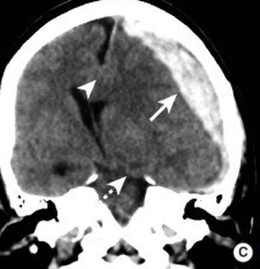
- Singulat girusun falx cerebri'nin serbest alt kenarını aşarak karşı hemisfere uzanması
- Septum pellucidumun ve üçüncü ventrikülün kitle etkisinin karşı yönüne yer değiştirmesi
- Kitle tarafındaki lateral ventrikül frontal hornu veya gövdesinde basılanma
- Foramen Monro tıkanıklığına bağlı tek taraflı ventrikül genişlemesi görülebilir.
- Kitle etkisine bağlı orta hat sapmasında korpus kallozum yer değiştirebilir; ancak tanıda belirleyici olan singulat girusun falx altı geçişidir.
- Kitle etkisi ve eşlik eden ödem nedeniyle komşu sulkuslarda daralma veya silinme görülebilir.
- Perikallozal arter dahil ön serebral arter dallarının basısına bağlı olarak singulat girus ve medial korpus kallozumda infarkt gelişebilir.
Normalde
Normalde falx cerebri interhemisferik fissür içinde orta hatta uzanır; singulat giruslar falxın iki yanında kalır, septum pellucidum ve üçüncü ventrikül de orta hatta yer alır. Kıyaslamada bu anatomik sınırın aşılmasını, ventrikül konturlarındaki taraf farkını ve septum pellucidumun falxa göre konumunu birlikte incele.
Normal BT ile kıyasla
Aynı bölgenin, kaynak veri setinde patoloji içermediği belirtilen bir BT incelemesini kesit kesit kaydırın; organ sınırlarını açarak anatomiyi hasta görüntüsüyle karşılaştırın. Küçük rastlantısal bulgular tümüyle dışlanmamıştır.
Kaynak: TotalSegmentator v2.01 veri seti (Wasserthal ve ark., Radiology: Artificial Intelligence 2023), CC BY 4.0, vaka s0103; organ sınırları veri setinin otomatik segmentasyonundan, birleştirilerek sadeleştirildi. Görüntüler 2,5 mm kesit aralığına yeniden örneklendi.
Ayırıcı tanı
- Fizyolojik ventrikül asimetrisi
- frontal horn biçimleri farklı olabilir, ancak falx altında girus geçişi ve kitle etkisi bulunmaz
- Serebral atrofi
- sulkuslar ve ventriküller birlikte genişler; komşu ventrikül basısı beklenmez
- Santral aşağı transtentoryal herniasyon
- baskın yer değiştirme diensefalon ve mezensefalonun aşağı yönündedir
- Unkal herniasyon
- medial temporal lob tentoryal açıklığa yaklaşır ve perimezensefalik sisternleri asimetrik daraltır
Tuzaklar
- Septum pellucidumun tek başına deviasyonu herniasyonu kanıtlamaz; singulat girusun falxın altından geçtiğini komşu kesitlerde doğrula.
- Başın tarayıcı yatağındaki rotasyonu orta hat sapmasını olduğundan farklı gösterebilir; falxı üçüncü ventrikül ve kafa tabanı orta hattıyla beraber izle.
- Karşı lateral ventrikül genişlemesi tek başına obstrüktif hidrosefali diye yorumlanabilir; karşı frontal horn genişlemesini kitle tarafındaki horn basısı ve foramen Monro düzeyiyle eşleştir.
- Orta hat kayması herniasyonun anatomik kanıtı değildir; raporda singulat girusun falx altından geçişini ayrıca tarif et.
Kendini dene
Tek taraflı kitle etkisinde falxın altından karşıya yer değiştiren yapı hangisidir?
Cevabı göster
Singulat girus. Subfalsin herniasyonda medial frontal lobun singulat girusu falxın serbest kenarının altına geçer. Parahipokampal girus unkal, vermis yukarı transtentoryal, tonsiller ise tonsiller herniasyonla ilişkilidir.
Kitle tarafındaki frontal horn basılı, karşı frontal horn geniş görünüyorsa hangi yapıdaki tıkanıklık düşünülür?
Cevabı göster
Foramen Monro. Foramen Monro tıkanması karşı lateral ventrikülün BOS çıkışını engelleyerek tek taraflı genişlemeye yol açabilir. Akuaduktus Sylvii üçüncü-dördüncü ventrikül arasındadır; Luschka ve Magendie açıklıkları dördüncü ventrikül çıkışlarıdır.
İlgili konular
- Travmatik serebral ödem/şişme
- Perihematomal ödem
- Unkal/descending transtentorial herniasyon
- Santral aşağı transtentorial herniasyon
Kaynaklar
Bu sayfadaki 35 cümle ve 2 quiz sorusu aşağıdaki kaynaklardan alıntılarla otomatik olarak karşılaştırıldı; 6 cümle bu karşılaştırmada düzeltildi (2026-10-09).
- Malignant middle cerebral artery infarction revealing atrial fibrillation: A Multimodal imaging case of M1 occlusion treated with decompressive craniectomypmc.ncbi.nlm.nih.gov
- Delayed Expanding Intracerebral Pneumatocele Following Severe Craniofacial Trauma: A Case Report With Serial Imaging Correlationpmc.ncbi.nlm.nih.gov
- Actinomyces urogenitalis and Peptoniphilus asaccharolyticus brain abscess in an immunocompetent patient: A case reportpmc.ncbi.nlm.nih.gov
- Traumatic Middle Meningeal Artery Pseudoaneurysm Managed With Preoperative Glue Embolization Followed by Decompressive Craniectomy: A Case Reportpmc.ncbi.nlm.nih.gov
- Middle Meningeal Artery Embolization as Standalone Therapy for Chronic Subdural Hematoma with Radiological Herniation Features: A Case Reportpmc.ncbi.nlm.nih.gov
- Massive cerebral involvement in fat embolism syndrome and intracranial pressure management.pubmed.ncbi.nlm.nih.gov
- Ethmoidal meningoencephalocele in a C57BL/6J mouse.pubmed.ncbi.nlm.nih.gov
- Magnetic resonance imaging signs of presumed elevated intracranial pressure in dogs.pubmed.ncbi.nlm.nih.gov
- Incidence and type of brain herniation associated with intracranial meningioma in dogs and cats.pubmed.ncbi.nlm.nih.gov
- Neurosurgical management of L-asparaginase induced haemorrhagic stroke.pubmed.ncbi.nlm.nih.gov
- Awake brain tumor resection during pregnancy: Decision making and technical nuances.pubmed.ncbi.nlm.nih.gov
- Rare case of intracranial hemorrhage associated with seoul virus infection diagnosed by metagenomic next-generation sequencing.pubmed.ncbi.nlm.nih.gov
- Contralateral Ventriculostomy for Intraoperative Brain Relaxation in Supratentorial Brain Tumors.pubmed.ncbi.nlm.nih.gov
- Fixed, Dilated, and Conversing-Unreactive Pupil With Preserved Consciousness Indicating Acutely Rising Intracranial Pressure due to Traumatic Intraparenchymal Contusions: Case Report and Review of the Literature.pubmed.ncbi.nlm.nih.gov
- Lateral Ventricle Solitary Fibrous Tumor: A Case Report and Review of the Literature.pubmed.ncbi.nlm.nih.gov
- Isolated neuroparacoccidioidomycosis as a pseudotumoral lesion in the absence of systemic disease.pubmed.ncbi.nlm.nih.gov
- Paradoxical Brain Herniation After Decompressive Craniectomy Provoked by Drainage of Subdural Hygroma.pubmed.ncbi.nlm.nih.gov
- Diagnostic Challenge of Diffusion Tensor Imaging in a Patient With Hemiplegia After Traumatic Brain Injury.pubmed.ncbi.nlm.nih.gov
- Rapid reversal of coagulopathy in warfarin-related intracranial haemorrhages with prothrombin complex concentrates.pubmed.ncbi.nlm.nih.gov
- Peritumoral MRI findings and brain herniations in epileptic dogs with prosencephalic brain tumors: a multicentre retrospective study.pubmed.ncbi.nlm.nih.gov
- Optimal Timing for Stereotactic Minimally Invasive Surgery in Supratentorial Spontaneous Intracerebral Hemorrhage With Tentorial Herniation: A Retrospective Study.pubmed.ncbi.nlm.nih.gov
- Acute Epidural Hematoma: From Injury to Death.pubmed.ncbi.nlm.nih.gov
- Multifocal intracranial hemorrhage on emergency brain CT in a child with end-stage nephrotic syndrome.pubmed.ncbi.nlm.nih.gov
- Brain metastases as a manifestation of late relapse in high-grade serous ovarian cancer: Diagnostic and therapeutic challenges.pubmed.ncbi.nlm.nih.gov
- <i>Actinomyces urogenitalis</i> and <i>Peptoniphilus asaccharolyticus</i> brain abscess in an immunocompetent patient: A case report.pubmed.ncbi.nlm.nih.gov
- Malignant middle cerebral artery infarction revealing atrial fibrillation: A Multimodal imaging case of M1 occlusion treated with decompressive craniectomy.pubmed.ncbi.nlm.nih.gov
- Point-of-Care Ultrasound Assessment of Ventriculoperitoneal Shunt Patency: A Case Report.pubmed.ncbi.nlm.nih.gov
- Brain Herniationpubmed.ncbi.nlm.nih.gov
- Anteromedial striatum and globus pallidus infarcts: Uncommon complications of cerebral herniation in the setting of acute subdural hemorrhage.pubmed.ncbi.nlm.nih.gov
- Minimally invasive interventions for intracranial pathologies using tubular retractors in the pediatric population: Safety, efficacy, technical aspects and outcomes.pubmed.ncbi.nlm.nih.gov
- Revisiting Hemicraniectomy: Late Decompressive Hemicraniectomy for Malignant Middle Cerebral Artery Stroke and the Role of Infarct Growth Rate.pubmed.ncbi.nlm.nih.gov
- Brain Midline Shift Measurement and Its Automation: A Review of Techniques and Algorithms.pubmed.ncbi.nlm.nih.gov
- Intraventricular giant cavernous malformation mimicking central neurocytoma characterized by digital subtraction angiography: illustrative case.pubmed.ncbi.nlm.nih.gov
- Imaging features of a frontal neuroglial cyst in an adult patient: A rare case report.pubmed.ncbi.nlm.nih.gov
- Central neurocytomas: Decision-making process of postoperative treatment. Lesson learned.pubmed.ncbi.nlm.nih.gov
- A Review of the Clinical Presentation, Causes, and Diagnostic Evaluation of Increased Intracranial Pressure in the Emergency Department.pubmed.ncbi.nlm.nih.gov
- Tumor inflammation-associated neurotoxicity.pubmed.ncbi.nlm.nih.gov
- Advances in Fetal Repair of Spina Bifida Integrating Prenatal Surgery, Stem Cells, and Biomaterials.pubmed.ncbi.nlm.nih.gov
- MDCT imaging of traumatic brain injury.pubmed.ncbi.nlm.nih.gov
- Imaging in Chronic Traumatic Encephalopathy and Traumatic Brain Injury.pubmed.ncbi.nlm.nih.gov
- Anatomy-Guided Microsurgical Resection of a Dominant Frontal Lobe Tumor Without Intraoperative Adjuncts: A Case Report from a Resource-Limited Context.pubmed.ncbi.nlm.nih.gov
- Neurological manifestations and neuroimaging findings in patients with SARS-CoV2-a systematic review.pubmed.ncbi.nlm.nih.gov
- Prenatal assessment of brain malformations on neuroimaging: an expert panel review.pubmed.ncbi.nlm.nih.gov
- MR insights into fetal brain development: what is normal and what is not.pubmed.ncbi.nlm.nih.gov
- Interhemispheric Approach.pubmed.ncbi.nlm.nih.gov
- Giant Right Sphenoid Wing Meningioma as a Reversible Frontal Network Lesion: A Pseudo-bvFTD Case with Venous-Sparing Skull-Base Resection.pubmed.ncbi.nlm.nih.gov
- Anatomical Progression of Neuropathology in FTLD-TDP Type C and Linkage to Annexin A11.pubmed.ncbi.nlm.nih.gov
- Clinicopathological characterization of vacuolar tauopathy associated with VCP D395G.pubmed.ncbi.nlm.nih.gov
- Characteristics of brain computed tomography in dementia with cardiovascular disease and psychological and behavioral symptoms.pubmed.ncbi.nlm.nih.gov
- Comparative CT Ventricular Morphometrics in Hydrocephalus, Stroke, and Traumatic Brain Injury: A Distortion-Controlled Analysis.pubmed.ncbi.nlm.nih.gov
- Long-term brain volume trajectories and lifestyle associations in cognitively normal adults: the BRAIN-STRIDE study.pubmed.ncbi.nlm.nih.gov
- Brain Sagging Dementia.pubmed.ncbi.nlm.nih.gov
- Uncal Herniationpubmed.ncbi.nlm.nih.gov
- Congenital transtentorial parahippocampal herniation presenting with new-onset seizures in a 14-year-old boy.pubmed.ncbi.nlm.nih.gov
- Brain Shift Patterns: Upward, Lateral and Downward Herniation, Its Correlation with Clinical Patterns in Acute Intracranial Pathologies and Neurosurgical Management.pubmed.ncbi.nlm.nih.gov
- Clinical applications of portable CT scanners.pubmed.ncbi.nlm.nih.gov
- Aggressive biological behavior of benign central nervous system neoplasms: The case of hemorrhagic subependymomas - An illustrated review.pubmed.ncbi.nlm.nih.gov
- Emergency Resection of a Large Third Ventricle Colloid Cyst: Ipsilateral vs. Contralateral Interhemispheric Approach Based on the Hydrocephalus Status.pubmed.ncbi.nlm.nih.gov
- Primary Ischemic Core Resection (Corectomy) with Immediate Bone Flap Replacement in Malignant Middle Cerebral Artery Infarction: A Case Series and Systematic Comparison with Decompressive Craniectomy Outcomes.pubmed.ncbi.nlm.nih.gov
- Imaging Evaluation of Acute Traumatic Brain Injury.pubmed.ncbi.nlm.nih.gov
- Imaging biomarkers for detection and longitudinal monitoring of ventricular abnormalities from birth to childhood.pubmed.ncbi.nlm.nih.gov
- Management of intracranial hypertension with and without invasive intracranial pressure monitoring.pubmed.ncbi.nlm.nih.gov
- Anatomically curated segmentation of human subcortical structures in high resolution magnetic resonance imaging: An open science approach.pubmed.ncbi.nlm.nih.gov
- Spontaneous ischaemic stroke lesions in a dog brain: neuropathological characterisation and comparison to human ischaemic stroke.pubmed.ncbi.nlm.nih.gov
- Case Report: Occlusion of the foramen of Monro treated with endoscopic septostomy and foraminotomy in a preterm neonate.pubmed.ncbi.nlm.nih.gov
- Movement Disorders in Neurocysticercosis: A Systematic Review.pubmed.ncbi.nlm.nih.gov
- Acute subdural hematoma in Anaplasma phagocytophilum infection: illustrative case.pubmed.ncbi.nlm.nih.gov
- Rapid spontaneous regression of a massive post-thrombolysis intracerebral hemorrhage: A case report.pubmed.ncbi.nlm.nih.gov
- An ABC for decision making.pubmed.ncbi.nlm.nih.gov
- When Stroke Leads to Hydrocephalus: A Case of Unilateral Ventricular Enlargement.pubmed.ncbi.nlm.nih.gov
- Surgical management of biventricular hydrocephalus caused by tuberculosis-induced bilateral obstruction of the foramen of Monro: illustrative case.pubmed.ncbi.nlm.nih.gov
- Technical considerations of neuroendoscopic management of obstructive hydrocephalus secondary to intraventricular hemorrhage.pubmed.ncbi.nlm.nih.gov
- Fourth Ventricle Epidermoid Cyst: Case Report of Precision Telovelar Microsurgery, Functional Preservation, and Lifelong Surveillance.pubmed.ncbi.nlm.nih.gov
- Dandy-Walker syndrome: an updated literature review.pubmed.ncbi.nlm.nih.gov
- Rare Complications of CSF Diversion: Paradoxical Neuroimaging Findings in a Double, Chiasmic Case Report.pubmed.ncbi.nlm.nih.gov
- Post-traumatic hydrocephalus in adults: the mechanisms of development, predictors of progression and management strategies. A narrative review and case series analysis.pubmed.ncbi.nlm.nih.gov
- Case Report: Reversible acquired Chiari-like state and upper cervical syringomyelia caused by a giant posterior tentorial tumor confined to the supratentorial compartment.pubmed.ncbi.nlm.nih.gov
- Paradoxical Brain Herniation: An Unexpected Diagnosis.pubmed.ncbi.nlm.nih.gov
- Large Parenchymal Perianeurysmal Cyst: A Case Report.pubmed.ncbi.nlm.nih.gov
- Distal Flow Diversion with Anti-Thrombotically Coated and Bare Metal Low-Profile Flow Diverters-A Comparison.pubmed.ncbi.nlm.nih.gov
Eğitim amaçlıdır; hasta başında tanı ve tedavi kararının yerine geçmez, tıbbi tavsiye değildir. Metinler yapay zekâ desteğiyle yazıldı ve otomatik bir adımda kaynak alıntılarıyla karşılaştırıldı; bu bir tıbbi doğrulama değildir ve içerik uzman radyolog incelemesinden geçmedi. Klinik kararlarda güncel kılavuzları, kurum protokollerini ve radyoloji raporunu esas alın.