Özet
Serebral amiloid anjiyopati (CAA), kortikal ve leptomeningeal küçük damarlardaki amiloid birikimiyle ilişkili olarak lobar parankim kanamasına yol açabilir; yaşlı erişkinde spontan lobar hematom bu olasılığı akla getirir. Kontrastsız BT akut lobar pıhtıyı, kortikal yüzeye ve sulkuslara uzanımını gösterir; seçilmiş olguda BTA makrovasküler taklitçileri araştırır. BT'de lobar hematom ve subaraknoid uzanım değerlendirilebilir; mikrokanama ve kortikal yüzeyel sideroz gibi CAA belirteçleri için duyarlılık ağırlıklı MRI sekansları gerekir. Lobar hematom CAA olasılığını düşündürebilir; CAA değerlendirmesinde alternatif nedenler de dışlanmalıdır.
Faz ve pencere
- Kontrastsız tanısal
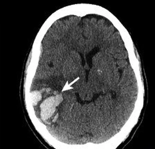
- Lobar kortiko-subkortikal hematom, düzensiz ya da lobüle hiperdens pıhtı olarak izlenebilir; parmak benzeri uzantılar ve komşu sulkusa/subaraknoid aralığa kan geçişi kaydedilir. CAA tanısı BT ile kesinleştirilmez.
- BTA
- Çift enerjili BT anjiyografi, özellikle neoplaziler, serebral venöz sinüs trombozu ve gelişimsel venöz anomalilerin erken saptanmasında ek tanısal değer sağlayabilir. BT anjiyografi ve BT venografi, CAA'yı sınıflandıran Edinburgh veya Boston ölçütlerinin yerine geçmez; bunlar ICH'nin makrovasküler nedenlerini araştırmak için kullanılır.
Önerilen pencereler: Beyin (G 80 / M 40), Anjiyo (G 600 / M 150).
BT bulguları
- Lobar kortiko-subkortikal merkez — frontal, parietal, temporal veya oksipital lobda gri-beyaz bileşkeye yakın pıhtı; bazal ganglion, talamus ve beyin sapı tutulumu CAA için tipik dağılım değildir.
- Lobüleasyonlu hematom konturu — BT'de hematomun şekil düzensizliği kaydedilebilir; bu görünüm tek başına CAA ile ilişkili kabul edilmemelidir.
- Parmak benzeri uzantılar — hematom kenarından dışarı uzayan ince, sivri parankimal çıkıntılar not edilir; bu görünüm CAA olasılığıyla ilişkilidir ancak tek başına tanısal değildir.
- Subaraknoid uzanım — hematomla bitişik sulkusta hiperdens kanın bulunması pial yüzeye geçişi düşündürür; sulkus kanaması tek başına neden belirlemez.
- İntraventriküler uzanım — pıhtı ventrikül yüzeyine ulaşıyorsa kanın lateral ventriküle geçişi ve pıhtı yükü incelenir.
- Perihematomal ödem ve kitle etkisi — hipodens çevre alan, komşu girusların silinmesi ve ventrikül/orta hat etkisi değerlendirilir.
- Eşlik eden eski hemorajik izler — BT'de eski lobar hematom kavitesi görülebilir; mikrokanama ve kortikal yüzeyel sideroz için MRI gerekir.
- Boston ölçütleri v2.0, olası CAA değerlendirmesinde klinik sunumla birlikte MR bulgularını kullanır; mikrokanama ve kortikal yüzeyel sideroz gibi duyarlılık bulgularını saptamada SWI, BT'den daha duyarlıdır.
Ölçütler ve sınıflamalar
- Basitleştirilmiş Edinburgh CT ölçütleri
- Lobar hematomda hem subaraknoid uzanım hem parmak benzeri projeksiyon varsa CAA olasılığı yüksek; yalnız biri varsa orta; ikisi de yoksa düşük olasılık grubudur. Bu olasılık sınıflaması MRI temelli Boston ölçütlerinin veya patolojik doğrulamanın yerine geçmez.
Normalde
Subaraknoid uzanım ve parmak benzeri projeksiyonlar basitleştirilmiş Edinburgh BT ölçütleriyle CAA olasılığını sınıflandırmaya yardımcı olabilir; bu sınıflama kesin tanı değildir ve klinik/MR değerlendirmesinin yerini tutmaz.
Normal BT ile kıyasla
Aynı bölgenin, kaynak veri setinde patoloji içermediği belirtilen bir BT incelemesini kesit kesit kaydırın; organ sınırlarını açarak anatomiyi hasta görüntüsüyle karşılaştırın. Küçük rastlantısal bulgular tümüyle dışlanmamıştır.
Kaynak: TotalSegmentator v2.01 veri seti (Wasserthal ve ark., Radiology: Artificial Intelligence 2023), CC BY 4.0, vaka s0103; organ sınırları veri setinin otomatik segmentasyonundan, birleştirilerek sadeleştirildi. Görüntüler 2,5 mm kesit aralığına yeniden örneklendi.
Ayırıcı tanı
- Arteriyovenöz malformasyon, lobar hematomun makrovasküler nedenleri arasında değerlendirilir; BT anjiyografi bu nedeni araştırmak için kullanılabilir.
- Anevrizma, lobar hematomun makrovasküler nedenleri arasında değerlendirilir; BT anjiyografi bu nedeni araştırmak için kullanılabilir.
- Kanamalı tümör, spontan intrakraniyal kanamanın altta yatan nedenlerinden biri olabilir; BT anjiyografi incelemesine ek olarak çift enerjili BT anjiyografi neoplastik kanamaların saptanmasına katkı sağlayabilir.
- Hemorajik infarkt, spontan intrakraniyal kanamanın alternatif nedenlerinden biridir ve iskemik inmenin hemorajik dönüşümü olarak değerlendirilir.
- Serebral venöz sinüs trombozu, intrakraniyal kanamanın altta yatan nedenlerinden biri olabilir; çift enerjili BT anjiyografi bu nedeni saptamaya katkı sağlayabilir.
Tuzaklar
- Lobar hematom CAA olasılığını düşündürür, ancak tek başına kesin tanı koydurmaz; alternatif nedenler de değerlendirilmelidir.
- Sulkustaki subaraknoid uzanımı hematomun parankim içindeki parmak benzeri projeksiyonlarından ayrı değerlendirin; subaraknoid uzanım, yüzeyel hematomun kortikal subaraknoid aralığa yayılımını ifade eder.
- Akut BT'de mikrokanamalar ve kortikal yüzeyel sideroz görünmeyebilir; bu negatiflik CAA'yı dışlamaz ve CAA tanısı için MRI temelli değerlendirme gerekir.
- Parmak benzeri uzantıların görülmemesi CAA'yı dışlamak için yeterli değildir; bu BT bulgularının yokluğu CAA'yı dışlamada sınırlı değer taşır.
Kendini dene
Yaşlı hastanın BT'sinde parietal lobda parmak benzeri pıhtı uzantıları ve bitişik sulkusta kan var. En doğru çıkarım hangisidir?
Cevabı göster
CAA olasılığı artar. Lobar hematomda subaraknoid uzanım ile parmak benzeri projeksiyonların birlikte bulunması, basitleştirilmiş Edinburgh BT ölçütlerinde yüksek CAA olasılığı kategorisine girer; bu BT bulguları tek başına kesin tanı koydurmaz. Bu BT bulguları CAA olasılığını düşündürür; olasılık ölçütleri tek başına kesin tanı koymaz.
CAA şüphesinde periferik duyarlılık artefaktlarını ve yüzeyel siderozu saptamak için kaynakta en duyarlı yöntem olarak belirtilen görüntüleme hangisidir?
Cevabı göster
MRI'de duyarlılık ağırlıklı görüntüleme (SWI). Kaynak, periferik duyarlılık artefaktları ve yüzeyel sideroz için duyarlılık ağırlıklı MRI görüntülemesini en duyarlı yöntem olarak bildirir.
İlgili konular
Kaynaklar
Bu sayfadaki 41 cümle ve 2 quiz sorusu aşağıdaki kaynaklardan alıntılarla otomatik olarak karşılaştırıldı; 22 cümle bu karşılaştırmada düzeltildi (2026-10-09).
- Fulminant Amyloid-β–Related Angiitis after Tenecteplase: A Biopsy-Confirmed Cause of Post-Thrombolysis Neurologic Deteriorationpmc.ncbi.nlm.nih.gov
- Sex differences in imaging features including cerebral amyloid angiopathy markers in intracerebral hemorrhagepmc.ncbi.nlm.nih.gov
- Visuospatial Neglect Caused by Microbleeds in a Patient With Cerebral Amyloid Angiopathy: A Case Reportpmc.ncbi.nlm.nih.gov
- Is FIB-4 Index an Independent Risk Factor for Hematoma Expansion in Acute Intracerebral Hemorrhage? A Retrospective Multicenter Observational Cohort Studypmc.ncbi.nlm.nih.gov
- Island Sign on Computed Tomography Is Associated With Cerebral Amyloid Angiopathy in Patients With Intracerebral Hemorrhagepmc.ncbi.nlm.nih.gov
- Cerebral Amyloid Angiopathy.pubmed.ncbi.nlm.nih.gov
- Plasma β-amyloid levels in cerebral amyloid angiopathy-associated hemorrhagic stroke.pubmed.ncbi.nlm.nih.gov
- Apolipoprotein E genotype predicts subarachnoid extension in spontaneous intracerebral haemorrhage.pubmed.ncbi.nlm.nih.gov
- Dementia risk after spontaneous intracerebral haemorrhage: a prospective cohort study.pubmed.ncbi.nlm.nih.gov
- Occipital Cortical Calcifications in Cerebral Amyloid Angiopathy.pubmed.ncbi.nlm.nih.gov
- Subarachnoid and Subdural Hemorrhages in Lobar Intracerebral Hemorrhage Associated With Cerebral Amyloid Angiopathy.pubmed.ncbi.nlm.nih.gov
- Remote Intracerebral Hemorrhage After Intravenous Thrombolysis: Results From a Multicenter Study.pubmed.ncbi.nlm.nih.gov
- Cerebral amyloid angiopathy-related inflammation: imaging findings and clinical outcome.pubmed.ncbi.nlm.nih.gov
- Body mass index and etiology of intracerebral hemorrhage.pubmed.ncbi.nlm.nih.gov
- Unraveling the risk factors for spontaneous intracerebral hemorrhage among West Africans.pubmed.ncbi.nlm.nih.gov
- Cerebral Amyloid Angiopathy and the Risk of Hematoma Expansion.pubmed.ncbi.nlm.nih.gov
- MRI and CT imaging biomarkers of cerebral amyloid angiopathy in lobar intracerebral hemorrhage.pubmed.ncbi.nlm.nih.gov
- Elderly-onset iatrogenic cerebral amyloid angiopathy presenting with lobar and deep haemorrhages.pubmed.ncbi.nlm.nih.gov
- Applicability of the Edinburgh CT Criteria for Lobar Intracerebral Hemorrhage Associated with Cerebral Amyloid Angiopathy.pubmed.ncbi.nlm.nih.gov
- Cerebral Amyloid Angiopathy Presenting as Lobar Intracerebral Hemorrhage with Cognitive Decline in an 80-Year-Old Patient: A Clinicoradiologic Case Report.pubmed.ncbi.nlm.nih.gov
- Visuospatial Neglect Caused by Microbleeds in a Patient With Cerebral Amyloid Angiopathy: A Case Report.pubmed.ncbi.nlm.nih.gov
- Following the curvature: Sulcal versus gyral cortical localization of cerebral microbleeds in cerebral amyloid angiopathy.pubmed.ncbi.nlm.nih.gov
- Minimally Invasive Evacuation for Spontaneous Supratentorial Intracerebral Hemorrhage After Contemporary Randomized Trials: Patient Selection, Timing, and Platform Choice.pubmed.ncbi.nlm.nih.gov
- AI Models for Surgical Decision Support in Spontaneous Intracerebral Hemorrhage: A Systematic Review in Relation to Trials and Guidelines.pubmed.ncbi.nlm.nih.gov
- Cortical superficial siderosis predicts early recurrent lobar hemorrhage.pubmed.ncbi.nlm.nih.gov
- Amyloid-related imaging abnormalities (ARIA) in anti-amyloid therapies for Alzheimer's disease: An update from the Alzheimer's Association ARIA workgroup.pubmed.ncbi.nlm.nih.gov
- Clinical Practice Recommendations of the Swiss Society for Neuroradiology: Neuroimaging Standards for Enrollment and Disease Monitoring in Anti-Amyloid Immunotherapies.pubmed.ncbi.nlm.nih.gov
- Drug-Induced Amyloid-Related Imaging Abnormalities: A Neurovascular Perspective on Risk Assessment.pubmed.ncbi.nlm.nih.gov
- Clinical Management of Cerebral Amyloid Angiopathy.pubmed.ncbi.nlm.nih.gov
- Cerebral Amyloid Angiopathy: Clinical Presentation, Sequelae and Neuroimaging Features-An Update.pubmed.ncbi.nlm.nih.gov
- Pediatric Intracerebral Hemorrhage Management-Consensus Statement of the International Pediatric Stroke Organization-Part 1: Acute Phase and Workup.pubmed.ncbi.nlm.nih.gov
- Updated imaging markers in cerebral amyloid angiopathy: What radiologists need to know.pubmed.ncbi.nlm.nih.gov
- Frequency of deep-seated cerebral microbleeds in patients with lobar hemorrhages and histopathological evidence for cerebral amyloid angiopathy.pubmed.ncbi.nlm.nih.gov
- Cerebral microbleeds: Causes, clinical relevance, and imaging approach - A narrative review.pubmed.ncbi.nlm.nih.gov
- Refining the Edinburgh Computed Tomography Criteria with a 30 mL Hematoma Volume Threshold.pubmed.ncbi.nlm.nih.gov
- Island Sign on Computed Tomography Is Associated With Cerebral Amyloid Angiopathy in Patients With Intracerebral Hemorrhage.pubmed.ncbi.nlm.nih.gov
- Anti-amyloid treatment in Alzheimer disease: updated implications for patients with cerebral amyloid angiopathy and other cerebrovascular conditions.pubmed.ncbi.nlm.nih.gov
- Extracorporeal Membrane Oxygenation in Massive Pulmonary Hemorrhage Secondary to Arteriovenous Malformation.pubmed.ncbi.nlm.nih.gov
- Prognostic value of nidus sphericity in brain AVMs treated with Gamma Knife Radiosurgery: a preliminary study.pubmed.ncbi.nlm.nih.gov
- Effect of tranexamic acid for acute spontaneous intracerebral haemorrhage: a systematic review and individual patient data meta-analysis.pubmed.ncbi.nlm.nih.gov
- Inside the Heterogeneity of Primary CNS Vasculitis: A Single-Center 40-Year Experience.pubmed.ncbi.nlm.nih.gov
- A vascular component to the selective vulnerability of the medial temporal lobe to tau pathology.pubmed.ncbi.nlm.nih.gov
- Congo Red Unmasks the Culprit Behind Gastrointestinal Bleeding.pubmed.ncbi.nlm.nih.gov
- Thalamocortical hodology to personalize electrical stimulation for focal epilepsy.pubmed.ncbi.nlm.nih.gov
- The relationship between lipids level and cerebral microbleeds in patients with spontaneous intracerebral hemorrhage.pubmed.ncbi.nlm.nih.gov
- The Role of the Claustrum in Parkinson's Disease and Vascular Parkinsonism: A Matter of Network?pubmed.ncbi.nlm.nih.gov
- Neurological manifestation in COVID-19 disease with neuroimaging studies.pubmed.ncbi.nlm.nih.gov
- Paediatric cranial ultrasound: assessment of the preterm brain.pubmed.ncbi.nlm.nih.gov
- Surgery for spontaneous supratentorial intracerebral haemorrhage.pubmed.ncbi.nlm.nih.gov
- Continuous irrigation with thrombolytics for intraventricular hemorrhage: case-control study.pubmed.ncbi.nlm.nih.gov
- Advances in Stereotactic Hematoma Evacuation for Hypertensive Intracerebral Hemorrhage and the Value of Multimodal Monitoring.pubmed.ncbi.nlm.nih.gov
- Risk factors and development of a prediction model for hematoma expansion in elderly patients with spontaneous intracerebral hemorrhage.pubmed.ncbi.nlm.nih.gov
- Acute care of spontaneous intracerebral hemorrhage.pubmed.ncbi.nlm.nih.gov
- Imaging for anti-amyloid therapy eligibility: Comparing GRE and SWI in detecting microhemorrhages and superficial siderosis.pubmed.ncbi.nlm.nih.gov
- Beyond Amyloid: Systemic and Brain Frailty as Determinants of Response to Anti-Amyloid Therapy in Alzheimer's Disease-A Conceptual Review.pubmed.ncbi.nlm.nih.gov
- Progressive Neurodegeneration From Anoxic Injury Following Perioperative Cardiac Arrest.pubmed.ncbi.nlm.nih.gov
- Congenital central nervous system malformations in piedmontese calves-part 1. structural brain disorders.pubmed.ncbi.nlm.nih.gov
- Brain morphological changes in acquired hearing loss: A surface-based morphometry study.pubmed.ncbi.nlm.nih.gov
- A systematic approach to detection of epileptogenic foci on imaging: protocols, entities, search pattern, and reporting tools.pubmed.ncbi.nlm.nih.gov
- Tissue clock-guided prediction and intervention of futile recanalization: towards precision therapy in mechanical thrombectomy.pubmed.ncbi.nlm.nih.gov
- Tumor-like Lesions in Primary Angiitis of the Central Nervous System: The Role of Magnetic Resonance Imaging in Differential Diagnosis.pubmed.ncbi.nlm.nih.gov
- A Case-Based Review of Cerebral Venous Infarcts With Perfusion Imaging and Comparison to Arterial Ischemic Stroke.pubmed.ncbi.nlm.nih.gov
- Asian Computed Tomography Criteria for Cerebral Amyloid Angiopathy-Related Intracerebral Hemorrhage: Magnetic Resonance Imaging and Pathology Validations.pubmed.ncbi.nlm.nih.gov
- Sex differences in imaging features including cerebral amyloid angiopathy markers in intracerebral hemorrhage.pubmed.ncbi.nlm.nih.gov
- Biomarkers for Alzheimer's disease across diverse biological domains: an umbrella review and evidence map.pubmed.ncbi.nlm.nih.gov
- Florbetapir Regional Distribution in Cerebral Amyloid Angiopathy and Alzheimer's Disease: A PET Study.pubmed.ncbi.nlm.nih.gov
- Bridging Scales in Alzheimer's Disease: Biological Framework for Brain Simulation With The Virtual Brain.pubmed.ncbi.nlm.nih.gov
- Microsurgical obliteration of a high-flow intraorbital arteriovenous malformation fed by the ophthalmic artery: Illustrative case.pubmed.ncbi.nlm.nih.gov
- Unique Presentation of an Arteriovenous Malformation (AVM) in the Hand: A Case Report.pubmed.ncbi.nlm.nih.gov
- Co-occurring scalp arteriovenous malformation and dural arteriovenous fistula with diploic vein involvement: illustrative case.pubmed.ncbi.nlm.nih.gov
- Expert consensus on intracranial vessel wall MRI in cerebrovascular disease: Society for Magnetic Resonance Angiography recommendations.pubmed.ncbi.nlm.nih.gov
- Spontaneous subarachnoid hemorrhage: A primer for acute care practitioners.pubmed.ncbi.nlm.nih.gov
- Spontaneous Non-Aneurysmal Convexity Subarachnoid Hemorrhage: A Scoping Review of Different Etiologies beyond Cerebral Amyloid Angiopathy.pubmed.ncbi.nlm.nih.gov
- Spectrum of Neuroimaging Findings in CNS Lymphoproliferative Disorders.pubmed.ncbi.nlm.nih.gov
- Spontaneous and Drug-Induced Amyloid-Related Imaging Abnormalities: Overlaps, Divergences, and Clinical Implications Across a Continuum Between Alzheimer's Disease and Cerebral Amyloid Angiopathy.pubmed.ncbi.nlm.nih.gov
- Cognition in Cardiovascular Disease.pubmed.ncbi.nlm.nih.gov
- Multiterritory Bihemispheric Ischemic Stroke in the Setting of Unilateral Internal Carotid Artery Disease.pubmed.ncbi.nlm.nih.gov
- Acute large vessel occlusion stroke as a neurovascular complication of immune thrombotic thrombocytopenic purpura: Mechanical thrombectomy and neurocritical care challenges.pubmed.ncbi.nlm.nih.gov
- Acute Computer Tomography Findings in Pediatric Accidental Head Trauma-Review.pubmed.ncbi.nlm.nih.gov
- Spectrum of Magnetic Resonance Imaging Findings in Acute Pediatric Traumatic Brain Injury - A Pictorial Essay.pubmed.ncbi.nlm.nih.gov
- Forensic Pathologist Testimony, Part 1: Common Questions and Considerations.pubmed.ncbi.nlm.nih.gov
- Intracranial venous thrombosis imaging by anatomical location: an educational review with mimics and diagnostic pitfalls.pubmed.ncbi.nlm.nih.gov
- Venous-significant intracranial hemorrhage in neonatal HIE treated with therapeutic hypothermia: imaging phenotypes, diagnostic pitfalls, and prognostic implications.pubmed.ncbi.nlm.nih.gov
- Cerebral venous disorders: clinical presentation, diagnostic strategy, and contemporary management.pubmed.ncbi.nlm.nih.gov
- Intraoperative Collapse During First-Trimester Abortion: Hyperleukocytosis Crisis, Thrombotic Pulmonary Embolism, or Amniotic Fluid Embolism?pubmed.ncbi.nlm.nih.gov
- Transcatheter Management of Severe Aortic Stenosis, Acute Pulmonary Embolism, and Gastrointestinal Bleeding.pubmed.ncbi.nlm.nih.gov
- Tumor-like cortical venous thrombosis: Case report and literature review.pubmed.ncbi.nlm.nih.gov
- Cerebral amyloid angiopathy-related acute lobar intra-cerebral hemorrhage: diagnostic value of plain CT.pubmed.ncbi.nlm.nih.gov
- 2025 Heart Disease and Stroke Statistics: A Report of US and Global Data From the American Heart Association.pubmed.ncbi.nlm.nih.gov
- 2024 Heart Disease and Stroke Statistics: A Report of US and Global Data From the American Heart Association.pubmed.ncbi.nlm.nih.gov
- A guide to neuromodulation in drug-resistant epilepsy.pubmed.ncbi.nlm.nih.gov
- Machine learning-based radiomics in neurodegenerative and cerebrovascular disease.pubmed.ncbi.nlm.nih.gov
- Revisiting the critical roles of reactive microglia in traumatic brain injury.pubmed.ncbi.nlm.nih.gov
- Long-term safety and effectiveness of the "OptEase" vena cava filter.pubmed.ncbi.nlm.nih.gov
- Reversible cerebral vasoconstriction syndrome: A narrative review.pubmed.ncbi.nlm.nih.gov
- European Stroke Organisation (ESO) and European Association of Neurosurgical Societies (EANS) guideline on stroke due to spontaneous intracerebral haemorrhage.pubmed.ncbi.nlm.nih.gov
- Radiological assessment of dementia: the Italian inter-society consensus for a practical and clinically oriented guide to image acquisition, evaluation, and reporting.pubmed.ncbi.nlm.nih.gov
- Delayed intracranial lesions following cardiac myxoma resection in a patient with concurrent cerebral embolism: a diagnostic challenge in the absence of pathological evidence.pubmed.ncbi.nlm.nih.gov
- Surgical strategy to minimize dominant hemisphere retraction in an asymmetric giant tuberculum sellae meningioma with contralateral anterior cerebral artery positioning: A case report.pubmed.ncbi.nlm.nih.gov
- Preliminary investigation of the clinical value of thoracic ultrasound in pediatric pleuropulmonary blastoma: a comparative analysis with computed tomography and pathology.pubmed.ncbi.nlm.nih.gov
- Stroke-Mimic: An Intracranial Dural Arteriovenous Fistula Revealed by New-Onset Seizures and Post-Ictal Deficit.pubmed.ncbi.nlm.nih.gov
- Health system learning enables generalist neuroimaging models.pubmed.ncbi.nlm.nih.gov
- Atypical presentation of cerebral amyloid angiopathy in a 42-year-old man with recurrent lobar hemorrhages and neuropsychiatric symptoms.pubmed.ncbi.nlm.nih.gov
- Fulminant Amyloid-β-Related Angiitis after Tenecteplase: A Biopsy-Confirmed Cause of Post-Thrombolysis Neurologic Deterioration.pubmed.ncbi.nlm.nih.gov
- Reporting Clinically Actionable Imaging Features of Intracerebral Hemorrhage: A Survey of Neuroradiologist Practice.pubmed.ncbi.nlm.nih.gov
- Clinical and neuroimaging features, and associations with early post-stroke cognition in patients with intracerebral hemorrhage in Taiwan.pubmed.ncbi.nlm.nih.gov
- Imaging studies for predicting hematoma expansion: from traditional imaging signs to artificial intelligence-based multimodal fusion.pubmed.ncbi.nlm.nih.gov
- Imaging Evaluation of Acute Traumatic Brain Injury.pubmed.ncbi.nlm.nih.gov
- State of the art: noninvasive imaging and management of neurovascular trauma.pubmed.ncbi.nlm.nih.gov
- Managing Antithrombotic Treatment in Intracerebral Hemorrhage and Ischemic Stroke Due to Cerebral Amyloid Angiopathy With Supporting Pathology.pubmed.ncbi.nlm.nih.gov
- Acute Spontaneous Lobar Cerebral Hemorrhages Present a Different Clinical Profile and a More Severe Early Prognosis than Deep Subcortical Intracerebral Hemorrhages-A Hospital-Based Stroke Registry Study.pubmed.ncbi.nlm.nih.gov
- Spatiotemporal Associations Between Cortical Microinfarcts and Cortical Superficial Siderosis in Cerebral Amyloid Angiopathy.pubmed.ncbi.nlm.nih.gov
- Early imaging in spontaneous intracranial hemorrhage: Practices and etiological diagnosis.pubmed.ncbi.nlm.nih.gov
- Spontaneous Intracerebral Hemorrhage in young patients from a Comprehensive Stroke Care Unit in MENA region, Hospital Based Study.pubmed.ncbi.nlm.nih.gov
- Dual-energy CT angiography in detecting underlying causes of intracerebral hemorrhage: an observational cohort study.pubmed.ncbi.nlm.nih.gov
Eğitim amaçlıdır; hasta başında tanı ve tedavi kararının yerine geçmez, tıbbi tavsiye değildir. Metinler yapay zekâ desteğiyle yazıldı ve otomatik bir adımda kaynak alıntılarıyla karşılaştırıldı; bu bir tıbbi doğrulama değildir ve içerik uzman radyolog incelemesinden geçmedi. Klinik kararlarda güncel kılavuzları, kurum protokollerini ve radyoloji raporunu esas alın.