Özet
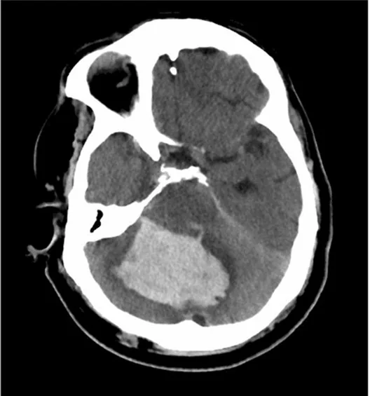
Serebellar parankim kanaması posterior fossada sınırlı hacimli bir alanda dördüncü ventriküle ve beyin sapına hızla bası yapabilir. Kontrastsız BT hematomun serebellar hemisfer veya vermisteki merkezini, ödemi ve ventrikül-sistern açıklığını gösteren ilk görüntüleme incelemesidir. Dördüncü ventrikülün daralması BOS akımını engelleyebilir; pons ve mezensefalon kompresyonu ayrıca değerlendirilmelidir. Yalnız hematomun görünür boyutuna odaklanıp ventrikül tıkanıklığı veya sisternal basıyı kaçırmak posterior fossa kitle etkisini olduğundan hafif gösterebilir.
Faz ve pencere
- Kontrastsız tanısal
- Serebellar hematom ve eşlik eden şişme, posterior fossada kitle etkisine, obstrüktif hidrosefaliye ve beyin sapı basısına yol açabilir.
- BTA
- Alışılmadık yerleşim, genç yaş veya şüpheli damar görünümünde anevrizma ve AVM gibi ikincil nedenler incelenebilir; parankim hematomunun ilk gösterimi kontrastsız kesitlerde yapılır.
Önerilen pencereler: Beyin (G 80 / M 40), Anjiyo (G 600 / M 150).
BT bulguları
- Serebellar hemisfer veya vermis merkezli pıhtı — hiperdens odağın folialar ve orta hat vermisiyle ilişkisi, karşı hemisferle kıyaslanarak tarif edilir.
- Perihematomal ödem — hematom çevresindeki hipodens alan folial sulkusları siler ve komşu serebellar parankim sınırını genişletir.
- Dördüncü ventrikül deformasyonu — ventrikülün öne/karşıya itilmesi, yarık biçiminde daralması veya seçilemez hale gelmesi kaydedilir.
- Akuadukt düzeyinde BOS yolu basısı — üst kesitlerde serebral akuaduktun açıklığı ve üçüncü ventrikül boyutları izlenir.
- Beyin sapı kompresyonu — ponsun arka konturundaki bası, yer değiştirme veya çevresindeki BOS aralığının kaybolması araştırılır.
- Posterior fossa sisternlerinin silinmesi — prepontin, serebellopontin ve perimezensefalik BOS boşluklarının açıklığı değerlendirilir.
- İntraventriküler uzanım — hematomun dördüncü ventriküle açılımı ve pıhtının akuadukt ya da daha üst ventriküllere uzanımı not edilir.
- Supratentoryal hidrosefali — lateral ventriküllerde ve temporal boynuzlarda genişleme, arka çukur tıkanıklığıyla bağlantılı olarak aranır.
Normalde
Karşı serebellar hemisferin folial aralıklarını referans alın; dördüncü ventrikülün biçimini, beyin sapı konturunu ve perimezensefalik sisternlerin devamlılığını aynı aksiyel düzeylerde kıyaslayın.
Normal BT ile kıyasla
Aynı bölgenin, kaynak veri setinde patoloji içermediği belirtilen bir BT incelemesini kesit kesit kaydırın; organ sınırlarını açarak anatomiyi hasta görüntüsüyle karşılaştırın. Küçük rastlantısal bulgular tümüyle dışlanmamıştır.
Kaynak: TotalSegmentator v2.01 veri seti (Wasserthal ve ark., Radiology: Artificial Intelligence 2023), CC BY 4.0, vaka s0103; organ sınırları veri setinin otomatik segmentasyonundan, birleştirilerek sadeleştirildi. Görüntüler 2,5 mm kesit aralığına yeniden örneklendi.
Ayırıcı tanı
- Hemorajik serebellar infarkt
- arteriyel veya venöz dağılımlı ödemli parankimde yama tarzı kan görülür; pıhtı folial alanları takip edebilir.
- Travmatik kontüzyon
- oksipital kemik kırığı, kafa derisi şişliği veya başka travmatik beyin kanamaları eşlik edebilir.
- Kanamalı metastaz
- ayrı nodüller, çevresinde orantısız ödem veya başka sistemik tümör odakları olasılığı artırır; MRI ile araştırılır.
- Kavernom
- küçük yuvarlak hemoraji odağına rağmen BT'de karakteristik iç yapı görülmeyebilir; susceptibility MRI ayırıcıdır.
- Subaraknoid kanama
- kan folial oluklarda veya sisternlerde yerleşir ve serebellar doku merkezli kitle oluşturmaz.
Tuzaklar
- Posterior fossa kemik artefaktı küçük pıhtıları örtebilir; beyin penceresinde ardışık kesitleri ve mümkünse ince rekonstrüksiyonları karşılaştırın.
- Dördüncü ventrikülün ince görünmesi tek başına normal kabul edilmemeli; hastanın diğer ventriküllerindeki genişleme ve prepontin ve perimezensefalik BOS boşluklarıyla birlikte okuyun.
- Serebellar folial subaraknoid kan, yüzeyel parankim hematomu izlenimi verebilir; kanın folial aralıkları takip edip etmediğini parankim konturuyla ayırın.
- Vermis merkezli hematom, iki hemisfer arasında simetrik yoğunluk sanılabilir; normalde vermis dokusu ile dördüncü ventrikül arasındaki sınır kesit kesit korunur.
Kendini dene
Serebellar dokudaki pıhtı dördüncü ventrikülü kapatıyor; lateral ventriküller genişlemiş. Hangi komplikasyon bulgusu vardır?
Cevabı göster
Obstrüktif hidrosefali. Dördüncü ventrikül tıkanmasıyla birlikte yukarı akım ventriküllerinin genişlemesi akut obstrüktif hidrosefaliyi destekler. Atrofi sulkus genişlemesi yapar; subdural higroma ekstraaksiyel sıvıdır; normal basınçlı hidrosefali kronik klinik bağlamda değerlendirilir.
Serebellar folialar boyunca ince hiperdens çizgiler var, ancak hemisfer parankiminde kitle yok. Kan en olası hangi kompartımandadır?
Cevabı göster
Subaraknoid aralık. Folial olukları izleyen çizgisel kan subaraknoid kompartımanla uyumludur. Kanamanın yerleştiği kompartıman (parankim içi, ekstraaksiyel veya vasküler) BT'deki görüntü biçimini belirler; örneğin venöz trombozlar dolum kusuru olarak görülür.
İlgili konular
Kaynaklar
Bu sayfadaki 37 cümle ve 2 quiz sorusu aşağıdaki kaynaklardan alıntılarla tek tek karşılaştırıldı; 4 cümle bu karşılaştırmada düzeltildi (2026-10-09).
- Risk factors of prognosis for spontaneous cerebellar hemorrhage: a systematic review and meta-analysispmc.ncbi.nlm.nih.gov
- Effectiveness and Safety of Minimally Invasive Surgery for Infratentorial Hemorrhages: A Meta-Analysis of Clinical and Technical Outcomespmc.ncbi.nlm.nih.gov
- Hemorrhagic meningioma with pulmonary metastasis: Case report and literature reviewpmc.ncbi.nlm.nih.gov
- A Narrative Review of Neurodevelopmental, Psychiatric, and Behavioral Consequences of Preterm Birthpmc.ncbi.nlm.nih.gov
- Cerebellar hemorrhage as a complication of spine surgerypmc.ncbi.nlm.nih.gov
- Cerebellar hypoplasia of prematurity: Causes and consequences.pubmed.ncbi.nlm.nih.gov
- Cerebellar hemorrhage as a complication of spine surgery.pubmed.ncbi.nlm.nih.gov
- Remote cerebellar hemorrhage in neurosurgery.pubmed.ncbi.nlm.nih.gov
- Neuroimaging of white matter injury, intraventricular and cerebellar hemorrhage.pubmed.ncbi.nlm.nih.gov
- Multislice Spiral CT Image Analysis and Meta-Analysis of Inspiratory Muscle Training on Respiratory Muscle Function.pubmed.ncbi.nlm.nih.gov
- Magnetic resonance and nuclear medicine imaging in ataxias.pubmed.ncbi.nlm.nih.gov
- Recurrent cerebellar hemorrhage: case report and review of the literature.pubmed.ncbi.nlm.nih.gov
- Diagnostic neuroimaging and early detection of germinal matrix hemorrhage-intraventricular hemorrhage.pubmed.ncbi.nlm.nih.gov
- Brain imaging in preterm infants <32 weeks gestation: a clinical review and algorithm for the use of cranial ultrasound and qualitative brain MRI.pubmed.ncbi.nlm.nih.gov
- Hemorrhagic meningioma with pulmonary metastasis: Case report and literature review.pubmed.ncbi.nlm.nih.gov
- Neuro-Behcet disease presenting as a solitary cerebellar hemorrhagic lesion: a case report and review of the literature.pubmed.ncbi.nlm.nih.gov
- Supratentorial subdural hemorrhage of a previous head injury and cerebellar hemorrhage after cervical spinal surgery: a case report and review of the literature.pubmed.ncbi.nlm.nih.gov
- Long-term neurodevelopmental outcomes and follow-up of germinal matrix-intraventricular hemorrhage in preterm infants.pubmed.ncbi.nlm.nih.gov
- Hypertension-Related Cerebral Microbleeds.pubmed.ncbi.nlm.nih.gov
- Acute fulminant cerebellitis in children: a case series and literature review on diagnostic challenges and management strategies.pubmed.ncbi.nlm.nih.gov
- Posterior fossa dynamic craniotomy utilizing NuCrani reversibly expandable bone flap fixation plates: patient series.pubmed.ncbi.nlm.nih.gov
- Vestibular schwannoma with hemorrhage in a 63-year-old female: A case report.pubmed.ncbi.nlm.nih.gov
- Is foramen magnum decompression for acquired Chiari I malformation like putting a finger in the dyke? - A simplistic overview of artificial intelligence in assessing critical upstream and downstream etiologies.pubmed.ncbi.nlm.nih.gov
- The Neuro-Ophthalmology of Tuberculosis.pubmed.ncbi.nlm.nih.gov
- A Critical Update of the Classification of Chiari and Chiari-like Malformations.pubmed.ncbi.nlm.nih.gov
- Syringomyelia in the Course of Resection of Foramen Magnum Pathology: A Case Report with an Intriguing Therapeutic Solution and Systematic Review.pubmed.ncbi.nlm.nih.gov
- Cardioembolic sources and stroke prevention: a systematic review.pubmed.ncbi.nlm.nih.gov
- Global Prevalence of Sleep-Disordered Breathing in Intracerebral Hemorrhage Survivors: A Meta-Analysis and Systematic Review.pubmed.ncbi.nlm.nih.gov
- Delayed multiple hemorrhagic brain metastases from atrial myxoma: a case report and literature review.pubmed.ncbi.nlm.nih.gov
- Case report and literature review: neuropsychiatric systemic lupus erythematosus presenting as massive intracerebral hemorrhage.pubmed.ncbi.nlm.nih.gov
- Non-contrast CT findings suggestive of secondary intracerebral haemorrhage.pubmed.ncbi.nlm.nih.gov
- Idiopathic intracranial hypertension: Imaging and clinical fundamentals.pubmed.ncbi.nlm.nih.gov
- Mechanisms of aortic dissection: From pathological changes to experimental and <i>in silico</i> models.pubmed.ncbi.nlm.nih.gov
- Magnetic resonance imaging signs of high intraventricular pressure--comparison of findings in dogs with clinically relevant internal hydrocephalus and asymptomatic dogs with ventriculomegaly.pubmed.ncbi.nlm.nih.gov
- Sympathetic hyperactivation following endoscopic third ventriculostomy: a rare and underrecognized postoperative complication. Illustrative case.pubmed.ncbi.nlm.nih.gov
- Novel endoscopic posterior fossa cyst fenestration via an occipital meningocele defect in neonatal Dandy-Walker malformation: illustrative case.pubmed.ncbi.nlm.nih.gov
- Anatomically Precise Microsurgical Resection of a Posterior Fossa Cerebellar Metastasis in an Elderly Patient with Preservation of Venous Outflow, Dentate Nucleus, and Cerebrospinal Fluid Pathways.pubmed.ncbi.nlm.nih.gov
- Fourth Ventricle Epidermoid Cyst: Case Report of Precision Telovelar Microsurgery, Functional Preservation, and Lifelong Surveillance.pubmed.ncbi.nlm.nih.gov
- Extra-ventricular application of flexible neuro-endoscopy in intracranial epidermoid cyst open resection: A novel technique.pubmed.ncbi.nlm.nih.gov
- Paediatric cranial ultrasound: assessment of the preterm brain.pubmed.ncbi.nlm.nih.gov
- Patient selection criteria and preliminary outcome of the first 20 endoscopic evacuation of intracerebral hematoma in a tertiary hospital center.pubmed.ncbi.nlm.nih.gov
- PITCH (pediatric infratentorial tumors - hydrocephalus-related complications) registry study design: observational, prospective, multicenter study evaluating the number of surgeries associated with the treatment of hydrocephalus secondary to infratentorial tumors in childhood and adolescence.pubmed.ncbi.nlm.nih.gov
- Paediatric cranial ultrasound: abnormalities of the brain in term neonates and young infants.pubmed.ncbi.nlm.nih.gov
- The management of space-occupying cyst in the tumor bed after brain tumor surgery: a case series integrated with literature synthesis.pubmed.ncbi.nlm.nih.gov
- Subarachnoid cisterns as surgical corridors: Integrating microsurgical anatomy and neuroimaging for intracranial navigation.pubmed.ncbi.nlm.nih.gov
- How I do it: pediatric lateral-type posterior fossa ependymoma resection.pubmed.ncbi.nlm.nih.gov
- Case Report: Structured MRI assessment of posterior thalamic infarction in a distribution compatible with posterior choroidal artery territory presenting as Déjerine-Roussy syndrome in an adolescent: differentiating arterial ischemia from venous thrombosis and thalamic neoplasm.pubmed.ncbi.nlm.nih.gov
- Multifocal intracranial hemorrhage on emergency brain CT in a child with end-stage nephrotic syndrome.pubmed.ncbi.nlm.nih.gov
- Admission Serum Albumin as a Predictor of Cardiovascular Complications in Non-Traumatic Intracerebral Hemorrhage.pubmed.ncbi.nlm.nih.gov
- Comparison of burr hole drainage and craniotomy for acute liquid epidural hematoma in pediatric patients.pubmed.ncbi.nlm.nih.gov
- Complications After Maternal Traumatic Brain Injury During Pregnancy: A Systematic Review.pubmed.ncbi.nlm.nih.gov
- Hypoglossal Nerve Neuropathies-Analysis of Causes and Anatomical Background.pubmed.ncbi.nlm.nih.gov
- Mesenchymal Nonmeningothelial Tumors of the CNS: Evolving Molecular Landscape and Implications for Neuroradiologists.pubmed.ncbi.nlm.nih.gov
- Metastatic brain lesion as the initial presentation of follicular thyroid carcinoma.pubmed.ncbi.nlm.nih.gov
- Metastasis in Adult Brain Tumors.pubmed.ncbi.nlm.nih.gov
- Pediatric Familial Cerebral Cavernous Malformation Associated With a Novel KRIT1 Initiation-Region Frameshift Variant.pubmed.ncbi.nlm.nih.gov
- Spontaneous hemorrhage of cavernous malformation in the region of the cochlear nucleus: A case report.pubmed.ncbi.nlm.nih.gov
- Posterior fossa cavernous malformation mimicking meningioma: A case report.pubmed.ncbi.nlm.nih.gov
- Remote cerebellar hemorrhage as a complication of lumbar spine surgery.pubmed.ncbi.nlm.nih.gov
- Where Does the Signal Go? Technical Challenges of CT Perfusion in Lacunar and Infratentorial Stroke.pubmed.ncbi.nlm.nih.gov
- Multimodal imaging in neuro-ophthalmology.pubmed.ncbi.nlm.nih.gov
- Neuroimaging of Acute Ischemic Stroke: Multimodal Imaging Approach for Acute Endovascular Therapy.pubmed.ncbi.nlm.nih.gov
- The Role of [<sup>18</sup>F]FDG PET Imaging for the Assessment of Vegetative State or Unresponsive Wakefulness Syndrome: A Systematic Review.pubmed.ncbi.nlm.nih.gov
- Imaging of brain tumors in children: the basics-a narrative review.pubmed.ncbi.nlm.nih.gov
- Consensus paper: radiological biomarkers of cerebellar diseases.pubmed.ncbi.nlm.nih.gov
- Arachnoid cyst in the pediatric patient: What the radiologist needs to know.pubmed.ncbi.nlm.nih.gov
- Exploring Rare Traumatic Injuries: A Miniseries of 4 Cases Discussing Epidural Hematomas Bridging the Infratentorial and Supratentorial Regions.pubmed.ncbi.nlm.nih.gov
- Surgical Management of Chiari Malformation Type I in the Pediatric Population: A Single-Center Experience.pubmed.ncbi.nlm.nih.gov
- Successfully treated delayed upward trans-tentorial herniation (UTTH) following ventriculoperitoneal shunt placement for an acute obstructive hydrocephalus secondary to midline posterior fossa mass in a low-resource setting: case report.pubmed.ncbi.nlm.nih.gov
- Treatment strategies, complications, and outcomes in spontaneous cerebellar hemorrhage: a swedish observational single-center study.pubmed.ncbi.nlm.nih.gov
- Delayed mass effect following endovascular coiling of posterior inferior cerebellar artery aneurysms: Mechanism and treatment strategy: A case report.pubmed.ncbi.nlm.nih.gov
- Postpartum Vertebral Artery Dissection After Neck Massage Resulting in a Right Cerebellar Infarct: A Case Report and Literature Review.pubmed.ncbi.nlm.nih.gov
- Isolated brainstem death: case presentation demonstrating preserved supratentorial perfusion on <sup>99m</sup>Tc-bicisate brain scintigraphy.pubmed.ncbi.nlm.nih.gov
- Vertebral Artery Dissection and PICA-Territory Stroke Associated with Chronic Atlantoaxial Structural Abnormality: A Case Report.pubmed.ncbi.nlm.nih.gov
- Regulation of brain fluid volumes and pressures: basic principles, intracranial hypertension, ventriculomegaly and hydrocephalus.pubmed.ncbi.nlm.nih.gov
- Comparative Study of Antemortem Computed Tomography (CT) Findings With Autopsy Findings in Head Injury Cases.pubmed.ncbi.nlm.nih.gov
- Emergence From Minimally Conscious State Following Addition of Zolpidem to Amantadine Therapy in a Patient With Preserved Cortical Metabolism After Subarachnoid Hemorrhage: A Case Report.pubmed.ncbi.nlm.nih.gov
- The retrosigmoid trans-middle cerebellar peduncle approach for pontine cavernous malformations: multicenter experience.pubmed.ncbi.nlm.nih.gov
- Leptomeninges: Anatomy, Mechanisms of Disease and Neuroimaging.pubmed.ncbi.nlm.nih.gov
- Automated Evans index measurement using deep learning in acute subarachnoid hemorrhage: reliability, agreement with experts, and association with external ventricular drainage.pubmed.ncbi.nlm.nih.gov
- A decision tree model for hematoma expansion prediction in women after spontaneous intracerebral hemorrhage.pubmed.ncbi.nlm.nih.gov
- Diagnostic yield and number needed to screen for cervical and concomitant spinal injuries in geriatric head trauma.pubmed.ncbi.nlm.nih.gov
- Intracranial venous thrombosis imaging by anatomical location: an educational review with mimics and diagnostic pitfalls.pubmed.ncbi.nlm.nih.gov
- When headache meets visual disturbance: An evidence-based review of common and critical entities.pubmed.ncbi.nlm.nih.gov
- Pseudo-Subarachnoid Hemorrhage After Spinal Myelography Mimicking Acute Subarachnoid Hemorrhage.pubmed.ncbi.nlm.nih.gov
- Cystic Cerebral Cavernous Malformations: Report of Five Cases and a Review of Literature.pubmed.ncbi.nlm.nih.gov
- Anatomy-Guided Microsurgical Resection of a Dominant Frontal Lobe Tumor Without Intraoperative Adjuncts: A Case Report from a Resource-Limited Context.pubmed.ncbi.nlm.nih.gov
- An Integrated Approach on the Diagnosis of Cerebral Veins and Dural Sinuses Thrombosis (a Narrative Review).pubmed.ncbi.nlm.nih.gov
- Fatal hemangioblastoma: a case report and literature review.pubmed.ncbi.nlm.nih.gov
- Posterior vascular anatomy of the encephalon: a comprehensive review.pubmed.ncbi.nlm.nih.gov
- Uncommon low-grade brain tumors.pubmed.ncbi.nlm.nih.gov
Eğitim amaçlıdır; hasta başında tanı ve tedavi kararının yerine geçmez. İçerik yapay zekâ destekli bir süreçle güncel literatüre göre cümle cümle kaynaklarla karşılaştırıldı, ancak uzman radyolog incelemesinden geçmedi. Klinik kararlarda güncel kılavuzları, kurum protokollerini ve radyoloji raporunu esas alın.