Özet
Günler veya haftalar önce başlamış nörolojik yakınması olan hastada subakut infarkt, ödemli korteksi ve kontrastlanmasıyla kitleyi taklit edebilir. Kontrastsız BT'de gri-beyaz cevher arayının silinmesi, kortikal sulkusların düzleşmesi ve damar yatağına uyum gösteren hipodensite izlenebilir; altıncı günden sonra lezyonun kenarları belirginleşip kitle etkisi artar. Lezyonun arter sulama alanına uyması, korteksi izlemesi ve zaman içindeki atenüite/kitle etkisi değişimi doğru yorum için önem taşır; MRI difüzyon ve klinik başlangıç zamanı ayrımı güçlendirir. Vasküler dağılım ve seri değişim göz ardı edilirse infarkt tümör sanılabilir veya kitleye bağlı akut kitle etkisi infarkt kabul edilerek gecikebilir.
Faz ve pencere
- Kontrastsız tanısal
- Akut evrede gri-beyaz sınırı silinmesi ve sulkus basısı görülürken; subakut evrede vazojenik ödem nedeniyle kitle etkisi ve hipodensite belirginleşir, lezyon kenarları netleşir.
- Kontrastlı
- Subakut evrede lezyonda kontrastlı MR ile boyanma/hiperintensite izlenebilir; ancak bu bulgu tek başına tümör kanıtı değildir ve arter alanına uyan dağılım ile birlikte değerlendirilmelidir.
Önerilen pencereler: Beyin (G 80 / M 40), İnme (G 40 / M 40).
BT bulguları
- Hipodensite, MCA, ACA veya PCA gibi bir arterin beslediği kortikal ve komşu subkortikal alana uyar.
- İnsular şerit veya lentiform çekirdekte silikleşme ve gri-beyaz ayrım kaybı eşlik edebilir.
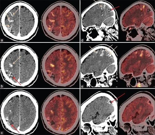
- Kontrast sonrası kıvrımları izleyen kortikal boyanma giral paterndedir; düzensiz kitle kenarı biçiminde olmak zorunda değildir.
- Etkilenen hemisferde sulkus silinmesi ve ventriküle bası, ödemin kitle etkisini gösterir; yayılımı damar alanı içinde kalabilir.
- İlerleyen dönemde sığalaşma ve doku kaybı gelişerek lezyon kalıcı olarak hipodens görünüm kazanır.
- İnfarkt alanında peteşiyal ya da parankimal kanama dönüşümü gelişebilir.
Normalde
Normal BT'de insular şerit, lentiform çekirdek ve kortikal gri-beyaz sınır belirgindir; sulkuslar simetrik açıklıktadır ve damar alanına uyan fokal hipodensite ya da giral kontrastlanma yoktur. Kıyaslamada şüpheli kortikal boyanmanın MCA/ACA/PCA sulama sahasını izleyip izlemediğine, aynı sahada erken iskemi işaretleri olup olmadığına ve komşu sulkusların basılıp basılmadığına bakılır.
Normal BT ile kıyasla
Aynı bölgenin, kaynak veri setinde patoloji içermediği belirtilen bir BT incelemesini kesit kesit kaydırın; organ sınırlarını açarak anatomiyi hasta görüntüsüyle karşılaştırın. Küçük rastlantısal bulgular tümüyle dışlanmamıştır.
Kaynak: TotalSegmentator v2.01 veri seti (Wasserthal ve ark., Radiology: Artificial Intelligence 2023), CC BY 4.0, vaka s0103; organ sınırları veri setinin otomatik segmentasyonundan, birleştirilerek sadeleştirildi. Görüntüler 2,5 mm kesit aralığına yeniden örneklendi.
Ayırıcı tanı
- Yüksek dereceli gliom
- damar sınırlarını aşan infiltratif kitle, düzensiz nekrotik merkez ve nodüler çevresel kontrastlanma gösterebilir.
- Beyin metastazı
- yuvarlak kitle ve belirgin vazojenik ödem oluşturabilir.
- Ensefalitte temporal lob tutulumu görülebilir; ayırım klinik bulgular, MRI ve gerektiğinde BOS incelemesiyle yapılır.
- Postiktal değişiklikler genellikle damar yatağı sınırlarını aşmayacak şekilde kortikal tutulum gösterir; nöbet öyküsü ve kısa aralıklı seri incelemelerde regresyon ayırıcı tanıyı destekler.
- Serebral venöz infarkt
- arter alanı dışına taşan ödem ve kanama ile birlikte venöz sinüs ya da kortikal ven trombozu aranır.
Tuzaklar
- Giral kontrastlanmayı tek başına tümör kabul etme; infarktla ilişkili kortikal boyanma günler-haftalar içinde ortaya çıkabilir.
- Subakut BT fogging döneminde infarkt atenüitesi silikleşebilir; önceki BT/MRI varsa doğrudan yan yana karşılaştır.
- Subakut evrede vazojenik ödem nedeniyle kitle etkisi ve herniasyon riski en üst düzeydedir; bu durum lezyonun benign veya yapışkan olmayan doğasını ortadan kaldırmaz.
- Tek bir zamanda çekilmiş kontrastlı BT, infarkt-tümör ayrımını kesinleştirmeyebilir; belirtilerin başlangıcı ve difüzyon MRI'sı tanısal bağlamı değiştirir.
Kendini dene
Yakınmaları bir haftadır süren hastada PCA alanına uyan kortikal hipodensite ve giral boyanma var. En olası açıklama nedir?
Cevabı göster
Subakut infarkt. Akut evre sitotoksik ödemin ardından gelen subakut evrede vazojenik ödem nedeniyle lezyon kenarları belirginleşir ve kitle etkisi artar; bu dönemde infarkt ile gliomalar iyi bilinen radyolojik taklitçilerdir.
İkinci haftada daha önce görülen damar alanı hipodensitesi BT'de daha az belirgin. Bu değişim nasıl adlandırılır?
Cevabı göster
BT fogging. Subakut dönemde infarkt atenüitesi geçici olarak normal beyin yoğunluğuna yaklaşabilir; buna BT fogging denir ve lezyon sınırını olduğundan küçük gösterebilir. Diğer seçenekler bu geçici atenüite değişimini tanımlamaz.
İlgili konular
Kaynaklar
Bu sayfadaki 35 cümle ve 2 quiz sorusu aşağıdaki kaynaklardan alıntılarla tek tek karşılaştırıldı; 10 cümle bu karşılaştırmada düzeltildi (2026-10-08).
- Imaging in Acute Strokepmc.ncbi.nlm.nih.gov
- Case Report: Structured MRI assessment of posterior thalamic infarction in a distribution compatible with posterior choroidal artery territory presenting as Déjerine-Roussy syndrome in an adolescent: differentiating arterial ischemia from venous thrombosis and thalamic neoplasmpmc.ncbi.nlm.nih.gov
- Middle meningeal artery embolization of meningioma en plaque misdiagnosed as subdural hematoma: illustrative casespmc.ncbi.nlm.nih.gov
- Glioblastoma Masquerading as a Presumed Ischemic Stroke: A Case of Delusional Jealousy and Fluctuating Psychosispmc.ncbi.nlm.nih.gov
- Computed tomography of renal emergencies: A comprehensive diagnostic guide for radiology residentspmc.ncbi.nlm.nih.gov
- Ruptured Intracranial Infectious Aneurysm Mimicking an Expansive Brain Tumor in the Setting of Subacute Infective Endocarditis.pubmed.ncbi.nlm.nih.gov
- Middle meningeal artery embolization of meningioma en plaque misdiagnosed as subdural hematoma: illustrative cases.pubmed.ncbi.nlm.nih.gov
- Case Report: Structured MRI assessment of posterior thalamic infarction in a distribution compatible with posterior choroidal artery territory presenting as Déjerine-Roussy syndrome in an adolescent: differentiating arterial ischemia from venous thrombosis and thalamic neoplasm.pubmed.ncbi.nlm.nih.gov
- Spectrum of Neuroimaging Findings in CNS Lymphoproliferative Disorders.pubmed.ncbi.nlm.nih.gov
- AI-Based Approaches for the Identification and Quantification of Traumatic Brain Injury in Computed Tomography Imaging: Systematic Review.pubmed.ncbi.nlm.nih.gov
- A 25-Year Retrospective of the Use of AI for Diagnosing Acute Stroke: Systematic Review.pubmed.ncbi.nlm.nih.gov
- Ischemic infarct detection, localization, and segmentation in noncontrast CT human brain scans: review of automated methods.pubmed.ncbi.nlm.nih.gov
- From stroke workup to mitochondrial disease: A case report of MELAS.pubmed.ncbi.nlm.nih.gov
- Characteristic MR Imaging Findings of Cerebral Air Embolism Infarcts: A Case Series.pubmed.ncbi.nlm.nih.gov
- Multidimensional analysis of cerebral iron and vascular changes in chronic unilateral middle cerebral artery stenosis with quantitative susceptibility mapping, arterial spin labeling and high-resolution vessel wall imaging-a pilot study.pubmed.ncbi.nlm.nih.gov
- Challenges, limitations, and pitfalls of PET and advanced MRI in patients with brain tumors: A report of the PET/RANO group.pubmed.ncbi.nlm.nih.gov
- 6th Annual Conference of the Neuropathology Society of India (NPSICON) - Meeting Abstracts: February 23-25, 2023.pubmed.ncbi.nlm.nih.gov
- Clinical and radiographic spectrum of pathologically confirmed tumefactive multiple sclerosis.pubmed.ncbi.nlm.nih.gov
- Biot's respiratory pattern under mechanical ventilation: Two case reports and literature review.pubmed.ncbi.nlm.nih.gov
- Amyloid-related imaging abnormalities (ARIA) in anti-amyloid therapies for Alzheimer's disease: An update from the Alzheimer's Association ARIA workgroup.pubmed.ncbi.nlm.nih.gov
- Tissue clock-guided prediction and intervention of futile recanalization: towards precision therapy in mechanical thrombectomy.pubmed.ncbi.nlm.nih.gov
- Intracerebral Hemorrhage in Aging: Pathophysiology, Clinical Challenges, and Future Directions.pubmed.ncbi.nlm.nih.gov
- Spontaneous and Drug-Induced Amyloid-Related Imaging Abnormalities: Overlaps, Divergences, and Clinical Implications Across a Continuum Between Alzheimer's Disease and Cerebral Amyloid Angiopathy.pubmed.ncbi.nlm.nih.gov
- Diagnostic performance of contrast-enhanced T2-FLAIR MRI in the detection of meningitis.pubmed.ncbi.nlm.nih.gov
- Machine learning-based prediction of cerebral artery territorial infarcts in acute ischemic stroke using non-contrast CT.pubmed.ncbi.nlm.nih.gov
- Moyamoya disease in a 10-year-old male patient in the Middle East with the outcome of the surgery: A case report and literature review.pubmed.ncbi.nlm.nih.gov
- Multimodality Imaging of Moyamoya Disease: A Practical Guide for Neuroradiologists Based on a Case Report.pubmed.ncbi.nlm.nih.gov
- Current state and advancements of imaging in acute ischemic stroke: a practical review.pubmed.ncbi.nlm.nih.gov
- A Novel Nomogram to Predict Symptomatic Intracranial Hemorrhage in Ischemic Stroke Patients After Intravenous Thrombolysis.pubmed.ncbi.nlm.nih.gov
- Evaluating the Safety and Efficacy of Intravenous Thrombolysis in Acute Ischemic Stroke Patients Without Perfusion Deficit: A Retrospective Analysis.pubmed.ncbi.nlm.nih.gov
- Case Report: longitudinal imaging in a patient with SMART syndrome after cranial radiotherapy.pubmed.ncbi.nlm.nih.gov
- Post-therapy imaging of adult brain tumours: patterns, pitfalls, and practical framework.pubmed.ncbi.nlm.nih.gov
- Malignant Posterior Reversible Encephalopathy Syndrome (PRES) After Allogeneic Hematopoietic Stem Cell Transplantation Requiring Emergency Decompressive Craniectomy: A Case Report.pubmed.ncbi.nlm.nih.gov
- Anatomically Precise Microsurgical Resection of a Posterior Fossa Cerebellar Metastasis in an Elderly Patient with Preservation of Venous Outflow, Dentate Nucleus, and Cerebrospinal Fluid Pathways.pubmed.ncbi.nlm.nih.gov
- Fetal intracranial hemorrhage and infarct: Main sonographic and MRI characteristics: A review article.pubmed.ncbi.nlm.nih.gov
- The early fogging effect in cerebral infarction - a case report and review of the literature.pubmed.ncbi.nlm.nih.gov
- Immune checkpoint blockade and thrombo-inflammatory remodeling in microsatellite instability-high intrahepatic cholangiocarcinoma with Trousseau syndrome: a case report.pubmed.ncbi.nlm.nih.gov
- Equivocal Dopamine Transporter Imaging: Integrating Visual Assessment, Quantification, and Clinical Follow-Up.pubmed.ncbi.nlm.nih.gov
- A Transient Left Atrial Appendage Filling Defect During Anticoagulation Interruption After Ischemic Stroke with Progressive Hemorrhagic Transformation: A Case Report.pubmed.ncbi.nlm.nih.gov
- Clinical Utility of Circulating Biomarkers for Diagnosis and Prognosis of Acute Stroke in the Emergency Department: A Narrative Review from Current Evidence to Clinical Implementation.pubmed.ncbi.nlm.nih.gov
- Comparison of HEmorrhage on CT versus MRI After ThrombEctomy: The HECATE study.pubmed.ncbi.nlm.nih.gov
- Evaluation of Nonlinear Pixel-Wise Squaring for Enhancing Low-Contrast Resolution in CT Imaging.pubmed.ncbi.nlm.nih.gov
- Molecular Mechanisms of Ischemic Stroke: A Review Integrating Clinical Imaging and Therapeutic Perspectives.pubmed.ncbi.nlm.nih.gov
- Diagnosis and treatment of Watershed strokes: a narrative review.pubmed.ncbi.nlm.nih.gov
- Spatiotemporal Deep Video-Phenomapping Decodes Microvascular Rarefaction in Middle-Aged and Elder Renovascular Hypertension: A Multi-Modal Study Integrating Spatial Transcriptomics and Mitochondrial Pyroptosis.pubmed.ncbi.nlm.nih.gov
- Semantic and Phonological Brain Networks in Older Adults: A Systematic Scoping Review.pubmed.ncbi.nlm.nih.gov
- Gamma Knife radiosurgery delivered to internal auditory canal glioblastoma under a presumed diagnosis of vestibular schwannoma: illustrative case.pubmed.ncbi.nlm.nih.gov
- Giant Calcified Frontal Lobe Metastasis as the Initial Manifestation of Pulmonary Large-Cell Neuroendocrine Carcinoma: Diagnostic and Surgical Insights From Intraoperative Diagnosis.pubmed.ncbi.nlm.nih.gov
- Delayed intracranial lesions following cardiac myxoma resection in a patient with concurrent cerebral embolism: a diagnostic challenge in the absence of pathological evidence.pubmed.ncbi.nlm.nih.gov
- Clinicopathological insights of supratentorial embryonal tumor with medulloblastoma-like histopathology in adulthood: A case report.pubmed.ncbi.nlm.nih.gov
- Primary central nervous system lymphoma: Imaging features and differential diagnosis.pubmed.ncbi.nlm.nih.gov
- Myelin oligodendrocyte glycoprotein antibody-related autoimmune encephalitis misdiagnosed as acute cerebral infarction: A case report.pubmed.ncbi.nlm.nih.gov
- Anti-NMDA-receptor encephalitis and MOGAD associated optic neuritis: a case series.pubmed.ncbi.nlm.nih.gov
- Herpes Simplex Virus Encephalitis With Cerebellar Infarction and Cortical Laminar Necrosis.pubmed.ncbi.nlm.nih.gov
- Seizure-Related Deterioration in Treated Glioma: A Narrative Review of Nonconvulsive Status Epilepticus, Peri-Ictal Pseudoprogression, Treatment-Related Change, and True Progression.pubmed.ncbi.nlm.nih.gov
- Mild encephalitis/encephalopathy with a reversible splenial lesion (MERS): A case report and review of the literature.pubmed.ncbi.nlm.nih.gov
- Vigabatrin-related MRI changes and tapering strategy in hemispherotomy for infantile spasms: illustrative case.pubmed.ncbi.nlm.nih.gov
- Perfusion imaging abnormalities in acute cerebral venous sinus thrombosis: A case series.pubmed.ncbi.nlm.nih.gov
- Cerebral venous sinus thrombosis in pregnancy and the postpartum period: epidemiology, management, and maternal mortality-a systematic review and meta-analysis.pubmed.ncbi.nlm.nih.gov
- Cerebral venous disorders: clinical presentation, diagnostic strategy, and contemporary management.pubmed.ncbi.nlm.nih.gov
- Acute Seizures and Aphasia in a Patient With Suspected Systemic Lupus Erythematosus: A Diagnostic Puzzle Unveiling Diffuse Hemispheric Glioma, H3 G34-Mutant.pubmed.ncbi.nlm.nih.gov
- Magnetic Resonance Imaging in Acute Ischemic Stroke.pubmed.ncbi.nlm.nih.gov
- Beyond Recanalization: Neurovascular Unit Protection and Precision Adjunctive Therapy for Ischemic Stroke in the Reperfusion Era.pubmed.ncbi.nlm.nih.gov
- Research Progress of Blood-Based Biomarkers for the Diagnosis and Prognostic Evaluation of Acute Ischemic Stroke.pubmed.ncbi.nlm.nih.gov
- Spectral CT and Photon-counting Detector CT: Principles and Applications in Neuroradiology.pubmed.ncbi.nlm.nih.gov
- A systematic approach to MR spectroscopy of the brain: Indications, protocol, metabolites, and spectral analysis in neurologic diseases.pubmed.ncbi.nlm.nih.gov
- CT Imaging in Neurocritical Care.pubmed.ncbi.nlm.nih.gov
- A systematic approach to detection of epileptogenic foci on imaging: protocols, entities, search pattern, and reporting tools.pubmed.ncbi.nlm.nih.gov
- Acute Ischemic Stroke Lesion Characterization: Comparative Analysis of Cortical and Basal Ganglia Lesions Using Time-Histogram Metrics.pubmed.ncbi.nlm.nih.gov
- Evaluating iron deposition in gray matter nuclei of patients with acute ischemic stroke using quantitative susceptibility mapping.pubmed.ncbi.nlm.nih.gov
- Case Report: Ischemic stroke revealing carotid fibromuscular dysplasia with multiple metabolic disturbances in a 42-year-old man.pubmed.ncbi.nlm.nih.gov
- Territorial CT Perfusion Burden and Outcome After Aneurysmal Subarachnoid Haemorrhage: A Retrospective Single-Centre Cohort Study.pubmed.ncbi.nlm.nih.gov
- Fulminant Amyloid-β-Related Angiitis after Tenecteplase: A Biopsy-Confirmed Cause of Post-Thrombolysis Neurologic Deterioration.pubmed.ncbi.nlm.nih.gov
- Timing of DOAC Initiation After Atrial Fibrillation-Related Acute Ischemic Stroke: A Unified Protocol Harmonizing Clinical Safety and Imaging-Based Precision in the Post-CATALYST Era.pubmed.ncbi.nlm.nih.gov
- Beyond the embolus: "do not miss" diffusion abnormalities of ischaemic and non-ischaemic neurological disease.pubmed.ncbi.nlm.nih.gov
- In vitro and in vivo models of cerebral ischemia show discrepancy in therapeutic effects of M2 macrophages.pubmed.ncbi.nlm.nih.gov
- Stroke or Seizure? Diagnostic Role of Neuroimaging in Acute Neurologic Mimics.pubmed.ncbi.nlm.nih.gov
- Neurological manifestations and neuroimaging findings in patients with SARS-CoV2-a systematic review.pubmed.ncbi.nlm.nih.gov
- Imaging in Acute Anterior Circulation Ischemic Stroke: Current and Future.pubmed.ncbi.nlm.nih.gov
- A double threat: CNS toxoplasmosis and CMV encephalitis in a heart transplant recipientpubmed.ncbi.nlm.nih.gov
- Stroke-Like Migraine Attacks After Radiation Therapy (SMART) Syndrome: A Case Report and Review of a Rare Delayed Complication of Cranial Irradiation.pubmed.ncbi.nlm.nih.gov
- Central nervous system vasculitis mimicking a brain tumor: A case report.pubmed.ncbi.nlm.nih.gov
- Neurological complications following cerebral angiography: a case of contrast-induced encephalopathy.pubmed.ncbi.nlm.nih.gov
- Occult Amyloid-β-Related Angiitis: Neuroimaging Findings at 1.5T, 3T, and 7T MRI.pubmed.ncbi.nlm.nih.gov
- Neurite orientation dispersion and density imaging in stroke: current applications, limitations, and future directions.pubmed.ncbi.nlm.nih.gov
- Moyamoya disease masquerading as CNS vasculitis: Diagnostic challenges and imaging insights.pubmed.ncbi.nlm.nih.gov
- Intracranial venous thrombosis imaging by anatomical location: an educational review with mimics and diagnostic pitfalls.pubmed.ncbi.nlm.nih.gov
- Posterior reversible encephalopathy syndrome in dengue virus infection with concurrent encephalitis and ischemic stroke: a case report.pubmed.ncbi.nlm.nih.gov
- A comparative study of large-vessel and small-vessel primary angiitis of the central nervous system: insights from a Chinese single-center retrospective cohort.pubmed.ncbi.nlm.nih.gov
- Anatomy-Guided Microsurgical Resection of a Dominant Frontal Lobe Tumor Without Intraoperative Adjuncts: A Case Report from a Resource-Limited Context.pubmed.ncbi.nlm.nih.gov
- Predicting Functional Prognosis in Supratentorial Stroke Using [¹⁸F]Fluoro-2-deoxy-D-glucose Positron Emission Tomography.pubmed.ncbi.nlm.nih.gov
- Multiscale photoacoustic imaging of stroke in preclinical models and future directions toward clinical translation [Invited].pubmed.ncbi.nlm.nih.gov
- Selective Kv1.3 inhibition promotes M2 microglial polarization, accelerates hematoma resolution, and improves neurological recovery following collagenase-induced spontaneous intracerebral hemorrhage in mice.pubmed.ncbi.nlm.nih.gov
- Detection of Hemorrhagic Transformation of Cerebral Infarcts with Ultra-low Field MRI.pubmed.ncbi.nlm.nih.gov
- Risk factors for hemorrhagic transformation after tenecteplase therapy in acute ischemic stroke: A retrospective observational study.pubmed.ncbi.nlm.nih.gov
- Arterial Spin Labeling technique and clinical applications of the intracranial compartment in stroke and stroke mimics - A case-based review.pubmed.ncbi.nlm.nih.gov
- A Diagnostic Challenge: From Suspected Ischemic Stroke to Confirmed Herpetic Encephalitis.pubmed.ncbi.nlm.nih.gov
- Complex Aortic Graft Infection with Giant Collection after Bentall-Bono Surgery: Diagnostic and Therapeutic Challenges.pubmed.ncbi.nlm.nih.gov
- Right-to-Left Shunt via a Patent Foramen Ovale Triggered by Constrictive Pericarditis With Right Heart Compression: A Case Report.pubmed.ncbi.nlm.nih.gov
- Middle cerebral artery dissection: pathophysiology, diagnosis, and therapeutic options.pubmed.ncbi.nlm.nih.gov
- Neurological Puzzles After Radiotherapy: Mysterious Clues of Smart Syndrome, Two Case Reports.pubmed.ncbi.nlm.nih.gov
- Leptomeningeal Masses or Masquerades: A Spectrum of Diseases with Leptomeningeal Enhancement and their Mimics.pubmed.ncbi.nlm.nih.gov
Eğitim amaçlıdır; hasta başında tanı ve tedavi kararının yerine geçmez. İçerik yapay zekâ destekli bir süreçle güncel literatüre göre cümle cümle kaynaklarla karşılaştırıldı, ancak uzman radyolog incelemesinden geçmedi. Klinik kararlarda güncel kılavuzları, kurum protokollerini ve radyoloji raporunu esas alın.