Özet
Kemoterapi ilişkili karaciğer hasarı rejime göre değişir: irinotekan steatoz/steatohepatitle, oksaliplatin sinüzoidal obstrüksiyon sendromuyla ilişkilendirilir. Hepatik yağlanma karaciğer atenüasyonunu azaltabilir; MASLD küçük karaciğer lezyonlarında BT duyarlılığını düşürebilir. Bu değişiklik özellikle kolorektal kanser hastasında tedavi öncesi ve sonrası lezyon yükü kıyaslanırken tanısal güveni etkiler. Kontrastsız BT'de karaciğer atenüasyonunu dalakla kıyaslamak yağlanmayı göstermenin pratik yoludur; fokal yağlanma veya yağdan korunmuş alan metastazı taklit edebilir. Arka plan parankim değişikliği tümör yanıtı sanılırsa gereksiz evre veya yanıt değişikliği kararı doğabilir.
Faz ve pencere
- Kontrastsız tanısal
- Kontrastsız BT'de karaciğer atenüasyonunun dalaktan en az 10 HU düşük olması steatoz lehine bir bulgudur. Fokal veya nodüler yağlanma ile yağdan korunmuş alanlar karaciğerde kitleyi taklit eden odaklar şeklinde görülebilir. Kontrastlı BT'de karaciğer atenüasyonunun dalaktan en az 20 HU düşük olması steatoz lehine kullanılabilir; bu eşik kontrastsız BT'deki 10 HU eşiğinden farklıdır.
Önerilen pencereler: Karaciğer (G 150 / M 30), Batın (G 400 / M 50).
BT bulguları
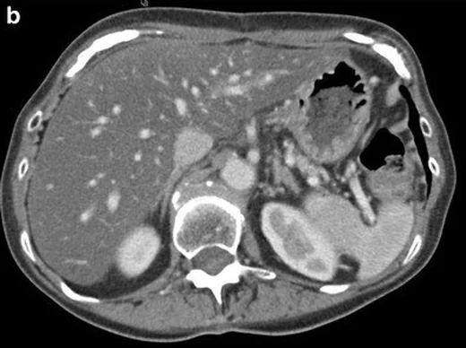
- Karaciğer parankiminin kontrastsız BT'de dalağa göre düşük atenüasyonda olması steatoz lehine olabilir.
- Kontrastsız BT'de karaciğer atenüasyonunun dalaktan en az 10 HU düşük olması steatozu destekler.
- Fokal yağlanma — karaciğer parankiminde coğrafi dağılım gösteren düşük atenüasyon alanı olarak görünür.
- Yağdan korunmuş parankim — yağlı karaciğerde nodüler odaklar şeklinde görülebilir ve metastazı taklit edebilir.
- Fokal veya nodüler steatoz, metastatik karaciğer hastalığını taklit edebilir.
- Tedaviyle parankim değişimi — tedavi öncesi ve sonrası BT incelemelerinde karaciğerde radyolojik steatoz gelişimini karşılaştırın.
- Metastaz görünürlüğü — kemoterapi, metastazların BT ile saptanma duyarlılığını azaltabilir.
Ölçütler ve sınıflamalar
- Karaciğer atenüasyonu dalağa göre düşük
- ≥10 HU daha düşük
- Karaciğer/dalak atenüasyon oranı
- <1
- Mutlak karaciğer atenüasyonu
- <40 HU (yalnız kontrastsız BT'de; HU eşikleri kontrastlı serilere aktarılmaz)
Normalde
Kontrastsız BT'de karaciğer atenüasyonunun dalaktan düşük olması steatoz lehine olabilir. Steatoz lehine eşik, karaciğerin dalaktan ≥10 HU düşük olmasıdır (karaciğer−dalak ≤−10 HU). Ölçümleri büyük damar ve fokal lezyonlardan uzak homojen parankimden alın.
Normal BT ile kıyasla
Aynı bölgenin, kaynak veri setinde patoloji içermediği belirtilen bir BT incelemesini kesit kesit kaydırın; organ sınırlarını açarak anatomiyi hasta görüntüsüyle karşılaştırın. Küçük rastlantısal bulgular tümüyle dışlanmamıştır.
Kaynak: TotalSegmentator v2.01 veri seti (Wasserthal ve ark., Radiology: Artificial Intelligence 2023), CC BY 4.0, vaka s0541; organ sınırları veri setinin otomatik segmentasyonundan, birleştirilerek sadeleştirildi. Görüntüler 2,5 mm kesit aralığına yeniden örneklendi.
Ayırıcı tanı
- Karaciğer metastazı
- yuvarlak veya lobüle kitle, kitle etkisi ve kontrastlı fazlarda lezyona özgü kontrastlanma gösterebilir; fokal yağ damarları itmez.
- Hemanjiyomlar en sık görülen benign karaciğer tümörleridir ve klasik görüntüleme özellikleriyle tanınabilir.
- Perfüzyon değişikliği
- segmental veya kama biçimli atenüasyon farklılıkları olarak görülebilir ve dinamik kontrast fazlarında karakterize edilebilir.
- İlaç ilişkili sinüzoidal hasar
- kontrastlı görüntülerde retiküler/yamalı heterojen perfüzyon ve portal hipertansiyon bulguları ön plandadır; kontrastsız düşük atenüasyon tek başına steatoz demek değildir.
Tuzaklar
- Kontrastlı BT'de kontrastsız BT için kullanılan ≥10 HU eşiğini uygulamayın; kaynakta kontrastlı BT için karaciğer atenüasyonunun dalaktan ≥20 HU düşük olması ölçütü kullanılmıştır.
- Fokal yağlanmanın kenarını kitle kapsülü sanma; coğrafi sınır, damarların kesintisiz içinden geçmesi ve kitle etkisinin olmaması yağ lehinedir.
- Dalak parenkim hastalıkları veya metabolik değişiklikleri dalak atenüasyonunu etkileyebilir; bu nedenle karaciğer ölçümü mutlak değerlerle ve önceki incelemelerle birlikte değerlendirilmelidir.
- Tedavi sonrası parankim yağlanması metastazı gizleyebilir; eski incelemedeki lezyon konumlarını mevcut parankim görünümüyle eşleştir.
- Oksaliplatin sonrası yamalı veya retiküler karaciğer atenüasyon değişikliği steatozla eş anlamlı değildir; sinüzoidal obstrüksiyon sendromu ve diğer tedavi ilişkili parankim değişiklikleri de düşünülmelidir.
Kendini dene
Karaciğer steatozu hangi görünümle başka multifokal karaciğer lezyonlarını taklit edebilir?
Cevabı göster
Multinodüler steatoz. Multinodüler hepatik steatoz nadir görülür, ancak karaciğerin diğer multifokal lezyonlarını taklit edebilir.
Karaciğer-dalak atenüasyon farkını steatoz için 10 HU eşiğiyle değerlendireceksiniz. Hangi BT serisini kullanmalısınız?
Cevabı göster
Kontrastsız BT. Bu kaynakta karaciğer atenüasyonunun dalaktan en az 10 HU düşük olması eşiği kontrastsız BT için tanımlanmıştır.
İlgili konular
Kaynaklar
Bu sayfadaki 40 cümle ve 2 quiz sorusu aşağıdaki kaynaklardan alıntılarla otomatik olarak karşılaştırıldı; 22 cümle bu karşılaştırmada düzeltildi (2026-10-09).
- Hepatic Steatosis After Neoadjuvant Chemotherapy for Pancreatic Cancer: Incidence and Implications for Outcomes After Pancreatoduodenectomypmc.ncbi.nlm.nih.gov
- CT Attenuation Thresholds for Grading Hepatic Steatosis in Adults with Overweight or Obesity and Metabolic Dysfunction-Associated Steatotic Liver Diseasepmc.ncbi.nlm.nih.gov
- Oxaliplatin-induced autoimmune-like hepatitis: A case report highlighting the role of clinical pharmacistspmc.ncbi.nlm.nih.gov
- Factors Affecting Temporal Changes in Ablated Liver Volume After Radiofrequency Ablation for Hepatocellular Carcinoma Evaluated by Three-Dimensional Volumetric Computed Tomographypmc.ncbi.nlm.nih.gov
- Sarcopenia does not affect total liver function measured using technetium-99m mebrofenin hepatobiliary scintigraphypmc.ncbi.nlm.nih.gov
- Consequences of chemotherapy on resection of colorectal liver metastases.pubmed.ncbi.nlm.nih.gov
- Impact of metabolic liver disease on colorectal cancer and liver metastases: Pathophysiology and implications for hepatobiliary surgery.pubmed.ncbi.nlm.nih.gov
- Hepatic Steatosis After Neoadjuvant Chemotherapy for Pancreatic Cancer: Incidence and Implications for Outcomes After Pancreatoduodenectomy.pubmed.ncbi.nlm.nih.gov
- Chemotherapy-associated steatohepatitis was concomitant with epicardial adipose tissue volume increasing in breast cancer patients who received neoadjuvant chemotherapy.pubmed.ncbi.nlm.nih.gov
- Comparison of Multimaterial Decomposition Fat Fraction with DECT and Proton Density Fat Fraction with IDEAL IQ MRI for Quantification of Liver Steatosis in a Population Exposed to Chemotherapy.pubmed.ncbi.nlm.nih.gov
- Risk factors for irinotecan-induced hepatotoxicity: a retrospective cohort study.pubmed.ncbi.nlm.nih.gov
- Post-pancreatectomy Liver Injury After Mayo Clinic Class Ia Celiac Axis Resection: Illustration of This Newly Described Entity with Delayed Hepatic Artery Revascularization.pubmed.ncbi.nlm.nih.gov
- Delaying surgery beyond six weeks after systemic therapy reduces postoperative morbidity without evidence of impaired oncologic outcomes in colorectal liver metastases.pubmed.ncbi.nlm.nih.gov
- Pattern-Based P53 Immunohistochemistry Predicts Recurrence in Colorectal Liver Metastases Without Complete Pathologic Response After Neoadjuvant Chemotherapy.pubmed.ncbi.nlm.nih.gov
- Artificial intelligence-driven body composition analysis and its association with survival in patients with colorectal liver metastases.pubmed.ncbi.nlm.nih.gov
- Preoperative CT-derived liver fat fraction and visceral adipose tissue index enhance postoperative recurrence risk stratification in colorectal cancer.pubmed.ncbi.nlm.nih.gov
- Automated Deep Learning Detection of Hepatic Steatosis on Noncontrast Computed Tomography Scans and Discrepancy With Scan Reports.pubmed.ncbi.nlm.nih.gov
- CT Attenuation Thresholds for Grading Hepatic Steatosis in Adults with Overweight or Obesity and Metabolic Dysfunction-Associated Steatotic Liver Disease.pubmed.ncbi.nlm.nih.gov
- Emerging quantitative CCTA imaging biomarkers for cardiovascular risk stratification: a narrative review.pubmed.ncbi.nlm.nih.gov
- Splenic Steatopathy: A Clinical and Experimental Framework for Lipid-Associated Splenic Pathology.pubmed.ncbi.nlm.nih.gov
- Breast cancer pharmacotherapy and imaging interpretation: pathophysiological perspectives and clinical application.pubmed.ncbi.nlm.nih.gov
- The association between epicardial adipose tissue thickness and diabetes mellitus, hyperlipidemia, hepatosteatosis, pancreatic steatosis and pancreatitis.pubmed.ncbi.nlm.nih.gov
- Association Between ADH1B rs1229984 Genetic Polymorphism and Hepatic Steatosis Assessed by CT Attenuation: A Mediation Analysis of Central Obesity in MASLD.pubmed.ncbi.nlm.nih.gov
- Gestational trophoblastic diseases - literature review and atypical case reports.pubmed.ncbi.nlm.nih.gov
- Review of COVID-19, part 1: Abdominal manifestations in adults and multisystem inflammatory syndrome in children.pubmed.ncbi.nlm.nih.gov
- The Malignant Transformation of Viral Hepatitis to Hepatocellular Carcinoma: Mechanisms and Interventions.pubmed.ncbi.nlm.nih.gov
- Advanced Imaging Techniques for Assessing Fat, Iron, and Fibrosis in Chronic Liver Disease.pubmed.ncbi.nlm.nih.gov
- Liver cancer segmentator: Metadata-guided confidence scoring for reliable segmentation of colorectal liver metastases in CT.pubmed.ncbi.nlm.nih.gov
- An Analysis of Age-Related Body Composition Changes and Metabolic Patterns in Korean Adults Using FDG-PET/CT Health Screening Data.pubmed.ncbi.nlm.nih.gov
- Multifocal fatty sparing areas in a steatotic liver mimicking hepatic lesions: a case report and review of the literature.pubmed.ncbi.nlm.nih.gov
- Smoldering Amiodarone-Induced Decompensated Cirrhosis: Cumulative Dose-Related Toxicity.pubmed.ncbi.nlm.nih.gov
- Imaging Evaluation for Steatotic Liver Disease.pubmed.ncbi.nlm.nih.gov
- Potential utility of postmortem CT-based classification as a practical pre-autopsy assessment tool for hepatic steatosis.pubmed.ncbi.nlm.nih.gov
- Factors Affecting Disparity Between Estimated Liver Volume and Actual Liver Volume in Living Donor Liver Transplantation.pubmed.ncbi.nlm.nih.gov
- Acute Liver Failure With Transient Liver Steatosis Following Multiple Hits Postoperatively in a Patient With Limb-Girdle Muscular Dystrophy: A Case Report.pubmed.ncbi.nlm.nih.gov
- Imaging features and management of focal liver lesions.pubmed.ncbi.nlm.nih.gov
- Focal incidental upper abdominal findings on unenhanced chest computed tomography that do not require further imaging: a roadmap for the thoracic radiologist.pubmed.ncbi.nlm.nih.gov
- Benign focal liver lesions: The role of magnetic resonance imaging.pubmed.ncbi.nlm.nih.gov
- International Harmonization of Nomenclature and Diagnostic Criteria (INHAND): Non-proliferative and Proliferative Lesions of the Non-human Primate ( M. fascicularis ).pubmed.ncbi.nlm.nih.gov
- Commentary: an eye on PET quantification.pubmed.ncbi.nlm.nih.gov
- Radiological Assessment After Pancreaticoduodenectomy for a Precision Approach to Managing Complications: A Narrative Review.pubmed.ncbi.nlm.nih.gov
- China Liver Cancer Guidelines for the Diagnosis and Treatment of Hepatocellular Carcinoma (2024 Edition).pubmed.ncbi.nlm.nih.gov
- Clinicopathological insights and management of liver metastases: Current advances and future perspectives.pubmed.ncbi.nlm.nih.gov
- Oxaliplatin-induced autoimmune-like hepatitis: A case report highlighting the role of clinical pharmacists.pubmed.ncbi.nlm.nih.gov
- Amiodarone-induced liver injury with bright liver sign: A case report.pubmed.ncbi.nlm.nih.gov
- Tirzepatide-Associated Severe Hepatocellular Injury: A Case Report and Diagnostic Considerations.pubmed.ncbi.nlm.nih.gov
- Multiparametric Abdominal Ultrasound as a Source of Imaging Biomarkers in Predictive and Personalized Medicine: A Narrative Review.pubmed.ncbi.nlm.nih.gov
- MASLD and Pancreatic Diseases: From Shared Mechanisms to Clinical Implications and Future Directions.pubmed.ncbi.nlm.nih.gov
- Diagnostic performance and dose reduction of virtual noncontrast imaging in pediatric computed tomography urography.pubmed.ncbi.nlm.nih.gov
- Triple-Phase Computed Tomography for Differentiating Canine Cutaneous and Subcutaneous Mast Cell Tumors From Benign Lesions: A Size-Based Evaluation.pubmed.ncbi.nlm.nih.gov
- Liver CT-based composite biomarkers can identify MASH and steatosis grade in people with obesity prior to bariatric surgery: a retrospective study.pubmed.ncbi.nlm.nih.gov
- Study on the combined application of selected virtual monoenergetic imaging, virtual non-contrast imaging, and adaptive statistical iterative reconstruction-V in head and neck CT angiography using 256-slice wide-body CT.pubmed.ncbi.nlm.nih.gov
- Coexistence of Small Duct Type and Biliary Adenofibroma-Associated Large Duct Type Intrahepatic Cholangiocarcinoma within a Single Lesion: A Case Report.pubmed.ncbi.nlm.nih.gov
- Eosinophilic Biliary Cystic Neoplasm of the Liver: A Case Report of an Unrecognized Subtype of Intrahepatic Biliary Neoplasm.pubmed.ncbi.nlm.nih.gov
- Practical Considerations in Abdominal MRI: Sequences, Patient Preparation, and Clinical Applications.pubmed.ncbi.nlm.nih.gov
- Differentiating hepatic hemangiomas from hepatocellular carcinomas using Sonazoid-enhanced ultrasonography: a proposed two-step diagnostic algorithm.pubmed.ncbi.nlm.nih.gov
- A rare site for a common tumor: Incidental hepatic lipoma detected during evaluation for perforated appendicitis.pubmed.ncbi.nlm.nih.gov
- Frequency and associated factors of hepatic hemangioma in systemic lupus erythematosus patients undergoing abdominal imaging: a retrospective cohort study.pubmed.ncbi.nlm.nih.gov
- Focal iron sparing mimicking liver lesion in post-transfusion iron overload: A case report.pubmed.ncbi.nlm.nih.gov
- Imaging-based fibrosis assessment and risk stratification in MASLD.pubmed.ncbi.nlm.nih.gov
- Aqueous Extract of <i>Siraitia grosvenorii</i> Alleviates MAFLD by Modulating Metabolism and Maintaining Gut Homeostasis in High-Fat Diet Fed Mice.pubmed.ncbi.nlm.nih.gov
- Massive bleeding from a Bricker ileal conduit during FOLFIRINOX therapy in noncirrhotic portal hypertension: suspected oxaliplatin-associated sinusoidal injury and presumed irinotecan-associated conduit mucositis: a case report.pubmed.ncbi.nlm.nih.gov
- Tegafur/Uracil-Induced Sinusoidal Obstruction Syndrome as a Suspected Cause of Liver Cirrhosis during Adjuvant Chemotherapy for Lung Adenocarcinoma: A Case Report.pubmed.ncbi.nlm.nih.gov
- Multiparametric Ultrasound in Chronic Viral Hepatitis: From Fibrosis and Portal Hypertension to Steatosis and Focal Lesion Characterisation.pubmed.ncbi.nlm.nih.gov
- MRI in MASLD: technical foundations and guideline-based applications.pubmed.ncbi.nlm.nih.gov
- Hepatocellular adenoma update: diagnosis, molecular classification, and clinical course.pubmed.ncbi.nlm.nih.gov
- A case report of multinodular hepatic steatosis mimicking pseudotumors of the liver.pubmed.ncbi.nlm.nih.gov
- Assessing Pancreatic Enhancement Variability in ICU Patients with CT Hypoperfusion Complex: A Retrospective Single-Center Study.pubmed.ncbi.nlm.nih.gov
- Case Report: Multimodality imaging features and serial metabolic changes on <sup>18</sup> F-FDG PET/CT in a dog with granulomatous hepatitis.pubmed.ncbi.nlm.nih.gov
- Nanoscale delivery systems for hepatocellular carcinoma: Functionalization, delivery strategies, and clinical challenges.pubmed.ncbi.nlm.nih.gov
- Macrophage: Biological Functions, Diseases, and Therapeutic Targets.pubmed.ncbi.nlm.nih.gov
- Liver Cancer in Methylmalonic and Propionic Acidemias: A Rare Complication? A Clinico-Pathological Study of 24 Livers.pubmed.ncbi.nlm.nih.gov
- Intraoperative Ultrasound in Hepatic Oncology Surgery: A Narrative Review of Its Impact on Surgical Strategy and Oncologic Outcomes.pubmed.ncbi.nlm.nih.gov
- Giant Atypical Hepatocellular Adenoma of Unknown Etiology in a Young Male: A Case Report.pubmed.ncbi.nlm.nih.gov
- Blood Pressure Trajectories in Young Adulthood and Midlife Steatotic Liver Disease.pubmed.ncbi.nlm.nih.gov
- Efficacy of CPAP Therapy on Liver Steatosis and Insulin Resistance in Fatty Liver Patients With Obstructive Sleep Apnea: A 4-Year Follow-Up Cohort Study.pubmed.ncbi.nlm.nih.gov
- Comparing ultrasound-derived fat fraction and CT for diagnosing hepatic steatosis: an MRI-PDFF reference study.pubmed.ncbi.nlm.nih.gov
- Imaging of MASLD: clinical standards and novel methods.pubmed.ncbi.nlm.nih.gov
- Effect of reduction in skeletal muscle quantity and sex differences on postoperative hepatic steatosis following total pancreatectomy.pubmed.ncbi.nlm.nih.gov
- Spectral CT: Current Liver Applications.pubmed.ncbi.nlm.nih.gov
- EACR 2025 Congress: Innovative Cancer Science, 16-19 June 2025.pubmed.ncbi.nlm.nih.gov
- Long-term safety and effectiveness of the "OptEase" vena cava filter.pubmed.ncbi.nlm.nih.gov
- The role of ceus in the characterization of indeterminate focal liver lesions at second-level imaging methods (INFOLIL STUDY).pubmed.ncbi.nlm.nih.gov
- Washout on Contrast-Enhanced Ultrasound of Benign Focal Liver Lesions-A Review on Its Frequency and Possible Causes.pubmed.ncbi.nlm.nih.gov
- Practical approach to diagnose and manage benign liver masses.pubmed.ncbi.nlm.nih.gov
- Pulmonary arteries: imaging of pulmonary embolism and beyond.pubmed.ncbi.nlm.nih.gov
- Splenic shear wave elastography beyond portal hypertension: a narrative review of non-portal applications.pubmed.ncbi.nlm.nih.gov
- The Dual-Faceted Role of Metal-Based Nanomaterials in Hepatic Fibrosis Therapy.pubmed.ncbi.nlm.nih.gov
- Medical hyperspectral imaging: an updated review of technology advancements and biomedical applications.pubmed.ncbi.nlm.nih.gov
- Ultrasound innovations in abdominal radiology: techniques and clinical applications in pediatric imaging.pubmed.ncbi.nlm.nih.gov
- Special Issue: EACR 2026 Congress: Innovative Cancer Science, 8-11 June 2026.pubmed.ncbi.nlm.nih.gov
- Pelvic Lymphangioma: The Purpose of a Clinical Case.pubmed.ncbi.nlm.nih.gov
- Diffusion-Weighted Imaging in the Musculoskeletal System: Evolving Role in Modern Imaging Practice.pubmed.ncbi.nlm.nih.gov
- A diagnostic approach to mediastinal masses in clinical practice.pubmed.ncbi.nlm.nih.gov
- Joint Consensus Statement of the Indian National Association for Study of the Liver and Indian Radiological and Imaging Association for the Diagnosis and Imaging of Hepatocellular Carcinoma Incorporating Liver Imaging Reporting and Data System.pubmed.ncbi.nlm.nih.gov
- Spontaneous bacteremia and spontaneous bacterial peritonitis share similar prognosis in patients with cirrhosis: a cohort study.pubmed.ncbi.nlm.nih.gov
- Multiomics Analysis-Based Biomarkers in Diagnosis of Polycystic Ovary Syndrome.pubmed.ncbi.nlm.nih.gov
- Immunoglobulin-G4 Related Disease: A Rare Entity with Many Clinical Faces - Literature Review and Case Illustration.pubmed.ncbi.nlm.nih.gov
- Gastrointestinal stromal tumor liver metastasis rupture following yttrium-90 radioembolization: A previously unreported complication.pubmed.ncbi.nlm.nih.gov
- Liver Imaging Reporting and Data System (LI-RADS): A Comprehensive Review.pubmed.ncbi.nlm.nih.gov
- Hepatic parenchymal hypoattenuation in dogs with diabetes mellitus on computed tomography consistent with hepatic steatosis.pubmed.ncbi.nlm.nih.gov
- WFUMB Review Paper. Incidental Findings in Otherwise Healthy Subjects, How to Manage: Liver.pubmed.ncbi.nlm.nih.gov
- Imaging of the chemotherapy-induced hepatic damage: Yellow liver, blue liver, and pseudocirrhosis.pubmed.ncbi.nlm.nih.gov
- Liver metastases: The role of magnetic resonance imaging.pubmed.ncbi.nlm.nih.gov
- Focal Hepatic Steatosis Mimicking a Hilar Tumor With Rapid Resolution After Glycemic Control: A Case Report.pubmed.ncbi.nlm.nih.gov
- Progressive hepatopathy after central division hepatectomy in a dog.pubmed.ncbi.nlm.nih.gov
- Xanthohumol: Mechanistic Actions and Emerging Evidence as a Multi-Target Natural Nutraceutical.pubmed.ncbi.nlm.nih.gov
- Photoacoustic Imaging for Liver Disease: The Systems and the Molecules.pubmed.ncbi.nlm.nih.gov
- A Deep Learning Algorithm for Liver Metastasis Detection at Contrast-enhanced Abdominal CT in Patients with Colorectal Cancer: A Comparative Study with Radiologists.pubmed.ncbi.nlm.nih.gov
- Advances in Artificial Intelligence-Based Liver-Related Semantic Segmentation Techniques and Applications Using CT Imaging.pubmed.ncbi.nlm.nih.gov
- Imaging insights into pediatric liver masses: A comprehensive minireview for hepatology practice.pubmed.ncbi.nlm.nih.gov
- Primary Posterior Neck Hydatid Cyst: A Case Report and Review of the Literature.pubmed.ncbi.nlm.nih.gov
- Case Report: Retroperitoneal bronchogenic cyst mimicking adrenal mass: Clinical implications and comprehensive literature analysis.pubmed.ncbi.nlm.nih.gov
- Use of the Term "Atypical Hepatic Hemangioma" in Radiology Reports: An Analysis of Diagnostic Precision.pubmed.ncbi.nlm.nih.gov
- Transarterial embolization with bleomycin-lipiodol for symptomatic multifocal hepatic hemangiomas: Concurrent nontarget lesion devascularization via permitted reflux.pubmed.ncbi.nlm.nih.gov
Eğitim amaçlıdır; hasta başında tanı ve tedavi kararının yerine geçmez, tıbbi tavsiye değildir. Metinler yapay zekâ desteğiyle yazıldı ve otomatik bir adımda kaynak alıntılarıyla karşılaştırıldı; bu bir tıbbi doğrulama değildir ve içerik uzman radyolog incelemesinden geçmedi. Klinik kararlarda güncel kılavuzları, kurum protokollerini ve radyoloji raporunu esas alın.