Özet
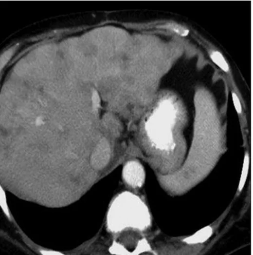
Oksaliplatin başta olmak üzere bazı kemoterapiler karaciğer sinüzoidlerinde hasar ve perfüzyon bozukluğu oluşturabilir; metastaz tedavisi gören hastalarda sonradan nodüler kontur ve hacim kaybı psödosiroz görünümü alabilir. Kontrastlı BT'de yamalı parankim kontrastlanması sinüzoidal obstrüksiyon sendromunu (SOS) düşündürürken, kapsüler çekinti ve lob hacimlerinin değişmesi tedavi sonrası psödosiroz lehinedir. Seri incelemede dalak büyümesi, asit ve kollateraller portal hipertansiyonu gösterir; bunlar klinik önem taşıyan eşlikçilerdir. Bulgular tedavi öncesi haritalamayla kıyaslanmazsa yeni parankim hasarı ya da değişen metastazlar tümör ilerlemesi sanılabilir.
Faz ve pencere
- Arteriyel tanısal
- Arteriyel fazda SOS, özellikle sağ lob periferinde mozaik veya yamalı heterojen parankim kontrastlanması yapabilir; normal parankim alanlarının çevresinde daha parlak, coğrafi perfüzyon bölgeleri izlenebilir. Fokal SOS taklitçisi, metastazdaki tipik çevresel tümör kontrastlanmasını göstermeyebilir.
- Portal tanısal
- Portal fazda SOS'a bağlı retiküler, coğrafi veya yamalı hipoatenüasyon; hepatomegali, periportal ödem ve asit görülebilir. Psödosirozda nodüler yüzey, segmental hacim kaybı, kapsüler retraksiyon ve kaudat lob/sol lobun göreceli büyümesi; portal hipertansiyon varsa splenomegali, kollateraller ve asit birlikte değerlendirilir.
Önerilen pencereler: Karaciğer (G 150 / M 30), Batın (G 400 / M 50), Yumuşak doku (G 400 / M 40).
BT bulguları
- Yamalı kontrastlanma — parankim tekdüze boyanmaz; periferik ve retiküler/coğrafi düşük atenüasyon alanları SOS ile ilişkili perfüzyon değişikliğini düşündürür.
- Mozaik veya pençe yaprağı biçimi — ana hepatik ven çevresindeki kontrastlanan alanlar lobüler sınırlar boyunca kıvrımlı şekil alabilir.
- Fokal SOS taklitçisi — sınırları belirsiz, heterojen hipoatenüan odak metastazı andırabilir; arteriyel/portal fazda belirgin tümör halkası bulunmaması ve eşlik eden yaygın parankim heterojenliği SOS lehinedir.
- Psödosirotik kontur — karaciğer yüzeyinde yeni nodülarite ve multifokal kapsüler çekinti gelişir; bu morfoloji kemoterapi öncesi görüntüden farklıdır.
- Lob hacim dağılımı — sağ lob veya segmentlerde atrofiye karşı kaudat lob ve sol lobda göreceli hipertrofi görülebilir.
- Portal hipertansiyon işaretleri — dalak büyümesi, asit, gastroözofageal/perisplenik kollateraller ve portal ven kalibresindeki değişimi birlikte kaydet.
- Metastazların seyri — parankim kontrastlanma bozukluğu içinde gerçek metastaz sınırları belirsizleşebilir; önceki faz eşleştirmesiyle lezyonların boyut ve sayısındaki değişimi ayır.
Normalde
Tedavi öncesi normal karaciğer yüzeyi düzgündür, sağ ve sol lob hacimleri korunmuştur ve portal faz parankim kontrastlanması homojendir; dalak normal boyutta, portal kollateraller ve asit yoktur. Önceki BT ile aynı aksiyel ve koronal düzlemlerde kapsüler konturu, segment hacmini, kaudat lobu ve parankim kontrast dağılımını karşılaştırın; ayrıca dalak, portal ven ve üst batın kollaterallerini gözden geçirin.
Normal BT ile kıyasla
Aynı bölgenin, kaynak veri setinde patoloji içermediği belirtilen bir BT incelemesini kesit kesit kaydırın; organ sınırlarını açarak anatomiyi hasta görüntüsüyle karşılaştırın. Küçük rastlantısal bulgular tümüyle dışlanmamıştır.
Kaynak: TotalSegmentator v2.01 veri seti (Wasserthal ve ark., Radiology: Artificial Intelligence 2023), CC BY 4.0, vaka s0541; organ sınırları veri setinin otomatik segmentasyonundan, birleştirilerek sadeleştirildi. Görüntüler 2,5 mm kesit aralığına yeniden örneklendi.
Ayırıcı tanı
- Metastatik ilerleme
- yeni yuvarlak/oval odaklar, büyüyen hedef lezyonlar veya yeni ekstrahepatik hastalık beklenir; SOS daha yaygın coğrafi parankim heterojenliği ve tipik tümör halkasının yokluğuyla ayrışabilir.
- Kronik siroz
- nodüler karaciğer ve portal hipertansiyon yapabilir; kemoterapi öncesi düzgün kontur ve tedaviyle başlayan kapsüler çekintiler psödosiroz lehine zaman ilişkisi sağlar.
- Portal ven trombozu
- portal ven lümeninde dolum defekti izlenebilir; fokal portal venöz obstrüksiyona bağlı bölgesel parankim kontrastlanma değişikliği de görülebilir.
- Konjestif hepatopati
- geniş IVC/hepatik venler, sistemik venöz konjesyon ve periferik mozaik kontrastlanma eşlik edebilir; kardiyak bulgular ve damar kalibresi SOS ayrımına yardım eder.
- Kemoterapi ilişkili steatoz
- kontrastsız BT'de karaciğer atenüasyonunun dalağa göre düşmesi öne çıkar; kapsüler retraksiyon ve retiküler kontrastlanma temel bulgusu değildir.
Tuzaklar
- Yamalı hipoatenüasyonu otomatik olarak yeni metastaz sayma; önceki incelemeyle aynı fazı kıyasla ve odakta gerçek kitle sınırı, kitle etkisi ya da tümör tipi kontrastlanma arayın.
- Psödosiroz terimi histolojik siroz kanıtı değildir; radyolojik kontur değişikliğini tedavi öyküsü, karaciğer hacim dağılımı ve portal hipertansiyon işaretleriyle tarif et.
- Asit ve splenomegaliyi yalnız tümör yüküne bağlama; tedavi sonrası portal hipertansiyon ve sinüzoidal hasar bu bulgularla ortaya çıkabilir.
- Tedaviye yanıt veren metastazların küçülmesi ve çevresindeki skarlaşma kapsüler retraksiyona yol açabileceğinden, bu değişimi önceki görüntülerle karşılaştırarak değerlendirin.
Kendini dene
Oksaliplatin alan hastanın BT'sinde sağ lob periferinde retiküler hipoatenüasyon, yamalı kontrastlanma ve küçük asit var; yeni sınırlı kitle yok. En olası süreç nedir?
Cevabı göster
Sinüzoidal obstrüksiyon. Oksaliplatin sonrası periferik retiküler/yamalı perfüzyon değişikliği ve asit SOS ile uyumludur; belirgin yeni kitle görünmemesi yaygın tümör ilerlemesini daha az olası kılar. Basit steatoz kontrastsız atenüasyon azalması yapar; hemanjiyom fokal nodüler dolum paterni gösterir.
Metastatik meme kanseri tedavisi sonrasında önceki düzgün karaciğer konturu nodülerleşmiş, segmentlerde çekinti ve splenomegali gelişmiş. Bu morfolojik tanım hangisidir?
Cevabı göster
Psödosiroz. Tedavi sonrası yeni yüzey nodülaritesi, kapsüler çekinti ve hacim yeniden dağılımı psödosiroz görünümünü tanımlar; splenomegali portal hipertansiyonu destekler. Fokal nodüler hiperplazi sınırlı bir parankim kitlesidir; akut hepatit ve kist bu kontur-hacim örüntüsünü oluşturmaz.
Kaynaklar
Bu sayfadaki 40 cümle ve 2 quiz sorusu aşağıdaki kaynaklardan alıntılarla otomatik olarak karşılaştırıldı; 2 cümle bu karşılaştırmada düzeltildi (2026-10-09).
- Metastatic carcinomatous cirrhosis from diffuse breast cancer liver metastasis: a case reportpmc.ncbi.nlm.nih.gov
- The pseudocirrhotic liver: Hepar lobatum carcinomatosum in a patient with triple-negative breast cancerpmc.ncbi.nlm.nih.gov
- Infiltrative liver conditions: a pictorial review of malignant and benign entitiespmc.ncbi.nlm.nih.gov
- Key CT and MRI findings of drug-associated hepatobiliary and pancreatic disorderspmc.ncbi.nlm.nih.gov
- Chronic drug-induced liver injury in a child with acute lymphoblastic leukemia: a case reportpmc.ncbi.nlm.nih.gov
- Key CT and MRI findings of drug-associated hepatobiliary and pancreatic disorders.pubmed.ncbi.nlm.nih.gov
- Successful staged endoscopic treatment of multiple gastrointestinal varices caused by oxaliplatin-induced sinusoidal obstruction syndrome.pubmed.ncbi.nlm.nih.gov
- A Case of Recurrent Refractory Stomal Variceal Bleeding Associated with Oxaliplatin-Induced Portal Hypertension.pubmed.ncbi.nlm.nih.gov
- Early-onset severe sinusoidal obstruction syndrome following adjuvant CAPOX: clinicoradiological diagnosis and favourable outcome after treatment with defibrotide.pubmed.ncbi.nlm.nih.gov
- Imaging side effects and complications of chemotherapy and radiation therapy: a pictorial review from head to toe.pubmed.ncbi.nlm.nih.gov
- Cancer therapy related complications in the liver, pancreas, and biliary system: an imaging perspective.pubmed.ncbi.nlm.nih.gov
- Portal Vein Invasion Secondary to Hepatic Hydatid Disease: A Rare Case Report.pubmed.ncbi.nlm.nih.gov
- Umbilical vein recanalization with a diameter greater than 6.48mm may promote deterioration of liver reserve function: A retrospective cohort study.pubmed.ncbi.nlm.nih.gov
- Magnetic Resonance-Guided Radiotherapy for Unresectable Hepatocellular Carcinoma with Bile Duct Tumor Thrombus: A Case Series and Review of Treatment Options.pubmed.ncbi.nlm.nih.gov
- Hepatectomy outperforms microwave ablation for colorectal cancer liver metastases, with anatomical location affecting ablation outcomes.pubmed.ncbi.nlm.nih.gov
- Imaging of Small Cell Lung Cancer: An Updated Overview of Current and Emerging Applications.pubmed.ncbi.nlm.nih.gov
- Ultrasound Evaluation of Fontan-Associated Liver Disease: A State-of-the-Art Review.pubmed.ncbi.nlm.nih.gov
- Imaging in Vascular Liver Diseases.pubmed.ncbi.nlm.nih.gov
- Budd-Chiari Syndrome: Presentation, Management, and Prognosis.pubmed.ncbi.nlm.nih.gov
- Splenoses and Other Ectopic and Heterotopic Splenic Tissue: The Use of Long-Lasting Enhancement in Contrast-Enhanced Ultrasound to Avoid Unnecessary Intervention.pubmed.ncbi.nlm.nih.gov
- Discontinuous peripheral enhancement of focal liver lesions on CT and MRI: outside the box of typical cavernous hemangioma.pubmed.ncbi.nlm.nih.gov
- Washout on Contrast-Enhanced Ultrasound of Benign Focal Liver Lesions-A Review on Its Frequency and Possible Causes.pubmed.ncbi.nlm.nih.gov
- Treatments and cancer: implications for radiologists.pubmed.ncbi.nlm.nih.gov
- Imaging of the chemotherapy-induced hepatic damage: Yellow liver, blue liver, and pseudocirrhosis.pubmed.ncbi.nlm.nih.gov
- Causes of hepatic capsular retraction: a pictorial essay.pubmed.ncbi.nlm.nih.gov
- False Liver Metastasis by Positron Emission Tomography/Computed Tomography Scan after Chemoradiotherapy for Esophageal Cancer-Potential Overstaged Pitfalls of Treatment.pubmed.ncbi.nlm.nih.gov
- Typical imaging finding of hepatic infections: a pictorial essay.pubmed.ncbi.nlm.nih.gov
- Yttrium-90 radioembolization of liver tumors: what do the images tell us?pubmed.ncbi.nlm.nih.gov
- Preoperative imaging of colorectal liver metastases: what the radiologist and the multidisciplinary team need to know.pubmed.ncbi.nlm.nih.gov
- The pseudocirrhotic liver: Hepar lobatum carcinomatosum in a patient with triple-negative breast cancer.pubmed.ncbi.nlm.nih.gov
- Radioembolization: Technical Tips and Follow-Up Imaging.pubmed.ncbi.nlm.nih.gov
- Fontan-Associated Liver Disease.pubmed.ncbi.nlm.nih.gov
- Multiphase Computed Tomography Scan Findings for Artificial Intelligence Training in the Differentiation of Hepatocellular Carcinoma and Intrahepatic Cholangiocarcinoma Based on Interobserver Agreement of Expert Abdominal Radiologists.pubmed.ncbi.nlm.nih.gov
- Liver Disease in Common Variable Immunodeficiency: Current Evidence and Knowledge Gaps.pubmed.ncbi.nlm.nih.gov
- Portal Steal During Liver Transplantation in a 31-Year-Old: When Absent Varices Signal Extensive Splenorenal Shunting.pubmed.ncbi.nlm.nih.gov
- Assessing Venous Congestion in Acute and Chronic Heart Failure: A Review of Splanchnic, Cardiac and Pulmonary Ultrasound: Part 1: Conventional B-Mode, Colordoppler, and Vexus Protocol.pubmed.ncbi.nlm.nih.gov
- Beyond the tumor microenvironment: a hierarchical immune-circuit model of immune checkpoint blockade resistance.pubmed.ncbi.nlm.nih.gov
- Precise delineation of radiation targets for thoracic tumors in the immunotherapy era: From immune mechanisms to clinical practice.pubmed.ncbi.nlm.nih.gov
- Spatially-resolved pharmaco-ecology of immunotherapy resistance in lung adenocarcinoma: Single-cell and spatial transcriptomics for mechanism-matched combination therapies.pubmed.ncbi.nlm.nih.gov
- Contrast-enhanced ultrasound of the small organs in children.pubmed.ncbi.nlm.nih.gov
- Transcatheter embolization therapy in liver cancer: an update of clinical evidences.pubmed.ncbi.nlm.nih.gov
- Long-term safety and effectiveness of the "OptEase" vena cava filter.pubmed.ncbi.nlm.nih.gov
- Decoding the Complexity of Hepatocellular Carcinoma: Clinical Challenges and Targeting HuR as a Novel Therapeutic Strategy.pubmed.ncbi.nlm.nih.gov
- Hot Topics in the Surgical Treatment of Intrahepatic Cholangiocarcinoma: A Narrative Review of Current Managements.pubmed.ncbi.nlm.nih.gov
- Redefining Liver Transplantation Indications for Hepatic Malignancies in the Era of Precision Transplant Oncology: An Up-to-Date Narrative Review.pubmed.ncbi.nlm.nih.gov
- Pseudocirrhosis due to liver metastasis from lung squamous cell carcinoma: A case report.pubmed.ncbi.nlm.nih.gov
- Segmental Atrophy of the Liver: An Increasingly Recognized Pseudotumor With Distinct Clinicopathologic and Imaging Features.pubmed.ncbi.nlm.nih.gov
- Isolated serosal liver infiltration mimicking cirrhosis in gastroesophageal junction adenocarcinoma: A report of 2 cases.pubmed.ncbi.nlm.nih.gov
- From congenital variants to critical clues: A radiologic review of inferior vena cava pathologies.pubmed.ncbi.nlm.nih.gov
- Diagnostic value of ultrasonography for post-liver transplant hepatic vein complications.pubmed.ncbi.nlm.nih.gov
- Endoportal Radiofrequency Ablation and Stent Placement in Patients with Portal Vein Tumor Thrombosis from Hepatocellular Carcinoma: A Study on Feasibility and Safety.pubmed.ncbi.nlm.nih.gov
- Fontan-associated Liver Disease in Adults: What a Cardiologist Needs to Know. A Comprehensive Review for Clinical Practitioners.pubmed.ncbi.nlm.nih.gov
- From congestive hepatopathy to hepatocellular carcinoma, how can we improve patient management?pubmed.ncbi.nlm.nih.gov
- Imaging of chemotherapy-induced liver toxicity: an illustrated overview.pubmed.ncbi.nlm.nih.gov
- From guidelines to radiology practice: navigating the 2023 ASCO guidelines for advanced gastroesophageal cancer and beyond.pubmed.ncbi.nlm.nih.gov
- Review of imaging modalities and radiological findings of calvarial lesions.pubmed.ncbi.nlm.nih.gov
- Multimodality Imaging Features of Various Splenic Lesions: Clinical and Histopathologic Correlation.pubmed.ncbi.nlm.nih.gov
- Sinusoidal injury and portal hypertension after hepatectomy for colorectal liver metastases: predictors, risk stratification, and clinical outcomes.pubmed.ncbi.nlm.nih.gov
- From mechanisms to practice: risk management and multidisciplinary decision-making for systemic treatment-related liver injury in HBV-associated hepatocellular carcinoma.pubmed.ncbi.nlm.nih.gov
- Development and Validation of a Nomogram Predictive Model for Massive Ascites After Hepatectomy in Patients with Primary Hepatocellular Carcinoma.pubmed.ncbi.nlm.nih.gov
- Anatomy-Guided Microsurgical Resection of a Dominant Frontal Lobe Tumor Without Intraoperative Adjuncts: A Case Report from a Resource-Limited Context.pubmed.ncbi.nlm.nih.gov
- Early detection of gallbladder cancer: Current status and future perspectives.pubmed.ncbi.nlm.nih.gov
- Pediatric Neuroimaging of Multiple Sclerosis and Neuroinflammatory Diseases.pubmed.ncbi.nlm.nih.gov
- Prenatal assessment of brain malformations on neuroimaging: an expert panel review.pubmed.ncbi.nlm.nih.gov
- Multimodality Imaging of Dementia: Clinical Importance and Role of Integrated Anatomic and Molecular Imaging.pubmed.ncbi.nlm.nih.gov
- Multifocal autoimmune pancreatitis complicated by pancreatic insufficiency and segmental portal hypertension: A case report and review of the literature.pubmed.ncbi.nlm.nih.gov
- Portal vein recanalization and transjugular intrahepatic portosystemic shunt in chronic portal vein thrombosis: a narrative review.pubmed.ncbi.nlm.nih.gov
- Early-onset extrahepatic cholangiocarcinoma in a young adult with a resected congenital biliary cyst: a rare case report.pubmed.ncbi.nlm.nih.gov
- Oxaliplatin-induced hepatic sinusoidal obstruction syndrome in a patient with gastric cancer: A case report.pubmed.ncbi.nlm.nih.gov
- Utility of Pediatric European Society for Blood and Marrow Transplantation Criteria as a Sensitive Tool for Identifying Pediatric Sinusoidal Obstruction Syndrome Associated With Nonrelapse Mortality: A Retrospective Single-Center Study.pubmed.ncbi.nlm.nih.gov
- Impact of defibrotide on survival in sinusoidal obstruction syndrome / veno-occlusive disease: a retrospective cohort study comparing two sequential treatment eras.pubmed.ncbi.nlm.nih.gov
- Multiparametric Ultrasound in Budd-Chiari Syndrome: From Venous Outflow Obstruction to Microcirculatory Dysfunction.pubmed.ncbi.nlm.nih.gov
- Gadoxetic Acid-Enhanced Hepatobiliary-Phase Magnetic Resonance Imaging for Pyrrolizidine Alkaloid-Induced Hepatic Sinusoidal Obstruction Syndrome and Association with Liver Function.pubmed.ncbi.nlm.nih.gov
- EACR 2025 Congress: Innovative Cancer Science, 16-19 June 2025.pubmed.ncbi.nlm.nih.gov
- Percutaneous Cholecystostomy and Hydrodissection in Radiofrequency Ablation of Liver Subcapsular Leiomyosarcoma Metastasis Adjacent to the Gallbladder: Protective Effect.pubmed.ncbi.nlm.nih.gov
- Pseudocirrhosis and portal hypertension in patients with metastatic cancers: a systematic review and meta-analysis.pubmed.ncbi.nlm.nih.gov
- Pseudocirrhosis in Breast Cancer - Experience From an Academic Cancer Center.pubmed.ncbi.nlm.nih.gov
- Metastatic carcinomatous cirrhosis from diffuse breast cancer liver metastasis: a case report.pubmed.ncbi.nlm.nih.gov
- Incidental Findings in Pediatric Patients: How to Manage Liver Incidentaloma in Pediatric Patients.pubmed.ncbi.nlm.nih.gov
- International Harmonization of Nomenclature and Diagnostic Criteria (INHAND): Non-proliferative and Proliferative Lesions of the Non-human Primate ( M. fascicularis ).pubmed.ncbi.nlm.nih.gov
- Beyond PET/CT in Hodgkin lymphoma: a comprehensive review of the role of imaging at initial presentation, during follow-up and for assessment of treatment-related complications.pubmed.ncbi.nlm.nih.gov
- MR imaging in liver cirrhosis: classical and new approaches.pubmed.ncbi.nlm.nih.gov
- Masked polycythemia vera presenting as non-cirrhotic portal hypertension with isolated gastric varices: a case report.pubmed.ncbi.nlm.nih.gov
- Massive bleeding from a Bricker ileal conduit during FOLFIRINOX therapy in noncirrhotic portal hypertension: suspected oxaliplatin-associated sinusoidal injury and presumed irinotecan-associated conduit mucositis: a case report.pubmed.ncbi.nlm.nih.gov
- Adult Hepatic Infarction for Internists: A Practical Diagnostic and Management Pathway to Avoid Misdiagnosis, Unnecessary Drainage, and Delayed Vascular Recognition.pubmed.ncbi.nlm.nih.gov
- Imaging findings of post-chemotherapy non-metastatic hepatic parenchymal changes.pubmed.ncbi.nlm.nih.gov
- Magnetic Resonance Imaging Features of Hepatic Hydatid Disease: A Pictorial Review with Emphasis on Atypical Presentations and Differential Diagnosis.pubmed.ncbi.nlm.nih.gov
- Chronic drug-induced liver injury in a child with acute lymphoblastic leukemia: a case report.pubmed.ncbi.nlm.nih.gov
- Diagnostic value/performance of radiological liver imaging during chemoterapy for gastrointestinal malignancy: a critical review.pubmed.ncbi.nlm.nih.gov
- Chemotherapy induced liver abnormalities: an imaging perspective.pubmed.ncbi.nlm.nih.gov
- Complete recovery from pseudocirrhosis caused by chemotherapy for diffuse liver metastases of breast cancer.pubmed.ncbi.nlm.nih.gov
- Pseudocirrhosis Due to Desmoplastic Response to Chemotherapy in Breast Cancer Liver Metastases.pubmed.ncbi.nlm.nih.gov
- Imaging features, diagnosis, and clinical management of hepatic involvement in hereditary hemorrhagic telangiectasia: a case series report and literature review.pubmed.ncbi.nlm.nih.gov
- Pneumatosis intestinalis revisited: associated imaging findings, clinical correlation, management, risk stratification and future perspectives.pubmed.ncbi.nlm.nih.gov
- Metastatic yolk sac tumor masquerading as multifocal hepatocellular carcinoma in a young adult: A case report.pubmed.ncbi.nlm.nih.gov
- Acute portal vein thrombosis postimage-guided liver biopsy in common variable immunodeficiency.pubmed.ncbi.nlm.nih.gov
- KILT syndrome revealed by right iliac fossa pain in an 11-year-old boy: A case report.pubmed.ncbi.nlm.nih.gov
- Case Report: Acute superior mesenteric vein and portal vein thrombosis complicated by small intestinal necrosis: a case of successful endovascular treatment.pubmed.ncbi.nlm.nih.gov
Eğitim amaçlıdır; hasta başında tanı ve tedavi kararının yerine geçmez, tıbbi tavsiye değildir. Metinler yapay zekâ desteğiyle yazıldı ve otomatik bir adımda kaynak alıntılarıyla karşılaştırıldı; bu bir tıbbi doğrulama değildir ve içerik uzman radyolog incelemesinden geçmedi. Klinik kararlarda güncel kılavuzları, kurum protokollerini ve radyoloji raporunu esas alın.