Özet
Periton ve omentumda nodül, kalınlaşma veya asit görülmesi metastatik yayılımı düşündürür; enfeksiyon, lenfoma ve iyi huylu mezenter hastalıkları da benzer BT görünümü oluşturabilir. Portal venöz fazlı kontrastlı BT, peritoneal yüzey biçimini, nodal dağılımı, mezenter damarları çevresindeki dokuyu ve olası primer odağı birlikte gösterir. Düzgün yüzey kalınlaşması, santral düşük atenüasyonlu lenf nodları veya mezenterik damarları çevreleyen kitle izlendiğinde, tanı yalnızca peritoneal implantlara dayanarak konulmamalı; klinik bağlam ve gerekirse doku örneklemesi gereklidir. Taklitçiyi karsinomatozis saymak gereksiz invaziv işlemlere, maligniteyi iyi huylu süreç sanmak ise tanıda gecikmeye neden olabilir.
Faz ve pencere
- Portal tanısal
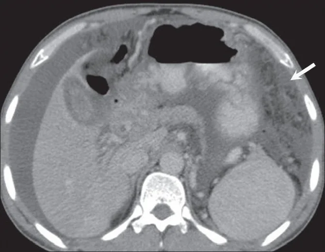
- Portal venöz fazda peritoneal kalınlaşmanın düzgün mü nodüler mi olduğu, mezenter ve omentumun infiltrasyon biçimi, lenf nodlarının santral atenüasyonu ve damarlarla ilişkisi değerlendirilir; olası primer solid organ ya da bağırsak kitlesi de taranır.
Önerilen pencereler: Batın (G 400 / M 50), Yumuşak doku (G 400 / M 40).
BT bulguları
- Düzgün ve yaygın peritoneal kalınlaşma ve kontrastlanma; yüzeyin nodüler ve düzensiz olduğu metastatik örüntüden farklı bir görünüm oluşturur.
- Santral düşük atenüasyonlu veya kalsifiye mezenterik-retroperitoneal lenf nodları, eşlik eden asit ve omental bulanıklıkla birlikte granülomatöz enfeksiyon olasılığını artırır.
- Mezenter kökünde hacimli nodal yumuşak doku, mezenter damarlarını iki yandan çevreleyebilir; damarlar kitle içinden geçerken lümenleri korunabilir.
- Splenomegali ve mezenter dışındaki yaygın lenfadenopati, izole peritoneal yüzey hastalığı yerine sistemik lenfoproliferatif süreci destekler.
- Apendiksten kaynaklanan düşük atenüasyonlu, kitle oluşturucu mukus birikimi ve karaciğer ile dalak yüzeylerinde dıştan basınçla oluşan çentiklenmeler, psödomiksoma peritonei bulgularıdır.
- Jejunal mezenter kökünde sınırlı bulanık yağ dansitesi, çevresel psödokapsül ve damar ya da küçük nod çevresinde korunmuş yağ halkası mezenterik pannükülit lehinedir.
- Primer peritoneal mezotelyoma, yaygın plak ve nodüllerle metastatik hastalığa benzeyebilir; plevral plaklar eşlik ediyorsa öykü ve göğüs bulguları önem kazanır.
Normalde
Normal periton BT'de çoğu kesitte ayrı bir yumuşak doku tabakası olarak seçilmez; ince bağırsak ansları arasında sıvı veya belirgin nodül bulunmaz. Mezenter kökünde damarlar yağlı planlar içinde dallanır; onları saran kitle, korunmuş yağ halkalı bulanıklık veya santral nekrozlu nod yoktur. Şüpheli alanı aynı düzeydeki karşı mezenter yaprakları ve karşı parakolik olukla; ayrıca dalak boyutu ve retroperitoneal nodlarla kıyaslayın.
Normal BT ile kıyasla
Aynı bölgenin, kaynak veri setinde patoloji içermediği belirtilen bir BT incelemesini kesit kesit kaydırın; organ sınırlarını açarak anatomiyi hasta görüntüsüyle karşılaştırın. Küçük rastlantısal bulgular tümüyle dışlanmamıştır.
Kaynak: TotalSegmentator v2.01 veri seti (Wasserthal ve ark., Radiology: Artificial Intelligence 2023), CC BY 4.0, vaka s0119; organ sınırları veri setinin otomatik segmentasyonundan, birleştirilerek sadeleştirildi. Görüntüler 2,5 mm kesit aralığına yeniden örneklendi.
Ayırıcı tanı
- Peritoneal tüberküloz
- düzgün yüzey kalınlaşması, santral nekroz ya da kalsifikasyon içeren nodlar ve ileoçekal hastalık birlikteliği tanıyı destekler; tek başına BT kesin ayrım sağlamaz.
- Peritoneal lenfomatozis
- büyük damarları saran mezenterik nodal kitleler, splenomegali ve başka istasyonlarda lenfadenopati görülebilir.
- Psödomiksoma peritonei
- apendiks ya da müsinöz tümör odağından yayılan jelatinöz içerik, septalı düşük atenüasyon ve organ yüzeylerinde dıştan çentiklenme yapar.
- Mezenterik pannükülit
- çoğunlukla jejunal mezenter kökünde sınırlı yağlı infiltrasyon, psödokapsül ve damar çevresinde korunmuş yağ görünür; bağımsız peritoneal nodüller beklenmez.
- Peritoneal malign mezotelyoma
- yaygın peritoneal kalınlaşma, nodüler kitle ve asitle karsinomatozisi taklit eder; plevral plak ve maruziyet öyküsü destekleyici olabilir.
Tuzaklar
- Düzgün kalınlaşma tüberkülozu destekler ama maligniteyi dışlamaz; klinik bağlam ve gerektiğinde sıvı ya da doku örneklemesi olmadan görüntüyü kesin tanı gibi raporlamayın.
- Mezenter damarlarını saran her doku lenfoma değildir; çevresinde yağ halkası korunmuş, sınırlı mezenterik bulanıklık pannükülit lehine ayrım sağlar.
- Psödomiksoma peritonei — apendiks müsinöz neoplazmalarından kaynaklanan düşük atenüasyonlu, septalı sıvı koleksiyonları ve karaciğer gibi viseral organ yüzeylerinde dıştan basıya bağlı çentiklenmeler gösterir.
- Peritoneal mezotelyoma ile metastatik karsinomun BT görünümleri örtüşür; görüntüleme, kökeni tek başına doğrulayamaz ve patolojik korelasyon gerekebilir.
Kendini dene
Asit ve peritoneal kalınlaşmaya eşlik eden merkezleri düşük atenüasyonlu nodlar ile ileoçekal duvar hastalığı görülüyor. En olası taklitçi hangisidir?
Cevabı göster
Peritoneal tüberküloz. Nekrotik nodlar, ileoçekal tutulum ve peritoneal hastalık birlikteliği tüberküloz peritonitini destekler. Lenfoma daha çok hacimli nodal doku ve sistemik lenfadenopatiyle; müsinöz yayılım organ yüzeylerinde bası etkisiyle öne çıkar. Mezotelyal tümör benzer peritoneal kitle yapabilir ancak bu nod merkezlerini ve ileoçekal örüntüyü tek başına açıklamaz.
Jejunal mezenter kökünde sınırlı yağ bulanıklığı var; damar çevresinde ince yağ korunmuş ve sınırda psödokapsül seçiliyor. En olası süreç nedir?
Cevabı göster
İnflamatuvar mezenter hastalığı. Sınırlı mezenterik yağ infiltrasyonu, damar çevresinde korunmuş yağ ve psödokapsül mezenterik pannükülit için tipiktir. Peritoneal ekim genellikle mezenter köküyle sınırlı kalmaz; desmoid tümör solid yumuşak doku kitlesidir, yağ nekrozu ise bu karakteristik mezenterik dağılımı açıklamaz.
Bu konunun yer aldığı turlar
İlgili konular
Kaynaklar
Bu sayfadaki 38 cümle ve 2 quiz sorusu aşağıdaki kaynaklardan alıntılarla tek tek karşılaştırıldı; 5 cümle bu karşılaştırmada düzeltildi (2026-10-08).
- Peritoneal Carcinomatosis and Its Mimics: Review of CT Findings for Differential Diagnosispmc.ncbi.nlm.nih.gov
- Peritoneal lymphomatosis: CT and PET/CT findings and how to differentiate between carcinomatosis and sarcomatosispmc.ncbi.nlm.nih.gov
- Mesenteric panniculitis: a clinical conundrumpmc.ncbi.nlm.nih.gov
- CT mimics of peritoneal carcinomatosispmc.ncbi.nlm.nih.gov
- Inflammatory Pseudotumor of the liver mimicking peritoneal Carcinomatosis: a diagnostic challengepmc.ncbi.nlm.nih.gov
- Evidence-based approach to diagnosis and management of abdominal tuberculosis.pubmed.ncbi.nlm.nih.gov
- CT imaging of peritoneal carcinomatosis and its mimics.pubmed.ncbi.nlm.nih.gov
- Pelvic Inflammatory Disease: Multimodality Imaging Approach with Clinical-Pathologic Correlation.pubmed.ncbi.nlm.nih.gov
- 18F-FDG PET/CT in Bilateral Breasts Metastasis From Ovarian Carcinoma.pubmed.ncbi.nlm.nih.gov
- Large Idiopathic Mesenteric Necrotizing Granuloma Mimicking Metastatic Disease on FDG PET/CT.pubmed.ncbi.nlm.nih.gov
- CT imaging review of uncommon peritoneal-based neoplasms: beyond carcinomatosis.pubmed.ncbi.nlm.nih.gov
- Non-neoplastic conditions mimicking peritoneal carcinomatosis at CT imaging.pubmed.ncbi.nlm.nih.gov
- CT mimics of peritoneal carcinomatosis.pubmed.ncbi.nlm.nih.gov
- Uremic tumoral calcinosis in the cervical spine: case report.pubmed.ncbi.nlm.nih.gov
- Peritoneal Carcinomatosis and Its Mimics: Review of CT Findings for Differential Diagnosis.pubmed.ncbi.nlm.nih.gov
- Growth of ovarian cancer xenografts causes loss of muscle and bone mass: a new model for the study of cancer cachexia.pubmed.ncbi.nlm.nih.gov
- Peritoneal tuberculosis mimicking advanced ovarian cancer case report: Laparoscopy as diagnostic modality.pubmed.ncbi.nlm.nih.gov
- Solitary fibrous tumor of the greater omentum, mimicking gastrointestinal stromal tumor of the small intestine: a case report.pubmed.ncbi.nlm.nih.gov
- MRI of Tumors and Tumor Mimics in the Female Pelvis: Anatomic Pelvic Space-based Approach.pubmed.ncbi.nlm.nih.gov
- Inflammatory Pseudotumor of the liver mimicking peritoneal Carcinomatosis: a diagnostic challenge.pubmed.ncbi.nlm.nih.gov
- Peritoneal Tuberculosis in a Female Patient Mimicking Peritoneal Carcinomatosis: A Case Report.pubmed.ncbi.nlm.nih.gov
- Occult Metastatic Invasive Lobular Breast Carcinoma Presenting as Gastric Outlet Obstruction and Peritoneal Carcinomatosis.pubmed.ncbi.nlm.nih.gov
- Pseudolipoma of the Glissonian capsule mimicking hepatic metastases after surgery for colorectal cancer: The role of dual-energy CT.pubmed.ncbi.nlm.nih.gov
- When Portal Hypertension Conceals a Gastric Neuroendocrine Tumor: A Rare Mimic of Decompensated Chronic Liver Disease.pubmed.ncbi.nlm.nih.gov
- Dual-Layer Spectral CT for Locoregional Assessment of Rectal Cancer: A Comparison with MRI in a Pilot Study.pubmed.ncbi.nlm.nih.gov
- Radiological Patterns of Abdominal Tuberculosis With Microbiological Confirmation: A Multi-Modal Prospective Analysis.pubmed.ncbi.nlm.nih.gov
- Computed tomography and magnetic resonance imaging features of struma ovarii: A retrospective case series.pubmed.ncbi.nlm.nih.gov
- Perforated Gastric Cancer: Epidemiology, Diagnosis, and Surgical Management Strategies.pubmed.ncbi.nlm.nih.gov
- Pregnancy Deciduosis Mimicking Abdominal Tuberculosis and Peritoneal Carcinomatosis: A Case Report.pubmed.ncbi.nlm.nih.gov
- Gynecologic imaging errors and pitfalls: how to avoid misdiagnosis on pelvic MRI.pubmed.ncbi.nlm.nih.gov
- Benign <i>struma ovarii</i> mimicking ovarian cancer in an afghan woman: an unusual case report and literature review.pubmed.ncbi.nlm.nih.gov
- Peritoneal tuberculosis mimicking peritoneal carcinomatosis in a patient with T-cell lymphoma: Diagnostic pitfalls and radiologic correlation.pubmed.ncbi.nlm.nih.gov
- Case report: Radiological features of a case of desmoplastic malignant mesothelioma of peritoneum.pubmed.ncbi.nlm.nih.gov
- Clinical and Radiological Parameters to Discriminate Tuberculous Peritonitis and Peritoneal Carcinomatosis.pubmed.ncbi.nlm.nih.gov
- Postpancreatitis extrapancreatic necrosis mimicking peritoneal carcinomatosis: A pediatric diagnostic pitfall.pubmed.ncbi.nlm.nih.gov
- Fibrothecoma with pseudo-carcinomatosis in a young woman: The radiologic pitfalls of Meigs syndrome and the importance of fertility-sparing surgery.pubmed.ncbi.nlm.nih.gov
- Cholecystogastric fistula masquerading as gastrointestinal stromal tumour: Unusual presentation of gallbladder adenomyomatosis - a case report.pubmed.ncbi.nlm.nih.gov
- A Rare Case of Multifocal Ileal and Mesenteric Gastrointestinal Stromal Tumors With Spontaneous Rupture.pubmed.ncbi.nlm.nih.gov
- Peritoneal Tuberculosis Mimicking Pseudomyxoma Peritonei: A Diagnostic Challenge.pubmed.ncbi.nlm.nih.gov
- Mesenteric Fat Necrosis Mimicking a Malignant Mesenteric Tumor: A Case Report.pubmed.ncbi.nlm.nih.gov
- Resection of a ganglioneuroma encasing major blood vessels using three-dimensional laparoscopy combined with organ suspension: A case report.pubmed.ncbi.nlm.nih.gov
- Feasibility of single-phase DECT as an alternative to triple-phase CT for imaging-defined risk factors assessment in neuroblastoma for dose reduction.pubmed.ncbi.nlm.nih.gov
- International Society of Paediatric Surgical Oncology (IPSO) Minimally Invasive Surgery (MIS) Guidelines.pubmed.ncbi.nlm.nih.gov
- Prolonged fever in ESBL-producing <i>Salmonella Typhi</i> in Lebanon: Diagnostic challenges and the possible role of occult low-level carbapenemase activity.pubmed.ncbi.nlm.nih.gov
- Rethinking Common Diagnoses: Idiopathic Multicentric Castleman Disease Presenting as TAFRO Syndrome: A Case Report.pubmed.ncbi.nlm.nih.gov
- Ultrasonographic Features and Associated Clinical Findings in 21 Dogs With Histologically Confirmed Abscessed and Non-Abscessed Splenitis.pubmed.ncbi.nlm.nih.gov
- A proposal for changing nomenclature from pseudomyxoma peritonei (PMP) to abdomino-peritoneal mucinous carcinoma (APM) based on its long journey and experience from tertiary oncology center in India.pubmed.ncbi.nlm.nih.gov
- From Diagnosis to Management; Mucocele of Stump Appendicitis, Extremely Rare Finding in an Uncommon Surgical Disease: Literature Review.pubmed.ncbi.nlm.nih.gov
- Causes of hepatic capsular retraction: a pictorial essay.pubmed.ncbi.nlm.nih.gov
- Mesenteric Abscess Secondary to Panniculitis: A Rare Case Report.pubmed.ncbi.nlm.nih.gov
- Mesenteric Panniculitis in a Patient with Ulcerative Colitis in Remission on Vedolizumab Therapy: A Case Report and Literature Review.pubmed.ncbi.nlm.nih.gov
- Computed tomography findings of peritoneal tumour in the absence of primary lesions: preliminary study.pubmed.ncbi.nlm.nih.gov
- Distinct epidemiologic patterns of mesothelioma: evidence from a 16-year provincial cohort in China.pubmed.ncbi.nlm.nih.gov
- Localised malignant mesothelioma presenting as an esophageal mass: An unusual manifestation.pubmed.ncbi.nlm.nih.gov
- An Unusual Presentation of Leiomyosarcoma Mimicking Parasitic Fibroids: A Diagnostic Dilemma.pubmed.ncbi.nlm.nih.gov
- Endometriosis as a Silent Emerging Malignant Threat in Young Women: A Case Report of Rare Rapid Transformation of Abdominal Wall Endometriosis Into Mixed High-Grade Serous and Clear Cell Carcinoma One Year Following Cesarean Section.pubmed.ncbi.nlm.nih.gov
- Caught in a Cocoon: Operative Release of Primary Sclerosing Encapsulating Peritonitis With Endoscopic Management of Early Postoperative Duodenojejunal Intussusception.pubmed.ncbi.nlm.nih.gov
- Elective Repair of Atypical Internal Hernia Following Roux-En-Y Gastric Bypass in a Patient With Recurrent Obstructive Symptoms: A Case Report.pubmed.ncbi.nlm.nih.gov
- Deep learning-assisted near-real-time identification of the inferior mesenteric artery and vein during laparoscopic rectal resection: a single-center proof-of-concept study.pubmed.ncbi.nlm.nih.gov
- Contrast-Enhanced CT of the Coelom in a Female Bald Eagle (<i>Haliaeetus leucocephalus</i>): Development of a Preliminary CT-Based Anatomical Segmentation Workflow.pubmed.ncbi.nlm.nih.gov
- Ileal tuberculosis presenting with severe unexplained anemia and unusual pseudo-tumoral imaging features: A case report.pubmed.ncbi.nlm.nih.gov
- CT and MR in peritoneal malignancies: pearls and pitfalls at preoperative examination.pubmed.ncbi.nlm.nih.gov
- Primary and secondary tumors of the peritoneum: key imaging features and differential diagnosis with surgical and pathological correlation.pubmed.ncbi.nlm.nih.gov
- Peritoneal Lymphomatosis: The Great Mimicker.pubmed.ncbi.nlm.nih.gov
- Umbilical hernia as the initial manifestation of pseudomyxoma peritonei: a case report.pubmed.ncbi.nlm.nih.gov
- Bilateral ovarian masses, massive ascites, and prior appendectomy: must it be ovarian cancer? --a case report of pseudomyxoma peritonei 24 years after appendectomy.pubmed.ncbi.nlm.nih.gov
- Spontaneous rupture of a mucinous borderline ovarian tumour in late pregnancy: a case report.pubmed.ncbi.nlm.nih.gov
- Mesenteric Panniculitis and the Gut-Mesentery-Metabolic Axis: A Hypothesis-Generating Narrative Review.pubmed.ncbi.nlm.nih.gov
- The Many Faces of the Angry Peritoneum.pubmed.ncbi.nlm.nih.gov
- Disseminated peritoneal leiomyomatosis post-laparoscopic myomectomy.pubmed.ncbi.nlm.nih.gov
- Case Report: Iparomlimab and Tuvonralimab-based therapy for epithelioid malignant peritoneal mesothelioma: a three-case series.pubmed.ncbi.nlm.nih.gov
- From Ascites to Malignant Peritoneal Mesothelioma: The Diagnostic Workup of a Rare Tumor.pubmed.ncbi.nlm.nih.gov
- A clinician's perspective on the new organ mesentery and non-vascular mesenteropathies.pubmed.ncbi.nlm.nih.gov
- Vascular contact with soft tissue: a sign of mesenteric masses at computed tomography.pubmed.ncbi.nlm.nih.gov
- Navigating Ovarian-Adnexal Reporting and Data System Magnetic Resonance Imaging (O-RADS MRI): A Review of Its Evolution, Current Advances, and Persistent Challenges in Ovarian Imaging.pubmed.ncbi.nlm.nih.gov
- Cystic lesions and their mimics involving the intrahepatic bile ducts and peribiliary space: diagnosis, complications, and management.pubmed.ncbi.nlm.nih.gov
- Mucocele of the appendix: what to expect.pubmed.ncbi.nlm.nih.gov
- Cystic epithelioid malignant mesothelioma of the mesentery with rapid postoperative metastasis: an unusual case report.pubmed.ncbi.nlm.nih.gov
- Localized peritoneal epithelioid clear cell subtype mesothelioma: a case report and literature review.pubmed.ncbi.nlm.nih.gov
- Updates on Imaging Assessment of Pancreatic Cancer for Determining Anatomic and Biologic Resectability.pubmed.ncbi.nlm.nih.gov
- Postoperative recurrence patterns and anatomy-stratified clinical target volume delineation in pancreatic cancer.pubmed.ncbi.nlm.nih.gov
- Use of imaging as staging and surgical planning for pancreatic surgery.pubmed.ncbi.nlm.nih.gov
- The use of MRI in the diagnosis of ovarian cancer.pubmed.ncbi.nlm.nih.gov
- <sup>68</sup>Ga-FAPI Avidity in Fat Necrosis and Adrenal Adenoma: Expanding the Spectrum of Benign Pitfalls in Oncologic Imaging.pubmed.ncbi.nlm.nih.gov
- Hidden in plain sight: Extrapulmonary manifestations of tuberculosis in Asia.pubmed.ncbi.nlm.nih.gov
- Ultrasound and computed tomography in the evaluation of mesenteric lesions: A pictorial review.pubmed.ncbi.nlm.nih.gov
- Imaging of Mischievous Intra-abdominal Fat Presenting with Abdominal Pain: A Pictorial Review.pubmed.ncbi.nlm.nih.gov
- Radiological Approach to Splenomegaly: Etiologies, Pathophysiologies, and Diagnostic Strategies.pubmed.ncbi.nlm.nih.gov
- Clinicopathological Pearls and Diagnostic Pitfalls in IgG4-Related Disease: Challenging Case Series and Literature Review.pubmed.ncbi.nlm.nih.gov
- Paradoxical ileocecal perforation during early anti-tuberculosis therapy: A rare case report.pubmed.ncbi.nlm.nih.gov
- An Integrated Review of Conventional and Emerging Diagnostic and Therapeutic Modalities to Reduce the Risk of Surgical Resections in Intestinal Tuberculosis.pubmed.ncbi.nlm.nih.gov
- Unravelling mesenteric fibrosis in small intestinal neuroendocrine tumours and implications for clinical management.pubmed.ncbi.nlm.nih.gov
- Cecal Perforation Presenting as Apparent Peritoneal Carcinomatosis: Distinguishing Acute Peritonitis From Malignant Mimicry.pubmed.ncbi.nlm.nih.gov
- Bilateral ovarian metastases from gastric signet-ring cell carcinoma in an 18-year-old: a case report and narrative review.pubmed.ncbi.nlm.nih.gov
- Computed tomographic findings in dogs with ovarian tumors: A tortuous ovarian artery consistently identifies ovarian origin in complex abdominal masses.pubmed.ncbi.nlm.nih.gov
- Characteristic CT Imaging Features of Atraumatic Peritoneal and Subperitoneal Pathologies in Emergency Settings: A Pictorial Review.pubmed.ncbi.nlm.nih.gov
- Case Report: Pelvic splenosis confused with malignancy-a reminder for differential diagnosis.pubmed.ncbi.nlm.nih.gov
- Radiology of the Mesentery.pubmed.ncbi.nlm.nih.gov
- Small Bowel Imaging from Stepchild of Roentgenology to MR Enterography, Part II: The Reliable Disclosure of Crohn's Disease and Non-Inflammatory Small Bowel Disorder Plot through MRI Findings.pubmed.ncbi.nlm.nih.gov
- Ovarian cancer reporting lexicon for computed tomography (CT) and magnetic resonance (MR) imaging developed by the SAR Uterine and Ovarian Cancer Disease-Focused Panel and the ESUR Female Pelvic Imaging Working Group.pubmed.ncbi.nlm.nih.gov
- International Society of Paediatric Surgical Oncology (IPSO) Surgical Practice Guidelines.pubmed.ncbi.nlm.nih.gov
- The development of fluorescence guided surgery for pancreatic cancer: from bench to clinic.pubmed.ncbi.nlm.nih.gov
- EACR 2025 Congress: Innovative Cancer Science, 16-19 June 2025.pubmed.ncbi.nlm.nih.gov
- Splenic pathologies: A pictorial review of common, wandering, twisting and rare presentations.pubmed.ncbi.nlm.nih.gov
- Signet-ring cell carcinoma of the appendix presenting as diffuse uterine involvement: a case report and literature review.pubmed.ncbi.nlm.nih.gov
- The genetics of cancer heterogeneity and mesothelioma.pubmed.ncbi.nlm.nih.gov
- The comparative diagnostic and therapeutic application value of FAPI PET/CT and <sup>18</sup>F-FDG PET/CT in oncology.pubmed.ncbi.nlm.nih.gov
- Fibroblast Activation Protein Inhibitor (FAPI) PET: A Scoping Review of Emerging Oncologic and Fibroinflammatory Applications.pubmed.ncbi.nlm.nih.gov
Eğitim amaçlıdır; hasta başında tanı ve tedavi kararının yerine geçmez. İçerik yapay zekâ destekli bir süreçle güncel literatüre göre cümle cümle kaynaklarla karşılaştırıldı, ancak uzman radyolog incelemesinden geçmedi. Klinik kararlarda güncel kılavuzları, kurum protokollerini ve radyoloji raporunu esas alın.