Özet
Bebek ve çocuklarda birden fazla kraniyal sütürün erken kemikleşmesi, kafatası büyümesini kısıtlayıp kompansatuvar şekil değişikliğine ve bazı sendromlarda yüz, kafa tabanı ve beyin eşlikçilerine yol açar. İnce kesit BT, hangi sütürlerin açık ya da füze olduğunu haritalar; üç boyutlu kemik görüntüleri deformiteyi ve kafa tabanı anatomisini cerrahi planlama için görünür kılar. Klinik bağlam sendromik kraniyosinostozu düşündürüyorsa venöz sinüs darlığı ve kollateral drenaj yolları ayrıca önem taşır. Füzyonun yaygınlığı veya venöz anatomi gözden kaçarsa intrakraniyal basınçla ilişkili bulgular ve cerrahi açıdan önemli drenaj varyantları eksik tanımlanabilir.
Faz ve pencere
- Kontrastsız tanısal
- İnce kesit kraniyal BT'nin kemik algoritmasında sütür boyunca kaybolan açıklık, perisutural skleroz ve kemik köprüleşmesi aranır; kompansatuvar kafa şekli, dijital izler, ventriküller ve eşlik eden intrakraniyal anatomi kontrastsız görüntüde değerlendirilir. BT ile kraniyosinostozun yerini ve yaygınlığını değerlendirmek mümkündür.
- Kemik algoritması tanısal
- Aksiyel kaynak kesitlere koronal ve sagittal çok düzlemli rekonstrüksiyonlar ile 3B kemik yüzeyi eklenir; 3B eşikleme artefaktı sütür açıklığını yanlış gösterebileceğinden sütür açıklığı iki boyutlu kaynak kesitlerde doğrulanır.
Önerilen pencereler G genişlik, M merkez; değerler Hounsfield birimi (HU)
- KemikG 1800 / M +400
- BeyinG 80 / M +40
- Yumuşak dokuG 400 / M +40
BT bulguları
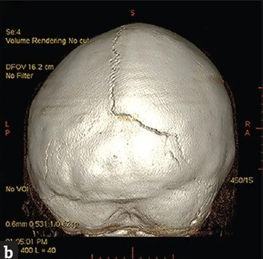
- Birden fazla sütürde kemik köprüleşmesi — sagittal, koronal, metopik veya lambdoid sütürlerden birden çoğunda fibroz aralığın silinmesi ve kemik devamlılığı.
- Kompansatuvar kalvaryal şekil — füzyon yerindeki dik büyüme kısıtlanırken açık sütürlere paralel bölgelerde genişleme veya çıkıntı oluşması.
- Kleeblattschädel biçimi — yaygın çoklu sütür tutulumunda öne, yana ve arkaya lobüle kalvaryum konturu.
- Kafa tabanı ve orbita değişikliği — kafa tabanı hipoplazisi, orta yüz geriliği, sığ orbita veya orbita çatısında asimetri; sendrom fenotipiyle birlikte tariflenir.
- İç kalvaryal tabuladaki girintiler bakır dövmesi görünümü olarak kronik kafa içi basınç artışıyla ilişkilendirilmiştir; ancak yenidoğan ve süt çocuklarında BT’de görülen dijital izler kafa içi basınç artışıyla ilişkili bulunmamış ve tek başına tanıda yararlı görülmemiştir.
- Venöz drenaj varyantı — seçilmiş kompleks olguda BT venografide daralmış dural sinüsler, genişlemiş emissary/transosseöz kollateraller veya persistan fetal sinüs yolları görülebilir.
- Eşlik eden beyin bulgusu — ventrikül boyutları, arka çukur ve kraniyoservikal bileşke; mevcut BT'nin kapsadığı alanda Chiari benzeri aşağı yer değiştirme veya hidrosefali açısından incelenir.
- Metopik sütür kapanması normal bir varyant olan metopik kret şeklinde görülebileceğinden, metopik sinostoz değerlendirmesinde trigonosefali ve orbitofrontal şekil değişikliği de dikkate alınır.
Normalde
Normal gelişimde metopik sütür dışında sagittal, koronal ve lambdoid sütürler genellikle erişkinliğe kadar açık kalır. Metopik sütür normal gelişimde yaşamın ilk aylarında, yaklaşık 7–9 aylıkken kapanabilir; bu kapanma metopik sinostozu tek başına göstermez ve metopik kret ile trigonosefali gibi morfolojik bulgularla birlikte değerlendirilir.
Normal BT ile kıyasla
Aynı bölgenin, kaynak veri setinde patoloji içermediği belirtilen bir BT incelemesini kesit kesit kaydırın; organ sınırlarını açarak anatomiyi hasta görüntüsüyle karşılaştırın. Küçük rastlantısal bulgular tümüyle dışlanmamıştır.
Kaynak: TotalSegmentator v2.01 veri seti (Wasserthal ve ark., Radiology: Artificial Intelligence 2023), CC BY 4.0, vaka s0103; organ sınırları veri setinin otomatik segmentasyonundan, birleştirilerek sadeleştirildi. Görüntüler 2,5 mm kesit aralığına yeniden örneklendi.
Ayırıcı tanı
- Pozisyonel plagiosefali
- sütürler açıktır; oksipital basıklık ve karşı taraf frontal çıkıntı görülebilse de sütür hattında kemik köprüleşmesi bulunmaz.
- Tek sütürlü kraniyosinostoz
- bir sütürde erken füzyon ve ona uyan şekil değişikliği vardır; diğer ana sütürler BT'de açıktır.
- İkincil sütür kapanması
- metabolik veya sistemik kemik hastalığı bağlamında birden fazla sütürde kapanma olabilir; klinik ve kemik mineralizasyon paterni primer sendromik sinostozdan ayrımı destekler.
- Gelişimsel varyant veya aksesuar sütür
- ince çizgi kortike ve interdigitasyonlu kalır, beklenen kemik birleşme sınırını izler ve çevresinde kalvaryal deformite oluşturmaz.
- Bakır dövmesi görünümü olmadan şekil bozukluğu
- deformasyonel baş şekli açık sütürlerle birlikteyse erken kapanma tanısı yalnız dış kontura dayanarak konmamalıdır.
Tuzaklar
- Üç boyutlu yeniden biçimlendirilmiş BT, klinik değerlendirme sonrasında kraniyosinostoz teşhisinde ve yönetiminde kullanılan önemli bir görüntüleme yöntemidir.
- BT değerlendirmesi kraniyosinostozun yerini ve yaygınlığını belirlemeyi, altta yatan beyin anomalilerini saptamayı ve cerrahi sonrası durumu değerlendirmeyi amaçlar.
- Bakır dövmesi girintileri tek başına intrakraniyal basınç tanısı koydurmaz; yaş, kraniyal şekil ve diğer BT bulgularıyla birlikte yorumlayın.
- Venöz kollateraller kontrastsız BT'de her zaman haritalanamaz; klinik olarak sendromik/kompleks anatomi şüphesi varsa venöz fazlı tetkik gereksinimi ayrı ele alınır.
Kendini dene
BT'de sagittal ve bilateral lambdoid sütür füzyonu saptanan çocuk için en uygun tanımlama hangisidir?
Cevabı göster
Çoklu sütürlü kraniyosinostoz. Sagittal ve bilateral lambdoid sütürlerin birlikte füzyonu çoklu sütürlü kraniyosinostozdur.
Kraniyosinostozda 3B BT'nin temel katkısı hangisidir?
Cevabı göster
Tanı ve yönetimde kraniyal morfolojiyi değerlendirmeye katkı sağlamak. 3B BT, kraniyosinostozun doğru tanı ve yönetiminde önemli bir rol oynar.
İlgili konular
- Aksesuar sütürler, wormian kemikler ve sütür taklitçileri
- Pediatrik kafatası kırıkları ve çökme kırığı
Kaynaklar
Bu sayfadaki 35 cümle ve 2 quiz sorusu aşağıdaki kaynaklardan alıntılarla otomatik olarak karşılaştırıldı; 10 cümle bu karşılaştırmada düzeltildi (2026-10-09).
- Syndromic craniosynostosis: current concepts in intracranial hypertension, cranial base pathology, and surgical managementpmc.ncbi.nlm.nih.gov
- Multisuture craniosynostosis and acquired Chiari malformation secondary to vitamin D deficiency rickets: illustrative casepmc.ncbi.nlm.nih.gov
- When Virchow's law fails: Crouzon syndrome as a test case for quantitative suture closure–endocranial shape relationshipspmc.ncbi.nlm.nih.gov
- Postoperative raised ICP after minimally invasive surgery for monosutural craniosynostosispmc.ncbi.nlm.nih.gov
- Treatment strategies and prognostic outcomes in craniosynostosis with concurrent hydrocephalus: a case seriespmc.ncbi.nlm.nih.gov
- Progressive Postnatal Pansynostosis.pubmed.ncbi.nlm.nih.gov
- Skull base development and craniosynostosis.pubmed.ncbi.nlm.nih.gov
- Squamosal Craniosynostosis: Defining the Phenotype and Indications for Surgical Management.pubmed.ncbi.nlm.nih.gov
- The Significance of Squamosal Suture Synostosis.pubmed.ncbi.nlm.nih.gov
- Pure Bilateral Lambdoid and Posterior Sagittal Synostosis (Mercedes-Benz Syndrome): Case Report and Literature Review.pubmed.ncbi.nlm.nih.gov
- Spring-Assisted Surgery in the Treatment of Complex Craniosynostosis.pubmed.ncbi.nlm.nih.gov
- Squamosal Suture Synostosis: An Under-Recognized Phenomenon.pubmed.ncbi.nlm.nih.gov
- Craniosynostosis: imaging review and primer on computed tomography.pubmed.ncbi.nlm.nih.gov
- Craniosynostosis in Siblings, an Extremely Rare Occurrence: A Case Report.pubmed.ncbi.nlm.nih.gov
- Quantitative evaluation of facial hypoplasia and airway obstruction in infants with syndromic craniosynostosis: relationship with skull base and splanchnocranium sutural pattern.pubmed.ncbi.nlm.nih.gov
- Delayed intracranial hypertension after cranial vault remodeling for nonsyndromic single-suture synostosis.pubmed.ncbi.nlm.nih.gov
- Correlation of Intracranial Volume With Head Surface Volume in Patients With Multisutural Craniosynostosis.pubmed.ncbi.nlm.nih.gov
- Incidence of Ventriculomegaly in Patients With Craniosynostosis.pubmed.ncbi.nlm.nih.gov
- Volumetric changes in cranial vault expansion: comparison of fronto-orbital advancement and posterior cranial vault distraction osteogenesis.pubmed.ncbi.nlm.nih.gov
- Multisuture craniosynostosis and acquired Chiari malformation secondary to vitamin D deficiency rickets: illustrative case.pubmed.ncbi.nlm.nih.gov
- When Virchow's law fails: Crouzon syndrome as a test case for quantitative suture closure-endocranial shape relationships.pubmed.ncbi.nlm.nih.gov
- Craniofacial Morphology and Dental Age Assessment in Pediatric Craniosynostosis: A Comparative Cross-Sectional Study.pubmed.ncbi.nlm.nih.gov
- Treatment strategies and prognostic outcomes in craniosynostosis with concurrent hydrocephalus: a case series.pubmed.ncbi.nlm.nih.gov
- Total Cranial Vault Remodeling Using Multidirectional Cranial Distraction Osteogenesis in Craniosynostosis.pubmed.ncbi.nlm.nih.gov
- Contralateral Coronal-Lambdoid Craniosynostosis Treated with Distraction Osteogenesis and Spring-Mediated Cranioplasty.pubmed.ncbi.nlm.nih.gov
- Case Report: Reversible acquired Chiari-like state and upper cervical syringomyelia caused by a giant posterior tentorial tumor confined to the supratentorial compartment.pubmed.ncbi.nlm.nih.gov
- Neuroimaging in Nonsyndromic Craniosynostosis: Key Concepts to Unlock Innovation.pubmed.ncbi.nlm.nih.gov
- Dural venous sinus anatomy in children with external hydrocephalus: analysis of a series of 97 patients.pubmed.ncbi.nlm.nih.gov
- Non-syndromic craniosynostosis: contemporary concepts in biology, diagnosis, and distraction-based operative strategy.pubmed.ncbi.nlm.nih.gov
- Brain Lymphatic Dysfunction in Subarachnoid Hemorrhage: Pathophysiology and Clinical Implications.pubmed.ncbi.nlm.nih.gov
- Reversible abducens nerve palsy following cranial vault expansion in the setting of multisutural craniosynostosis: illustrative case.pubmed.ncbi.nlm.nih.gov
- Clinical and genetic features of syndromic craniosynostosis in 18 Chinese probands: novel candidate genes and phenotypes of known pathogenic genes.pubmed.ncbi.nlm.nih.gov
- From Bench to Bone: Translational Pathways of Nanomaterial-Based Therapies in Craniomaxillomandibulofacial and Cranial Vault Reconstruction.pubmed.ncbi.nlm.nih.gov
- Cranial nerves involvement in craniosynostosis: a systematic review.pubmed.ncbi.nlm.nih.gov
- Hidden Hollows of the Fetal Cranium-Lacunar Skull in Isolation.pubmed.ncbi.nlm.nih.gov
- Detection of intracranial hemorrhage using ultralow-dose brain computed tomography with deep learning reconstruction versus conventional-dose computed tomography.pubmed.ncbi.nlm.nih.gov
- Pulmonary Involvement in Proteus Syndrome: Clinical and Imaging Correlates in a Rare Case.pubmed.ncbi.nlm.nih.gov
- Can Craniosynostosis be Diagnosed on Physical Examination? A Retrospective Review.pubmed.ncbi.nlm.nih.gov
- Exploring the role of <i>TWIST1</i> in malocclusion and craniofacial morphology.pubmed.ncbi.nlm.nih.gov
- Craniosynostosis : Updates in Radiologic Diagnosis.pubmed.ncbi.nlm.nih.gov
- Prenatal diagnosis of Apert syndrome caused by a de novo FGFR2 mutation in the second trimester: a case report.pubmed.ncbi.nlm.nih.gov
- Severe Cloverleaf Skull Deformity in c.1061C>G (p.Ser354Cys) Mutated Fibroblast Growth Factor Receptor 2 Gene in Crouzon Syndrome.pubmed.ncbi.nlm.nih.gov
- Zero-TE MRI: principles and applications in the head and neck.pubmed.ncbi.nlm.nih.gov
- Fibroblast Growth Factor Receptor 2 ( FGFR2 ) Mutation Related Syndromic Craniosynostosis.pubmed.ncbi.nlm.nih.gov
- Measurement of orbital volume after enucleation and orbital implantation.pubmed.ncbi.nlm.nih.gov
- Syndromic craniosynostosis: current concepts in intracranial hypertension, cranial base pathology, and surgical management.pubmed.ncbi.nlm.nih.gov
- Endoscope-assisted release for pancraniosynostosis with delayed single-suture refusion managed with endoscopic re-release in the same patient: illustrative case.pubmed.ncbi.nlm.nih.gov
- Phenotypic heterogeneity in Crouzon syndrome: A case of severe intracranial hypertension from isolated temporal squamosal suture synostosis with a de novo FGFR2 p.Cys342Tyr mutation.pubmed.ncbi.nlm.nih.gov
- Clinical Significance of Venous Anomalies in Syndromic Craniosynostosis.pubmed.ncbi.nlm.nih.gov
- The Genetics of Chiari 1 Malformation.pubmed.ncbi.nlm.nih.gov
- Is foramen magnum decompression for acquired Chiari I malformation like putting a finger in the dyke? - A simplistic overview of artificial intelligence in assessing critical upstream and downstream etiologies.pubmed.ncbi.nlm.nih.gov
- A Critical Update of the Classification of Chiari and Chiari-like Malformations.pubmed.ncbi.nlm.nih.gov
- Automated Diagnosis of Infantile Skull Fractures From X-Ray Images Using an Ensemble Deep Learning Model.pubmed.ncbi.nlm.nih.gov
- Clinical and molecular characterization of TCF12 variants in an Asian pediatric cohort with craniosynostosis.pubmed.ncbi.nlm.nih.gov
- Enhancing Predictive Tools for Skeletal Growth and Craniofacial Morphology in Syndromic Craniosynostosis: A Focus on Cranial Base Variables.pubmed.ncbi.nlm.nih.gov
- Surgical Outcomes in Chiari 1 and Chiari 1.5 Malformation Treated by Posterior Fossa Reconstruction: A Comprehensive Analysis of 110 Pediatric Cases and Literature Review.pubmed.ncbi.nlm.nih.gov
- Rhinosinusitis: Evidence and experience - 2024.pubmed.ncbi.nlm.nih.gov
- Calvarial Bloom Biomechanics: Dural Tension Governing Cranial Form.pubmed.ncbi.nlm.nih.gov
- Differential regulation of coronal and lambdoid suture patency by PTHLH and HHIP activity in mice.pubmed.ncbi.nlm.nih.gov
- The Evolution of the Role of Imaging in the Diagnosis of Craniosynostosis: A Narrative Review.pubmed.ncbi.nlm.nih.gov
- Embryodysgenesis of the Lids and Orbit.pubmed.ncbi.nlm.nih.gov
- Acute, chronic, and non-fracture-related compartment syndrome in children. A current concepts review.pubmed.ncbi.nlm.nih.gov
- Unossified Membranous Strips of the Parietal Bone Mimicking a Fracture in a Child Presenting With Scalp Swelling: A Case Report.pubmed.ncbi.nlm.nih.gov
- Anatomy of the Middle Meningeal Artery: Implications for Middle Meningeal Artery Embolization in Chronic Subdural Hematoma.pubmed.ncbi.nlm.nih.gov
- Sagittal craniosynostosis with Moebius syndrome; case illustration: first case in the literatüre.pubmed.ncbi.nlm.nih.gov
- Imaging Modalities in Craniosynostosis: A Systematic Review and Proposal of the ARCANA Protocol for Multimodal Radiation-Free Assessment.pubmed.ncbi.nlm.nih.gov
- Single-center experience with aortopexy in pediatric tracheomalacia: a case series.pubmed.ncbi.nlm.nih.gov
- Divergent patterns of cranial suture fusion in marsupial and placental mammals.pubmed.ncbi.nlm.nih.gov
- From FGFR2 mutations to precision management: a review of prenatal diagnosis and multidisciplinary interventions in apert syndrome.pubmed.ncbi.nlm.nih.gov
- Review of imaging modalities and radiological findings of calvarial lesions.pubmed.ncbi.nlm.nih.gov
- Clinical and Genetic Studies of the First Monozygotic Twins with Pfeiffer Syndrome.pubmed.ncbi.nlm.nih.gov
- Acute dural venous sinus injuries: Imaging and challenges.pubmed.ncbi.nlm.nih.gov
- Current perspective in acute ischemic stroke: net water uptake and imaging-guided selection as bridges to personalized, tissue-based care.pubmed.ncbi.nlm.nih.gov
- The Initial Diagnostic Approach to Adult Congenital Heart Disease: A Practical Imaging Pathway for General Cardiologists and Cardiac Imagers.pubmed.ncbi.nlm.nih.gov
- Compression fracture of the T12 vertebra combined with diaphragmatic hernia after low-energy trauma: a case report and literature review.pubmed.ncbi.nlm.nih.gov
- Evaluation of midpalatal suture maturation in young adults using cone-beam computed tomography.pubmed.ncbi.nlm.nih.gov
- Arthroscopic Latarjet using double suture-button fixation: learning curve, graft positioning, and complications - A two-center study of 80 patients.pubmed.ncbi.nlm.nih.gov
- Premature closure of the caudal calvarial midline suture is associated with scaphocephaly and ventriculomegaly in Boxer dogs.pubmed.ncbi.nlm.nih.gov
- Transformer-based models in dentistry: a systematic review.pubmed.ncbi.nlm.nih.gov
- Progressive Insights into 3D Bioprinting for Corneal Tissue Restoration.pubmed.ncbi.nlm.nih.gov
- Micro-CT for Biological and Biomedical Studies: A Comparison of Imaging Techniques.pubmed.ncbi.nlm.nih.gov
- Postoperative raised ICP after minimally invasive surgery for monosutural craniosynostosis.pubmed.ncbi.nlm.nih.gov
- Neurocognitive outcomes in children with craniosynostosis after surgical correction: a narrative review.pubmed.ncbi.nlm.nih.gov
- Prevalence of motor development delay in children with craniosynostosis: a systematic review and meta-analysis.pubmed.ncbi.nlm.nih.gov
- Atypical Presentation of Undiagnosed Crouzon's Syndrome With Pansinusitis, Orbital Cellulitis, and Altered Mentation Successfully Managed With Functional Endoscopic Sinus Surgery: A Case Report.pubmed.ncbi.nlm.nih.gov
- Diagnostic performance of digital markings on head computed tomography for evaluating intracranial hypertension in neonates and infants.pubmed.ncbi.nlm.nih.gov
- Insights into craniosynostosis management in low- and middle-income countries: A narrative review of outcomes, shortcomings and paediatric neurosurgery capacity.pubmed.ncbi.nlm.nih.gov
- Morphologic Severity and Age at Surgery Are Associated with School-Age Neurocognitive Outcomes in Metopic Craniosynostosis.pubmed.ncbi.nlm.nih.gov
- Comparison of an unsupervised machine learning algorithm and surgeon diagnosis in the clinical differentiation of metopic craniosynostosis and benign metopic ridge.pubmed.ncbi.nlm.nih.gov
- Deep learning-based craniosynostosis classification via suture segmentation and mask-weighted classification.pubmed.ncbi.nlm.nih.gov
- The biomechanics of chewing and suckling in the infant: A potential mechanism for physiologic metopic suture closure.pubmed.ncbi.nlm.nih.gov
- Eye and Orbital Anatomy in Metopic Synostosis.pubmed.ncbi.nlm.nih.gov
- Craniofacial development: genetics, signaling pathways, teratogenic mechanisms, and clinal significance.pubmed.ncbi.nlm.nih.gov
- Genetic Disorders Detectable by Fetal MRI: A Review.pubmed.ncbi.nlm.nih.gov
- Allometry of human calvaria bones during development from birth to 8 years of age shows a nonlinear growth pattern.pubmed.ncbi.nlm.nih.gov
- The 27 Facial Sutures: Timing and Clinical Consequences of Closure.pubmed.ncbi.nlm.nih.gov
- Divergent Mechanisms of Cranial Suture Ossification in Normal Development and Pathologic Fusion.pubmed.ncbi.nlm.nih.gov
- A combined machine learning and finite element modelling tool for the surgical planning of craniosynostosis correction.pubmed.ncbi.nlm.nih.gov
- Visual outcome in 5-year-old children operated for non-syndromic craniosynostosis.pubmed.ncbi.nlm.nih.gov
- Evaluating compliance with postoperative cranial remolding orthotic treatment in infants with single suture craniosynostosis: A comparative study of objective and subjective approaches.pubmed.ncbi.nlm.nih.gov
- TGFβ-mediated dural progenitor cell migration into the coronal suture is crucial for preventing craniosynostosis.pubmed.ncbi.nlm.nih.gov
- Modern technology complements classic technique: 3D-printed inter-locking cutting guides for frontal sinus osteoma resection and reconstruction with split-calvarial bone graft.pubmed.ncbi.nlm.nih.gov
Eğitim amaçlıdır; hasta başında tanı ve tedavi kararının yerine geçmez, tıbbi tavsiye değildir. Metinler yapay zekâ desteğiyle yazıldı ve otomatik bir adımda kaynak alıntılarıyla karşılaştırıldı; bu bir tıbbi doğrulama değildir ve içerik uzman radyolog incelemesinden geçmedi. Klinik kararlarda güncel kılavuzları, kurum protokollerini ve radyoloji raporunu esas alın.