Özet
Bebek ve çocukta ilerleyen ventrikül genişlemesi; BOS dolaşımındaki tıkanma, emilim bozukluğu veya daha seyrek başka bir mekanizmayla ilişkili olabilir. Kontrastsız BT, hızlı değerlendirme gereken durumda ventrikül kalibrasyonunu, bası bulgularını ve olası tıkanma nedenini gösterebilir; açık fontaneli bebekte ultrason ve ayrıntılı etiyoloji değerlendirmesinde MR da önem taşır. Ventrikülomegaliyi tek başına hidrosefali saymayın: yaşa uygun görünüm, parankim hacmi, basınç etkisi ve önceki incelemelerdeki değişim tanısal yorumu belirler. İlerleyen basınç etkisi veya BOS yolundaki engel fark edilmezse periventriküler doku hasarı ve nörolojik kötüleşme gözden kaçabilir.
Faz ve pencere
- Kontrastsız tanısal
- Konvansiyonel görüntüleme, ventrikül genişlemesinin derecesini ve BOS yolundaki tıkanma noktasını gösterir; transependimal sıvı ekzosyonu ise şant endikasyonu açısından önemlidir. Konvansiyonel BT ve MR, ventrikül genişlemesinin derecesini ve BOS yolundaki tıkanma noktasını göstermek için kullanılır; detaylı anatomik değerlendirme için gelişmiş MR teknikleri tercih edilir.
Önerilen pencereler: Beyin (G 80 / M 40).
BT bulguları
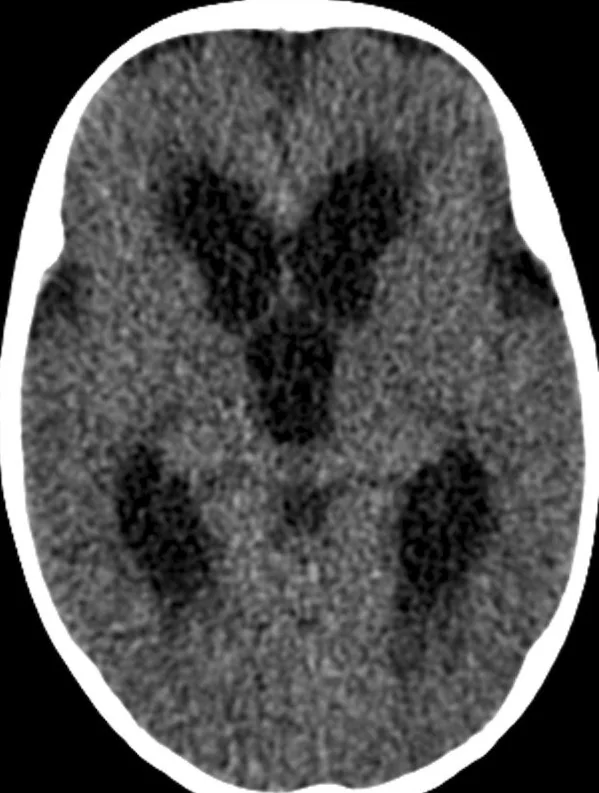
- Lateral ventrikül genişlemesi — frontal ve oksipital boynuzların birlikte dolgunlaşması, karşı taraf ve önceki tetkikle kıyaslanır.
- Lateral ventriküllerde ilerleyen genişleme, seri görüntülemede objektif olarak değerlendirilmelidir; kaynaklar, ventriküler büyümenin zaman içindeki değişiminin hidrosefali progresyonunu değerlendirmede önemli olduğunu belirtir.
- Üçüncü ventrikül genişlemesi — lateral ventriküllerle birlikte genişleme, BOS yolunun üst bölümündeki basınç birikimini gösteren paternin parçası olabilir.
- Periventriküler hipodensite, ventriküler genişleme ile ilişkilidir ve transependimal sıvı ekzosyonu veya periventriküler ödemle açıklanabilir.
- Akuadükte bağlı tıkanıklık, üçüncü ventriküle kadar yayılan supratentoryal hidrosefali paterni oluşturur; konvansiyonel görüntülemede tıkayıcı lezyonun doğrudan görülmemesi mümkündür.
- Sulkus ve sisterna basısı — geniş ventriküllere eşlik eden konveksite sulkuslarında silinme veya bazal sisternalarda daralma basınç etkisini destekler.
- Obstrüksiyona yol açan lezyon — akuadukt çevresi, foramen Monro veya posterior fossadaki kitle, kist ya da kan ürünleri ventriküler paternle birlikte aranır.
- Ventrikül konturunda seri değişim — aynı hastanın önceki incelemesine göre boynuzların ve ventrikül gövdelerinin genişlemesi, tek zamanlı ölçümden daha anlamlı olabilir.
Ölçütler ve sınıflamalar
- Obstrüktif ve komünikan hidrosefali
- Obstrüktif tipte BOS akım yolu içinde veya çıkışında bir engel bulunur; komünikan tipte belirgin intraventriküler tıkanma olmaksızın dolaşım ya da emilim bozulur. BT ventrikül paternini ve görünür engeli gösterebilir; ince BOS yolu ve akımın karakterizasyonu için MR daha duyarlıdır.
Normalde
Yaşa uygun normal BT'de lateral ventriküllerin frontal ve temporal boynuzları, üçüncü ve dördüncü ventrikül belirgin bası oluşturmadan seçilir; periventriküler beyaz cevher atenüasyonu düzgündür. Aynı yaş grubundaki veya hastanın önceki tetkikindeki benzer aksiyel düzeylerde boynuzların dolgunluğunu, üçüncü ventrikülün genişliğini, konveksite sulkuslarının açıklığını ve bazal sisternaları yan yana kıyaslayın.
Normal BT ile kıyasla
Aynı bölgenin, kaynak veri setinde patoloji içermediği belirtilen bir BT incelemesini kesit kesit kaydırın; organ sınırlarını açarak anatomiyi hasta görüntüsüyle karşılaştırın. Küçük rastlantısal bulgular tümüyle dışlanmamıştır.
Kaynak: TotalSegmentator v2.01 veri seti (Wasserthal ve ark., Radiology: Artificial Intelligence 2023), CC BY 4.0, vaka s0103; organ sınırları veri setinin otomatik segmentasyonundan, birleştirilerek sadeleştirildi. Görüntüler 2,5 mm kesit aralığına yeniden örneklendi.
Ayırıcı tanı
- Ventrikülomegali ex vacuo
- ensefalomalazi veya yaygın hacim kaybı ile birlikte sulkuslar da belirginleşir; bası paterni beklenmez.
- Benign genişlemiş subaraknoid boşluk
- sıvı artışı özellikle frontal konveksitede ve interhemisferik fissürde görünür; ventriküller normal ya da hafif geniştir.
- Tek taraflı ventrikül genişlemesi genellikle patolojiktir; kaynaklar, bir lateral ventrikülün diğerinden belirgin şekilde geniş olmasının foramen Monro düzeyinde obstrüksiyon veya komşu parankim hacim kaybı gibi altta yatan bir nedene işaret ettiğini belirtir.
- Porensefalik kavite
- parankim içindeki BOS yoğunluklu boşluk ventrikülle veya subaraknoid alanla devam edebilir; çevresinde hacim kaybı ve gliozis eşlik edebilir.
- Dıştan bası yapan kitle
- ventrikül genişlemesinden çok yer değiştirme, fokal daralma ve komşu sisternaların itilmesi görülür.
Tuzaklar
- Çocuğun yaşını hesaba katmadan erişkin ventrikül ölçütü uygulamak yanlış sınıflamaya yol açar; yaşa uygun referans ve önceki görüntüleme tercih edilir.
- Frontal boynuz çevresindeki hipodensiteyi tek başına transependimal geçiş saymayın; kronik beyaz cevher hasarı ve doku kaybı ventrikül çevresini de değiştirebilir.
- Geniş ventrikülleri otomatik olarak yüksek basınçla eşitlemeyin; parankim kaybı, sulkusların durumu ve seri değişim ayrımı destekler.
- Akuadukt BT'de çoğu kez doğrudan seçilemez; ventriküllerin yukarı-akım paterninden şüphelenmek, anatomik darlığı kesin gösterdiğini iddia etmekten daha doğrudur.
Kendini dene
Baş ağrısı ve kusması olan çocukta lateral ve üçüncü ventriküller geniş; frontal boynuz çevresinde yeni hipodensite var. En olası yorum nedir?
Cevabı göster
Transependimal BOS geçişi. Ventrikül kenarına komşu yeni beyaz cevher hipodensitesi, geniş ventriküllerle birlikte transependimal BOS geçişini destekler. Ek vakuo genişlemede parankim hacim kaybı ve sulkus genişliği beklenir; porensefalik kavite parankim içinde sınırlı bir boşluktur; benign dış BOS alanı ise konveksite boyunca yerleşir.
BT'de lateral ve üçüncü ventriküller geniş, dördüncü ventrikül küçük görünüyor. Hangi BOS geçiş bölgesi öncelikle şüphelidir?
Cevabı göster
Serebral akuadukt. Lateral ve üçüncü ventriküller genişken dördüncü ventrikülün korunması akuadukt düzeyindeki engel paternine uyar. Foramen magnum veya Luschka açıklığı tıkanması dördüncü ventrikülün de etkilenebileceği farklı bir dağılım oluşturur; superior sagittal sinüs ventriküller arasındaki geçiş yolu değildir.
İlgili konular
Kaynaklar
Bu sayfadaki 41 cümle ve 2 quiz sorusu aşağıdaki kaynaklardan alıntılarla tek tek karşılaştırıldı; 6 cümle bu karşılaştırmada düzeltildi (2026-10-09).
- Pre- and post-cranioplasty hydrocephalus in patients following decompressive craniectomy for ischemic stroke: a systematic review and meta-analysispmc.ncbi.nlm.nih.gov
- Subcortical and white matter alterations in canine ventriculomegaly: an observational volumetric and voxel-based-morphometry studypmc.ncbi.nlm.nih.gov
- Prenatal 2D/3D ultrasound in fetal dural sinus malformations: Case series and prognostic insightspmc.ncbi.nlm.nih.gov
- Operation of a giant occipital encephalocele in an infant: A surgical case reportpmc.ncbi.nlm.nih.gov
- Complications of ventriculoperitoneal shunts: Infection and exposure in hydrocephalus patients: A case seriespmc.ncbi.nlm.nih.gov
- Hydrocephalus Research.pubmed.ncbi.nlm.nih.gov
- Noncommunicating Hydrocephalus.pubmed.ncbi.nlm.nih.gov
- Hydrocephalus in Spina Bifida.pubmed.ncbi.nlm.nih.gov
- Hydrocephalus in children.pubmed.ncbi.nlm.nih.gov
- Posttraumatic hydrocephalus.pubmed.ncbi.nlm.nih.gov
- Subarachnoid Hemorrhage and Hydrocephalus.pubmed.ncbi.nlm.nih.gov
- [Treatment of hydrocephalus].pubmed.ncbi.nlm.nih.gov
- Neuroimaging in Pediatric Hydrocephalus.pubmed.ncbi.nlm.nih.gov
- Imaging diagnosis of ventriculomegaly: fetal, neonatal, and pediatric.pubmed.ncbi.nlm.nih.gov
- The idiopathic normal-pressure hydrocephalus Radscale: a radiological scale for structured evaluation.pubmed.ncbi.nlm.nih.gov
- Giant Colloid Cyst.pubmed.ncbi.nlm.nih.gov
- Radiologic evaluation of pediatric hydrocephalus.pubmed.ncbi.nlm.nih.gov
- Imaging of Ventricular Shunts.pubmed.ncbi.nlm.nih.gov
- Prevalence of idiopathic normal-pressure hydrocephalus.pubmed.ncbi.nlm.nih.gov
- Cerebrospinal fluid dysregulation and post-traumatic hydrocephalus after paediatric traumatic brain injury.pubmed.ncbi.nlm.nih.gov
- Progress in the diagnosis and treatment of post-traumatic hydrocephalus.pubmed.ncbi.nlm.nih.gov
- Endoscopic third ventriculostomy for diencephalosynapsis-related hydrocephalus in MPDZ gene syndrome: a case report and review of the literature.pubmed.ncbi.nlm.nih.gov
- Acute Neurological Deterioration in a Child with Shunt-Dependent Post-Hemorrhagic Hydrocephalus: A Case Report.pubmed.ncbi.nlm.nih.gov
- Incidental Diagnosis of Complete Agenesis of the Corpus Callosum in a 62-Year-Old Man Presenting With Dizziness.pubmed.ncbi.nlm.nih.gov
- Cranial POCUS in Neonates and Infants: Structured Evidence Synthesis and Proposal of the KORE Brain POCUS Protocol.pubmed.ncbi.nlm.nih.gov
- Differential diagnosis of non-hydrocephalus ventricular dilation and hydrocephalus.pubmed.ncbi.nlm.nih.gov
- Continuum of Irreversible and Potentially Reversible Traumatic Brain Injury Sequelae: Axonal Injury and Hydrocephalus.pubmed.ncbi.nlm.nih.gov
- When Stroke Leads to Hydrocephalus: A Case of Unilateral Ventricular Enlargement.pubmed.ncbi.nlm.nih.gov
- Contents under pressure: vascular proliferation and gliosis in the brains of Cavalier King Charles Spaniels with syringomyelia.pubmed.ncbi.nlm.nih.gov
- Anatomically Precise Microsurgical Resection of a Posterior Fossa Cerebellar Metastasis in an Elderly Patient with Preservation of Venous Outflow, Dentate Nucleus, and Cerebrospinal Fluid Pathways.pubmed.ncbi.nlm.nih.gov
- Radiological Predictors of Cognitive Impairment in Paediatric Brain Tumours Using Multiparametric Magnetic Resonance Imaging: A Review of Current Practice, Challenges and Future Directions.pubmed.ncbi.nlm.nih.gov
- Cystic Cerebral Cavernous Malformations: Report of Five Cases and a Review of Literature.pubmed.ncbi.nlm.nih.gov
- Ventricular size on term magnetic resonance imaging in extremely preterm infants with and without germinal matrix-intraventricular haemorrhage.pubmed.ncbi.nlm.nih.gov
- Cortical folding and thickness characteristics of gray matter in fetuses with non-severe ventriculomegaly.pubmed.ncbi.nlm.nih.gov
- Advances in temporizing treatment strategies and associated complications for posthemorrhagic hydrocephalus in preterm infants.pubmed.ncbi.nlm.nih.gov
- Jugular Venous Compression via Soft Cervical Collar in Shunt-Refractory Low-Pressure Hydrocephalus: A Report of Two Cases.pubmed.ncbi.nlm.nih.gov
- TachoSil<sup>®</sup> as a ventricular sealant after glioma resection: A single-center retrospective cohort study.pubmed.ncbi.nlm.nih.gov
- Post-traumatic hydrocephalus in adults: the mechanisms of development, predictors of progression and management strategies. A narrative review and case series analysis.pubmed.ncbi.nlm.nih.gov
- Case Report: Giant acute hemorrhagic colloid cyst in the septum pellucidum-foramen of Monro region with obstructive hydrocephalus in an 8-month-old infant.pubmed.ncbi.nlm.nih.gov
- Post-Hemorrhagic Hydrocephalus with Secondary Cerebrospinal Fluid Pathway Obstruction in an Extremely Premature Infant: A Case Report.pubmed.ncbi.nlm.nih.gov
- A Case of <i>Listeria monocytogenes</i> Meningitis (Complicated) with Hydrocephalus and Occipital Lobe Infarction.pubmed.ncbi.nlm.nih.gov
- Magnetic resonance imaging of primary central nervous system neoplasms of dogs and cats: Basic concepts for diagnostic pathologists.pubmed.ncbi.nlm.nih.gov
- Non-Oncological Neuroradiological Manifestations in NF1 and Their Clinical Implications.pubmed.ncbi.nlm.nih.gov
- Fluid transport in the brain.pubmed.ncbi.nlm.nih.gov
- Comprehensive review of the cerebral aqueduct: anatomy, embryology, imaging, genetics, molecular, and pathology.pubmed.ncbi.nlm.nih.gov
- Hyponatremia Following Endoscopic Third Ventriculostomy in an Adolescent with an Aqueductal Web: A Case Report.pubmed.ncbi.nlm.nih.gov
- Beyond Aqueductal Stenosis: A Case of an Atypical Teratoid/Rhabdoid Tumor Presenting With Neonatal Hydrocephalus.pubmed.ncbi.nlm.nih.gov
- Stretching the Limits: A Rare Case Report of Perimesencephalic Subarachnoid Hemorrhage During Yoga.pubmed.ncbi.nlm.nih.gov
- Optic nerve sheath diameter in patients undergoing intracranial tumour resection: Perioperative changes and contributing factors - A prospective observational study.pubmed.ncbi.nlm.nih.gov
- Development and validation of a prediction model for postoperative cerebral edema progression in emergency traumatic brain injury patients.pubmed.ncbi.nlm.nih.gov
- CT ventriculographic identification and neuroendoscopic treatment of acquired hydrocephalus due to cerebral aqueduct lesions: a two-case report.pubmed.ncbi.nlm.nih.gov
- Treatment of a Large Hemorrhagic Midbrain Cavernoma Within the Silvian Aqueduct in a Five-Year-Old Girl-A Case Report.pubmed.ncbi.nlm.nih.gov
- <i>Streptococcus suis</i> meningitis complicated with communicating hydrocephalus: complete clinical and radiological resolution with non-neurosurgical management-A case report.pubmed.ncbi.nlm.nih.gov
- Diffuse leptomeningeal glioneuronal tumor mimicking chronic inflammatory and tuberculous meningitis.pubmed.ncbi.nlm.nih.gov
- Diagnostic performance of the main pulmonary artery-to-ascending aorta diameter ratio for pulmonary hypertension defined by mean pulmonary arterial pressure in children with left-to-right shunt congenital heart disease.pubmed.ncbi.nlm.nih.gov
- Ultrasound Assessment of the Third Ventricle Diameter: Agreement With CT Results in the Pediatric Population.pubmed.ncbi.nlm.nih.gov
- Paediatric cranial ultrasound: abnormalities of the brain in term neonates and young infants.pubmed.ncbi.nlm.nih.gov
- An ABC for decision making.pubmed.ncbi.nlm.nih.gov
- An automated approach to extracting head and brain circumference from MRI datasets.pubmed.ncbi.nlm.nih.gov
- Reassessment of two finite-element studies of infant head trauma: limits of the modelling evidence for shaking-induced bridging-vein rupture and protection by enlarged subarachnoid spaces.pubmed.ncbi.nlm.nih.gov
- Leptomeninges: Anatomy, Mechanisms of Disease and Neuroimaging.pubmed.ncbi.nlm.nih.gov
- <i>Wnt</i> and <i>Nodal</i> asymmetries stratify mouse laterality phenotypes in the absence of node flow.pubmed.ncbi.nlm.nih.gov
- Heart Transplant for Noncompaction Cardiomyopathy in NONO-Related Syndromic Intellectual Disability.pubmed.ncbi.nlm.nih.gov
- The Correlation Between Lateral Ventricle Asymmetry and Cerebral Blood Flow: Implications for Stroke Risk.pubmed.ncbi.nlm.nih.gov
- Life-threatening exacerbation of a chronic porencephalic cyst in an adult: A case report and literature review.pubmed.ncbi.nlm.nih.gov
- COL4A1-Related Leukoencephalopathy and Microangiopathy: A Case Series of Two Palestinian Siblings.pubmed.ncbi.nlm.nih.gov
- Case Report: Imaging and pathological features of compartmentalized subdural empyema and pachymeningitis within pre-existing porencephaly in a dog.pubmed.ncbi.nlm.nih.gov
- Delayed Diagnosis and Multimodality Management of a Primary Myxoid Chondrosarcoma of the Skull Base: A Case Report.pubmed.ncbi.nlm.nih.gov
- Imaging features of a frontal neuroglial cyst in an adult patient: A rare case report.pubmed.ncbi.nlm.nih.gov
- Ventricular Anatomy Across CT and MRI in Hydrocephalus: A Retrospective Study.pubmed.ncbi.nlm.nih.gov
- Postmenstrual age-adjusted brain volumes and their association with early neurodevelopmental outcomes in very low birth weight infants.pubmed.ncbi.nlm.nih.gov
- Comprehensive Morphometric MRI Assessment in Children with Breath-Holding Spells: Integration of Automated (Vol2Brain) and Semi-Automated (3D Slicer) Segmentation Methods.pubmed.ncbi.nlm.nih.gov
- Secondary parkinsonism associated with focal brain lesions.pubmed.ncbi.nlm.nih.gov
- Secondary Brain Injury Following Neonatal Intraventricular Hemorrhage: The Role of the Ciliated Ependyma.pubmed.ncbi.nlm.nih.gov
- The differential diagnosis and treatment of normal-pressure hydrocephalus.pubmed.ncbi.nlm.nih.gov
- Ablation of the choroid plexus attenuates hydrocephalus-induced parenchymal edema with moderate reduction of postnatal hippocampal neurogenesis.pubmed.ncbi.nlm.nih.gov
- Neuroendoscopic Diagnosis and Treatment of Adolescent-Onset Aqueductal Stenosis: A Report of Two Cases Demonstrating the Utility of Endoscopic Third Ventriculostomy.pubmed.ncbi.nlm.nih.gov
- Individualized retrograde endoscopic transoccipital-fourth ventricular-midbrain aqueduct to third and lateral ventriculoperitoneal shunt for complex multilocular hydrocephalus with isolated fourth ventricle: a case report.pubmed.ncbi.nlm.nih.gov
- Endoscopic and intrathecal management of intraventricular fungal infections: a case report of Cladophialophora bantiana and systematic review.pubmed.ncbi.nlm.nih.gov
- Acute decompensation of chronic hydrocephalus following elective abdominal surgery presenting as paroxysmal sympathetic hyperactivity: illustrative case.pubmed.ncbi.nlm.nih.gov
- Fruquintinib-Induced Cerebellar Hemorrhage in Left-Sided Descending Metastatic Colorectal Adenocarcinoma: A Case Report and Risk Assessment.pubmed.ncbi.nlm.nih.gov
- Topographical Patterns of Intraventricular Hemorrhage as a Novel Prognostic Imaging Biomarker in Intracerebral Hemorrhage.pubmed.ncbi.nlm.nih.gov
- Clinical and imaging findings after antiviral treatment in cats with presumptive neurologic feline infectious peritonitis: 9 cases (2020-2024).pubmed.ncbi.nlm.nih.gov
- Point-of-Care Ultrasound Assessment of Ventriculoperitoneal Shunt Patency: A Case Report.pubmed.ncbi.nlm.nih.gov
- Congenital central nervous system malformations in piedmontese calves-part 1. structural brain disorders.pubmed.ncbi.nlm.nih.gov
- Variability in the circulation of cerebrospinal fluid: causes and clinical implications for intraventricular drug delivery.pubmed.ncbi.nlm.nih.gov
- Technical considerations of neuroendoscopic management of obstructive hydrocephalus secondary to intraventricular hemorrhage.pubmed.ncbi.nlm.nih.gov
- Surgical management of biventricular hydrocephalus caused by tuberculosis-induced bilateral obstruction of the foramen of Monro: illustrative case.pubmed.ncbi.nlm.nih.gov
- Revisiting the disabling potential of calcified ventriculoperitoneal shunt tubing causing intractable neck pain with a novel technique for operative excision: illustrative case.pubmed.ncbi.nlm.nih.gov
- Prospective measurement of the width of cerebrospinal fluid spaces by cranial ultrasound in neurologically healthy children aged 0-19 months.pubmed.ncbi.nlm.nih.gov
- Benign enlargement of the subarachnoid spaces and subdural collections-when to evaluate for abuse.pubmed.ncbi.nlm.nih.gov
- Benign Enlargement of Subarachnoid Space in Infancy: "A Review with Emphasis on Diagnostic Work-Up".pubmed.ncbi.nlm.nih.gov
- Bilateral persistence of the proatlantal intersegmental arteries in moyamoya syndrome: illustrative case.pubmed.ncbi.nlm.nih.gov
- Cerebrospinal Fluid Shunts: An Updated Radiologic Review of Devices, Malfunctions, and Complications.pubmed.ncbi.nlm.nih.gov
- Treatment strategies and prognostic outcomes in craniosynostosis with concurrent hydrocephalus: a case series.pubmed.ncbi.nlm.nih.gov
- How should we interpret excessive left ventricular trabeculation? Update on controversies from the cardiac imaging perspective.pubmed.ncbi.nlm.nih.gov
- Diffuse bilateral cerebral calcification caused by Vein of Galen aneurysmal malformation in a two-year-old child: A rare presentation.pubmed.ncbi.nlm.nih.gov
- PHACE syndrome: a systematic literature review and illustrative case report of a patient with severe cerebrovascular and neurodevelopmental sequelae.pubmed.ncbi.nlm.nih.gov
- Neuroradiological Correlation of the Lateral Third Periventricular, Pituitary Gland and Stalk, Pineal Gland, Cerebral Aqueduct, and Foramen Magendie and Luschka With Intraventricular Neuroendoscopic Images: A Case Series.pubmed.ncbi.nlm.nih.gov
- Outcomes and Surgical Considerations for Neurosurgical Patients Hospitalized with COVID-19-A Multicenter Case Series.pubmed.ncbi.nlm.nih.gov
- Strengthening the argument for rapid brain MR imaging: estimation of reduction in lifetime attributable risk of developing fatal cancer in children with shunted hydrocephalus by instituting a rapid brain MR imaging protocol in lieu of Head CT.pubmed.ncbi.nlm.nih.gov
- Hydrocephalus Indian scenario - A review.pubmed.ncbi.nlm.nih.gov
- Cerebellar Hemorrhagepubmed.ncbi.nlm.nih.gov
- Spontaneous regression of a large lateral ventricle cyst: illustrative case.pubmed.ncbi.nlm.nih.gov
- Surgical strategy to minimize dominant hemisphere retraction in an asymmetric giant tuberculum sellae meningioma with contralateral anterior cerebral artery positioning: A case report.pubmed.ncbi.nlm.nih.gov
- From shunt dependency to shunt freedom: Endoscopic third ventriculostomy in ventriculoperitoneal shunt-dependent communicating tetraventricular hydrocephalus: A case report.pubmed.ncbi.nlm.nih.gov
Eğitim amaçlıdır; hasta başında tanı ve tedavi kararının yerine geçmez. İçerik yapay zekâ destekli bir süreçle güncel literatüre göre cümle cümle kaynaklarla karşılaştırıldı, ancak uzman radyolog incelemesinden geçmedi. Klinik kararlarda güncel kılavuzları, kurum protokollerini ve radyoloji raporunu esas alın.