
Özet
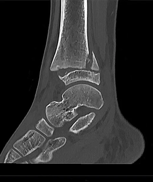
Açık büyüme plağı bulunan çocukta ekleme yakın travma, fizis boyunca veya komşu epifiz-metafizden geçen kırığa yol açabilir. İnce kesit kontrastsız BT, özellikle eklem yüzüne uzanan kırık çizgisini, kemik fragmanlarını ve deplasmanı düzlemler boyunca gösterir; basit fizis ayrışmasında ilk inceleme çoğunlukla radyografidir. Fizis hasarının gözden kaçması büyüme bozukluğu ve açısal deformite gibi sekellere yol açabilir; eklem içi Tip III ve IV kırıklarda eklem uyumsuzluğu da görülebilir. Salter–Harris tipi kırık yolunu tarif eder; tek başına bireysel büyüme sonucunu güvenilir biçimde öngörmez.
Faz ve pencere
- Kontrastsız tanısal
- İnce kesit kemik pencereli BT'de fizis boyunca düşük dansiteli ayrışma, metafiz veya epifiz korteksinden geçen keskin kırık hattı, deplasman ve eklem yüzü basamağı aranır; iyotlu kontrast ve HU eşiği tanı için gerekli değildir.
- Kemik algoritması tanısal
- Koronal ve sagittal rekonstrüksiyonlar kırığın fizis-metafiz-epifiz ilişkisini ve eklem içi fragmanların yerini gösterir; kompresyon tipi hasarda belirgin çizgi olmayabileceğinden normal BT fizis yaralanmasını dışlamaz.
Önerilen pencereler: Kemik (G 1800 / M 400).
BT bulguları
- Fizis boyunca ayrışma — epifiz ile metafiz arasında genişleme, basamak veya hizalanma kaybı.
- Metafizer Thurston–Holland fragmanı — kırık fizisten metafize çıkar ve üçgen metafiz parçası oluşturur.
- Epifiz içi kırık — Tip III'te kırık eklem yüzünden başlar, epifizden geçerek fizise ulaşır.
- Transfiziyel hat — aynı kırık epifiz, fizis ve metafizi keser; eklem yüzü de etkilenebilir.
- Kompresyon yaralanması — fizis ezilmesi belirgin ayrışma veya kortikal kırık oluşturmadan kalabilir.
- Deplasman ve eklem uyumsuzluğu — fragmanların ötelemesi, rotasyonu ya da eklem yüzü basamağı kaydedilir.
- Eşlik eden metafiz kırıkları — çocuklarda kalın periyost ve esnek kemik yapısı nedeniyle yeşil ağaç kırığı veya torus gibi örüntüler gözlenebilir.
Ölçütler ve sınıflamalar
- Salter–Harris
- Tip I: yalnız fizis; Tip II: fizis ve metafiz; Tip III: fizis ve epifiz; Tip IV: metafiz, fizis ve epifiz; Tip V: kompresyonla fizis ezilmesi. Tip II en sık örüntüdür; tip tanımı prognozu tek başına belirlemez.
Normalde
Normal açık fizis, MR'de kemik boyunca uzanan düzgün, homojen yüksek sinyalli bir çizgi olarak izlenir. Travmalı tarafı karşı tarafın aynı kemiği ve düzeyiyle kıyaslayarak fizis aralığında asimetri, çizginin fizisten epifize/metafize çıkışı ve eklem basamağını arayın.
Normal BT ile kıyasla
Aynı bölgenin, kaynak veri setinde patoloji içermediği belirtilen bir BT incelemesini kesit kesit kaydırın; organ sınırlarını açarak anatomiyi hasta görüntüsüyle karşılaştırın. Küçük rastlantısal bulgular tümüyle dışlanmamıştır.
Kaynak: TotalSegmentator v2.01 veri seti (Wasserthal ve ark., Radiology: Artificial Intelligence 2023), CC BY 4.0, vaka s0685; organ sınırları veri setinin otomatik segmentasyonundan, birleştirilerek sadeleştirildi. Görüntüler 2,5 mm kesit aralığına yeniden örneklendi.
Ayırıcı tanı
- Gelişimsel fizis görünümü
- düzgün ve beklenen anatomik konumlu kıkırdak aralık vardır; travmatik kortikal hat ya da fragman yoktur.
- Apofiz avulsiyonu
- çekme kuvveti kas-iskelet birimine veya ligamente uygulanır; tutunma yerinin yakınında küçük bir kemik fragmanı görülebilir.
- Metafizer buckle (torus) kırığı
- çocuklarda görülen stabil bir kırık örüntüsüdür ve greenstick kırığından ayrılır.
- Osteokondral lezyon
- değerlendirmede lezyonun boyutu, stabilitesi, fragmantasyonu ve subkondral tutulum dikkate alınır.
Tuzaklar
- Tip I ayrışması BT'de veya radyografide silik kalabilir; klinik şüphe sürüyorsa normal BT'yi fizis bütünlüğünün kesin kanıtı saymayın.
- Tip V kompresyonunda belirgin kırık çizgisi beklemek yanılgı yaratır; fizis yüksekliği ve karşı taraf simetrisi ile birlikte değerlendirin.
- Kısmi fizis kapanması fizyolojik olabilir; kapanma çizgisini travmatik köprü veya düzensiz genişleme diye sınıflandırmadan yaş ve karşı tarafı göz önüne alın.
Kendini dene
Çocukta distal tibia kırık hattı fizisten geçip metafizde üçgen parça oluşturuyor. Bu hangi Salter–Harris tipidir?
Cevabı göster
Tip II. Tip II'de hat fizis boyunca ilerleyip metafize çıkar ve Thurston–Holland fragmanı bırakır. Tip I fizisle sınırlıdır; Tip III epifize, Tip IV ise metafizden epifiz üzerinden ekleme uzanır.
Aksiyel yüklenme sonrası fizis ezilmiş görünüyor; belirgin kırık hattı ve fragman yok. En uygun sınıflama hangisidir?
Cevabı göster
Tip V. Tip V, kompresyonun fiziste ezilme oluşturduğu örüntüdür ve BT'de doğrudan çizgi silik olabilir. Diğer seçeneklerde kırık fizis boyunca ya da komşu epifiz/metafize uzanır.
İlgili konular
Kaynaklar
Bu sayfadaki 38 cümle ve 2 quiz sorusu aşağıdaki kaynaklardan alıntılarla otomatik olarak karşılaştırıldı; 10 cümle bu karşılaştırmada düzeltildi (2026-10-09).
- Imaging of Physeal Injurypmc.ncbi.nlm.nih.gov
- Ankle fractures in children.pubmed.ncbi.nlm.nih.gov
- Paediatric Ankle Fractures: Guidelines to Management.pubmed.ncbi.nlm.nih.gov
- Detection of early changes after growth plate injury using MRI.pubmed.ncbi.nlm.nih.gov
- Pediatric physeal ankle fracture.pubmed.ncbi.nlm.nih.gov
- Distal femoral fractures in children.pubmed.ncbi.nlm.nih.gov
- Pediatric Ankle Fractures: Concepts and Treatment Principles.pubmed.ncbi.nlm.nih.gov
- Proximal clavicle physeal fracture with posterior displacement: diagnosis, treatment, and prevention.pubmed.ncbi.nlm.nih.gov
- Open reduction and internal fixation for displaced Salter-Harris type II fractures of the distal tibia: a retrospective study of sixty-five cases in children.pubmed.ncbi.nlm.nih.gov
- Distal femoral physeal fixation: are smooth pins really safe?pubmed.ncbi.nlm.nih.gov
- Open Reduction and Internal Screw Fixation of Transitional Ankle Fractures in Adolescents.pubmed.ncbi.nlm.nih.gov
- Ankle fractures in children.pubmed.ncbi.nlm.nih.gov
- Vertebral growth modulation by hemicircumferential electrocoagulation: an experimental study in pigs.pubmed.ncbi.nlm.nih.gov
- Outcome of distal tibial physeal injuries.pubmed.ncbi.nlm.nih.gov
- Salter-Harris type II fracture of the femoral bone in a 14-year-old boy - case report.pubmed.ncbi.nlm.nih.gov
- Pediatric physeal fractures and growth disturbance: current concepts and surgical approaches.pubmed.ncbi.nlm.nih.gov
- Imaging of pediatric musculoskeletal trauma: Age-specific considerations and challenges.pubmed.ncbi.nlm.nih.gov
- An Anatomic Reduction Technique for Salter-Harris Type III Medial Femoral Condyle Fractures.pubmed.ncbi.nlm.nih.gov
- Fixed Flexion Deformity due to Displaced Hoffa Fracture: A Case Report.pubmed.ncbi.nlm.nih.gov
- Conservative Treatment of Pediatric Fractures-Narrative Review of Acceptable Deformity and Remodeling Potential.pubmed.ncbi.nlm.nih.gov
- Isolated Distal Ulnar Salter-Harris IV Fracture in an Adolescent Male With Distal Radioulnar Joint Injury: A Case Report and Review of the Literature.pubmed.ncbi.nlm.nih.gov
- Reliability and clinical utility of paediatric ankle fracture classifications : a systematic review.pubmed.ncbi.nlm.nih.gov
- Deep Learning-Based Multi-Class Pediatric Wrist Fracture Subtype Classification: A Pilot Study Comparing Convolutional Neural Network Architectures.pubmed.ncbi.nlm.nih.gov
- Physiological mechanisms and organism response in youth sports activities-related musculoskeletal injuries.pubmed.ncbi.nlm.nih.gov
- Management of pediatric ankle fractures: comparison of biodegradable PLGA implants with traditional metal screws.pubmed.ncbi.nlm.nih.gov
- The iliotibial band revisited: surgical anatomy and imaging-based guidelines for lateral tenodesis in ACL reconstruction.pubmed.ncbi.nlm.nih.gov
- Pediatric musculoskeletal ultrasound of the elbow: a pictorial review.pubmed.ncbi.nlm.nih.gov
- Management of pediatric hip infections - A narrative review.pubmed.ncbi.nlm.nih.gov
- Emergency Medicine Management of Distal Radius and Ulna Fractures: Narrative Review.pubmed.ncbi.nlm.nih.gov
- Evaluation of factors affecting development of complications in the early surgical treatment of distal tibial epiphyseal fractures.pubmed.ncbi.nlm.nih.gov
- A Systematic Approach to the Evaluation of Acute Musculoskeletal Injuries in the Emergency Department.pubmed.ncbi.nlm.nih.gov
- Three-Dimensional Planning and Surgical Correction of Bilateral Developmental Valgus Deformity of the Manus in a Dog.pubmed.ncbi.nlm.nih.gov
- Ultrasound-guided closed reduction of distal radius Salter-Harris I and II fracture in children.pubmed.ncbi.nlm.nih.gov
- The quiet flame: whole-body MRI and the underestimated burden of pediatric chronic non-bacterial osteomyelitis (CNO).pubmed.ncbi.nlm.nih.gov
- Diagnostic Pitfalls and Management of Transphyseal Fractures of the Distal Humerus: A Retrospective Review of 25 Cases.pubmed.ncbi.nlm.nih.gov
- Growth Plate Injury Leading to Madelung-Type Deformity After ESIN in Children: A Case Report and a Narrative Review of the Literature.pubmed.ncbi.nlm.nih.gov
- Utility of computed tomography in children's ankle fractures from classification to surgical planning.pubmed.ncbi.nlm.nih.gov
- A Review of Pediatric Orthopedic Disorders: Diagnosis and Treatment Updates.pubmed.ncbi.nlm.nih.gov
- Explainable machine learning-based prediction of early and mid-term postoperative complications in adolescent tibial fractures.pubmed.ncbi.nlm.nih.gov
- Guidelines on the Use of Therapeutic Apheresis in Clinical Practice-Evidence-Based Approach From the Writing Committee of the American Society for Apheresis: The Tenth Special Issue.pubmed.ncbi.nlm.nih.gov
- Exploring skeletal disorders in cattle and sheep: a WGS-based framework for diagnosis and classification.pubmed.ncbi.nlm.nih.gov
- Stop Searching under the Streetlight! A Primer and Practical Guide to the Diagnosis of Joint Pain and Inflammation.pubmed.ncbi.nlm.nih.gov
- Magnetic resonance imaging of the elbow.pubmed.ncbi.nlm.nih.gov
- Avulsion injuries: an update on radiologic findings.pubmed.ncbi.nlm.nih.gov
- Pediatric stress fractures: a pictorial essay.pubmed.ncbi.nlm.nih.gov
- Management of pediatric distal radius fractures - A systematic review and meta-analysis.pubmed.ncbi.nlm.nih.gov
- Management and Outcomes of Osteochondritis Dissecans of the Knee in the Pediatric and Adolescent Population.pubmed.ncbi.nlm.nih.gov
- Longitudinal In Vivo 3T MRI of Naturally Occurring Early Osteochondrosis Lesions in the Piglet Humeral Epiphyseal Cartilage and Growth Plate.pubmed.ncbi.nlm.nih.gov
- Magnetic Resonance Imaging Patterns of Common Injuries in Pediatric and Adolescent Athletes.pubmed.ncbi.nlm.nih.gov
- Outcome of Open Distal Tibial Physeal Fractures: A Descriptive Report of Two Cases.pubmed.ncbi.nlm.nih.gov
- Distal metaphyseal horizontal tibial fractures in children.pubmed.ncbi.nlm.nih.gov
- Type 4 Tibial Tuberosity Avulsion Fractures: Surgical Treatment Early Outcomes and a Presentation of the Distal Cortical Fixation.pubmed.ncbi.nlm.nih.gov
- Intraosseous tumours of the knee.pubmed.ncbi.nlm.nih.gov
- Modified Stoppa Approach for ORIF of a Paediatric Transverse Acetabular Fracture: Case Report and Systematic Review of Internal Fixation in Children.pubmed.ncbi.nlm.nih.gov
- Extensor Retinaculum Syndrome of the Ankle: An Adult Case Series.pubmed.ncbi.nlm.nih.gov
- Pathological and bacteriological findings in sows, finisher pigs, and piglets, being culled for lameness.pubmed.ncbi.nlm.nih.gov
- Management of Pediatric Elbow Fractures and Dislocations.pubmed.ncbi.nlm.nih.gov
- Application of biodegradable implants in pediatric orthopedics: shifting from absorbable polymers to biodegradable metals.pubmed.ncbi.nlm.nih.gov
- Management of Pediatric Ankle Fractures.pubmed.ncbi.nlm.nih.gov
- Distal ulna physeal fracture Salter-Harris type IV versus cleft ulnar epiphysis: Two pediatric case report.pubmed.ncbi.nlm.nih.gov
- Health-Related Quality of Life after Fractures of the Distal Forearm in Children and Adolescents-Results from a Center in Switzerland in 432 Patients.pubmed.ncbi.nlm.nih.gov
- Interventions for treating wrist fractures in children.pubmed.ncbi.nlm.nih.gov
- The Unique Features of Pediatric Sports Injuries: The Hip.pubmed.ncbi.nlm.nih.gov
- Imaging of pediatric foot disorders.pubmed.ncbi.nlm.nih.gov
- Recent Advances in the Diagnosis and Management of Pediatric Orthopedic Disorders: A Comprehensive Review.pubmed.ncbi.nlm.nih.gov
- 3D-Printed Orthoses vs Conventional Immobilization Methods for Pediatric Distal Radius Fractures.pubmed.ncbi.nlm.nih.gov
- The most common bone tumors of the upper extremity in childhood and adolescence and their treatment: a review of the current literature.pubmed.ncbi.nlm.nih.gov
- Understanding Osteochondritis Dissecans: A Narrative Review of the Disease Commonly Affecting Children and Adolescents.pubmed.ncbi.nlm.nih.gov
- Uniquely pediatric upper extremity injuries.pubmed.ncbi.nlm.nih.gov
- Abuse as a Cause of Childhood Fractures.pubmed.ncbi.nlm.nih.gov
- Trash Lesions Around the Elbow: A Review of Approach to Diagnosis and Management.pubmed.ncbi.nlm.nih.gov
- Development and National Validation of a Musculoskeletal Emergency Medicine Assessment Tool.pubmed.ncbi.nlm.nih.gov
- Pediatric Tibial Shaft Fractures.pubmed.ncbi.nlm.nih.gov
- Comparative outcomes of Kirschner wire and lag screw fixation in pediatric medial malleolar fractures.pubmed.ncbi.nlm.nih.gov
- Pediatric lateral Busch-Hoffa fracture associated with lateral meniscus tear and incarcerated popliteus tendon: systematic review of literature and case report.pubmed.ncbi.nlm.nih.gov
- Comparison of Arthroscopic Reduction and Percutaneous Fixation Versus Open Reduction for Pediatric Intra-articular Epiphyseal Ankle Fractures.pubmed.ncbi.nlm.nih.gov
- Arthroscopic management of pediatric ankle fractures: a systematic review.pubmed.ncbi.nlm.nih.gov
- Transphyseal Proximal Humeral Aneurysmal Bone Cyst with Pathologic Fracture in a Child: A Case Report.pubmed.ncbi.nlm.nih.gov
- Post-infection physeal bar resection of the proximal tibia: a case report and narrative review of the literature.pubmed.ncbi.nlm.nih.gov
- Radiocapitellar Alignment in Suspected Pediatric Monteggia Lesions: Narrative Review and Imaging Interpretation Framework.pubmed.ncbi.nlm.nih.gov
- Challenges in Reconstruction and Limb Lengthening Surgery for Congenital Lower Limb Deficiencies.pubmed.ncbi.nlm.nih.gov
Eğitim amaçlıdır; hasta başında tanı ve tedavi kararının yerine geçmez, tıbbi tavsiye değildir. Metinler yapay zekâ desteğiyle yazıldı ve otomatik bir adımda kaynak alıntılarıyla karşılaştırıldı; bu bir tıbbi doğrulama değildir ve içerik uzman radyolog incelemesinden geçmedi. Klinik kararlarda güncel kılavuzları, kurum protokollerini ve radyoloji raporunu esas alın.