Özet
Koroner BT anjiyografide yoğun kalsifiye plak, gerçek damar duvarı sınırından daha geniş ve parlak görünerek kontrastlı lümeni kısmen örtebilir. Parsiyel hacim etkisi ve ışın sertleşmesi nedeniyle oluşan blooming artefaktı, koroner lümeni örterek darlık derecesini olduğundan ağır gösterebilir. Yüksek uzaysal çözünürlük sağlayan ince kesitler ve gelişmiş rekonstrüksiyon teknikleri blooming artefaktını azaltarak değerlendirmeyi iyileştirse de, yoğun kalsifiye plaklar darlığın olduğundan ağır tahmin edilmesine neden olabilir. Yoğun kalsifiye plakların blooming etkisi, koroner lümeni örterek darlık derecesinin olduğundan ağır tahmin edilmesine yol açar.
Okuma sırası
- Proksimal sol ön inen arter. Sol ön inen arterin proksimal seyrini bul ve kalsifiye odağın lümen içindeki yerini kaynak kesitte saptayarak incelemeye başla.
- Kalsifiye plak kenarı. Parlak kalsiyumun damar duvarından lümene doğru bulanık biçimde genişleyip genişlemediğini takip et.
- Plak düzeyindeki lümen. Kalsiyumun arkasında kalan kontrastlı yarığı komşu aksiyel kesit ve damara dik düzlemde ara.
- Proksimal ve distal koroner arter. Plak öncesi ve sonrası damar opaklaşmasını izleyip gerçek kesintiyi artefaktla örtülmüş lümenden ayır.
Faz ve pencere
- Kontrastsız
- Koroner duvar boyunca çok yüksek atenüasyonlu kalsifik odaklar kalsiyum yükünü gösterir; lümen kontrastla dolmadığından bu seri tek başına darlık yüzdesini belirlemez.
- BTA tanısal
- Kontrastla opaklaşmış koroner lümen, parlak kalsiyumun yanından veya içinden izlenir; kalsiyum kenarı damar içine taşmış izlenimi verip gerçek açıklığı daraltabilir.
Önerilen pencereler: Anjiyo (G 600 / M 150), Mediasten (G 350 / M 50), Yumuşak doku (G 400 / M 40).
BT bulguları
- Kalsifiye plak komşuluğundaki parlak sınır, damar duvarına göre kalın ve bulanık görünür; bu blooming lümen alanını örtebilir.
- Kalsiyum kenarından dışarı uzanan koyu bantlar beam-hardening etkisini düşündürür ve düşük atenüasyonlu plakla karıştırılabilir.
- MIP görüntüsü boyunca üst üste binen parlak kalsiyum odakları küçük kontrastlı lümen adacıklarını kapatarak darlık izlenimini güçlendirir.
- Çok düzlemli yeniden yapılandırmalar ve kaynak kesitler, darlık derecesinin intravasküler görüntüleme bulgularıyla karşılaştırmalı olarak güvenilir biçimde değerlendirilmesine olanak tanır.
- Kalsiyumlu segmentte lümen sınırı pencere genişliği ve rekonstrüksiyon keskinliği değiştikçe yer değiştiriyorsa ölçülen stenoz artefakta duyarlıdır.
- Kalsifikasyonun hemen proksimal ve distalindeki opak lümen devamlılığı, odak çevresinde tam tıkanıklık varsayımını sınar.
Normalde
Normal koroner BT anjiyografide sol ana, ön inen, sirkumfleks ve sağ koroner arter kontrastla kesintisiz dolar; damar duvarı boyunca lümene taşan yoğun plak bulunmaz. Şüpheli segmenti aynı damarın plak dışındaki proksimal ve distal bölümüyle, ayrıca kalsiyumsuz yan dal lümeniyle kıyaslayarak görünür açıklığın rekonstrüksiyon değişince ne ölçüde değiştiğine bakın.
Normal BT ile kıyasla
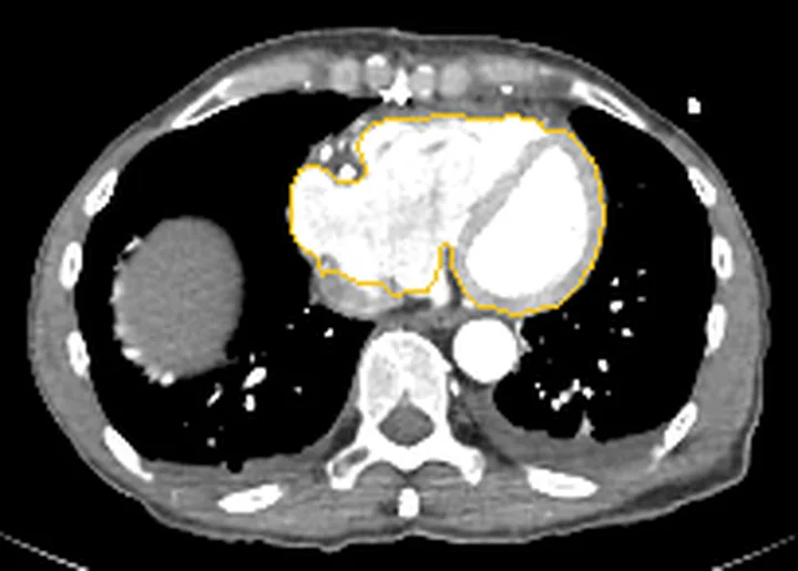
Aynı bölgenin, kaynak veri setinde patoloji içermediği belirtilen bir BT incelemesini kesit kesit kaydırın; organ sınırlarını açarak anatomiyi hasta görüntüsüyle karşılaştırın. Küçük rastlantısal bulgular tümüyle dışlanmamıştır.
Kaynak: TotalSegmentator v2.01 veri seti (Wasserthal ve ark., Radiology: Artificial Intelligence 2023), CC BY 4.0, vaka s0977; organ sınırları veri setinin otomatik segmentasyonundan, birleştirilerek sadeleştirildi. Görüntüler 2,5 mm kesit aralığına yeniden örneklendi.
Ayırıcı tanı
- Gerçek kalsifiye stenoz
- daralma damar eksenine dik düzlemlerde ve ardışık kaynak kesitlerde sürer.
- Kalsifiye olmayan plak
- kontrastlı lümen kenarında yumuşak doku atenüasyonlu duvar kalınlaşması oluşturur; parlak kemiksi odak gibi görünmez.
- Koroner hareket artefaktı
- damar konturunda basamaklanma veya çift kenar yapar ve komşu kesitlerde konum değiştirir.
- Kısmi hacim etkisi
- kalsiyum ile lümen aynı vokselde örneklendiği için sınır bulanıklaşır; daha ince rekonstrüksiyonda görünüm değişebilir.
Tuzaklar
- MIP’de üst üste binen kalsiyum lümeni bütünüyle kapalı gösterebilir; stenozu MIP’ye dayanarak derecelendirmeyin, ince kaynak kesite dönün.
- Daha keskin kernel kalsiyum kenarını ve küçük lümeni farklı gösterebilir; tek rekonstrüksiyondaki sınırı anatomik gerçek kabul etmeyin.
- Kalsiyumun yanında görülen koyu halo lümen içi yumuşak plak sanılabilir; dik düzlemde ve çevre kesitlerde damar duvarı ilişkisini kontrol edin.
Kendini dene
Koroner BT anjiyografide parlak plak lümene taşmış gibi; dik rekonstrüksiyonda sınır değişiyor. En olası açıklama?
Cevabı göster
Kalsiyum blooming. Yüksek yoğunluklu kalsiyumun parsiyel hacimle geniş görünmesi lümeni olduğundan dar gösterebilir. Diseksiyon intimal flep, yumuşak plak duvar atenüasyonlu doku, spazm ise segmental daralma görünümü verir.
MIP’de damar kapalı gibi, ince kaynak kesitlerde kontrastlı lümen plak yanından sürüyor. Hangi yorum en uygundur?
Cevabı göster
MIP kaynaklı yalancı ağır darlık. MIP, aynı izdüşümdeki yoğun kalsiyumu üst üste bindirip küçük lümen açıklığını örtebilir. Tam oklüzyonda kaynak kesit ve dik düzlemlerde de lümen devamlılığı kaybolur; diğer seçenekler bu görüntü davranışını açıklamaz.
Kaynaklar
Bu sayfadaki 34 cümle ve 2 quiz sorusu aşağıdaki kaynaklardan alıntılarla tek tek karşılaştırıldı; 4 cümle bu karşılaştırmada düzeltildi (2026-10-08).
- High-Z Contrast Media for Coronary Photon-counting Detector CT Angiography: Improved Quantification of Calcified Stenoses.pubmed.ncbi.nlm.nih.gov
- Diagnostic performance and clinical impact of photon-counting detector computed tomography in coronary artery disease.pubmed.ncbi.nlm.nih.gov
- Simulation of calcium blooming artifacts in coronary CT angiography from digital phantoms.pubmed.ncbi.nlm.nih.gov
- Spectral computed tomography characterization of coronary atherosclerotic plaque: principles, imaging biomarkers, and clinical significance.pubmed.ncbi.nlm.nih.gov
- Diagnostic and prognostic value of coronary artery calcium scoring and computed tomography angiography in coronary artery disease: a systematic review and meta-analysis.pubmed.ncbi.nlm.nih.gov
- Coronary artery calcium score-emerging role for opportunistic screening on thoracic CT and novel applications.pubmed.ncbi.nlm.nih.gov
- Beyond Angiography: Cardiac CT for Planning Complex PCI in Calcified Coronary Lesions.pubmed.ncbi.nlm.nih.gov
- Coronary Computed Tomography Angiography for the Diagnosis and Revascularization Guidance of Coronary Bifurcation Lesions: A Contemporary Review.pubmed.ncbi.nlm.nih.gov
- Understanding Photon-Counting CT: Physics, Detector Technology, and Image Reconstructions.pubmed.ncbi.nlm.nih.gov
- Left main coronary artery stenosis assessment on coronary CT angiography.pubmed.ncbi.nlm.nih.gov
- Photon-Counting CT in Cardiovascular Imaging: Clinical Applications.pubmed.ncbi.nlm.nih.gov
- Computed Tomography Coronary Angiography as a Gatekeeper for Invasive Coronary Assessment Before Transcatheter Aortic Valve Implantation.pubmed.ncbi.nlm.nih.gov
- Feasibility of deep learning-based image reconstruction using TrueFidelity for coronary calcium scoring.pubmed.ncbi.nlm.nih.gov
- Advanced quantitative CT for coronary artery disease: integrating stenosis quantification, plaque quantification, and fractional flow reserve-CT.pubmed.ncbi.nlm.nih.gov
- Plaque-Adapted Virtual Monoenergetic Image Selection for Carotid Plaque Visualization Using Photon-Counting CT.pubmed.ncbi.nlm.nih.gov
- Clinical benefits and current challenges of photon-counting detector CT in vascular imaging.pubmed.ncbi.nlm.nih.gov
- Spectral Computed Tomography Angiography in Visceral Artery Aneurysms: Technical Principles and Clinical Applications.pubmed.ncbi.nlm.nih.gov
- Consensus document on the role of cardiac computed tomography for pre-procedural planning in minimally invasive percutaneous coronary intervention from the Japanese association of cardiovascular intervention and therapeutics.pubmed.ncbi.nlm.nih.gov
- Coronary CT Angiography for PCI Planning and Guidance: A Comprehensive Narrative Review.pubmed.ncbi.nlm.nih.gov
- Coronary stenosis assessment: AI-based CT quantification, visual analysis of invasive angiography, and quantitative coronary angiography.pubmed.ncbi.nlm.nih.gov
- Exploring double low-dose CT technology for achieving coronary mixed reality radiation dose reduction: a preliminary study in coronary artery disease patients.pubmed.ncbi.nlm.nih.gov
- Cardiac Computed Tomography in Structural Heart Interventions: From Preprocedural Planning to Procedural Strategy.pubmed.ncbi.nlm.nih.gov
- Identification of coronary calcified nodules on noninvasive coronary CT: A two-case report with intravascular imaging correlation.pubmed.ncbi.nlm.nih.gov
- Bilateral Coronary Artery-to-Pulmonary Artery Fistula Treated Using Minimally Invasive Coils and Vascular Embolization Devices.pubmed.ncbi.nlm.nih.gov
- Case Report: Angiographic Remodeling of a Chronic Total Occlusion Four Years After Extraplaque Wiring: Implications for Procedural Reattempt.pubmed.ncbi.nlm.nih.gov
- Coil-Assisted Onyx Embolization of an Ophthalmic Artery-Supplied Anterior Cranial Fossa Dural Arteriovenous Fistula Mimicking an Intracranial Aneurysm.pubmed.ncbi.nlm.nih.gov
- Stress CT myocardial perfusion imaging after arterial switch operation for transposition of the great arteries.pubmed.ncbi.nlm.nih.gov
- Integrating Invasive and Noninvasive Imaging for Coronary Atherosclerosis: A Systematic Review With Pragmatic Algorithms From Anatomy to Physiology.pubmed.ncbi.nlm.nih.gov
- Clinical Applications of Artificial Intelligence in Cardiac CT: From Coronary CT Angiography to CT-Derived Fractional Flow Reserve and Myocardial Perfusion Imaging.pubmed.ncbi.nlm.nih.gov
- Transfemoral TAVR in Chronic Type A Aortic Dissection and Severe Aortic Stenosis.pubmed.ncbi.nlm.nih.gov
- Pictorial review: a step-by-step guide to coronary CT angiography with photon-counting detector CT.pubmed.ncbi.nlm.nih.gov
- Coronary Computed Tomography Angiography as a Method for Diagnosing a Thrombotic Occlusion of a Giant Right Coronary Artery Aneurysm in a Patient with Difficulty in Visualizing the Right Coronary Artery During Invasive Coronary Angiography.pubmed.ncbi.nlm.nih.gov
- Association of Low-Attenuation Noncalcified Plaque Shape and Degree of Intraplaque Embeddedness Using Atherosclerotic Imaging-enabled Quantitative CT with Acute Coronary Syndrome.pubmed.ncbi.nlm.nih.gov
- South Asian Cardiovascular Risk: Role of Coronary Artery Calcium Scoring and Computed Tomography Angiography.pubmed.ncbi.nlm.nih.gov
- Artificial Intelligence in Cardiovascular MRI: From Imaging to Biomechanics and Diagnosis.pubmed.ncbi.nlm.nih.gov
- Clinical Impact of CT-Based FFR in Everyday Cardiology: Bridging Computation and Decision-Making.pubmed.ncbi.nlm.nih.gov
- Four-dimensional computed tomography of the left ventricle, Part I: Motion artifact reduction.pubmed.ncbi.nlm.nih.gov
- Commentary: an eye on PET quantification.pubmed.ncbi.nlm.nih.gov
- International Harmonization of Nomenclature and Diagnostic Criteria (INHAND): Non-proliferative and Proliferative Lesions of the Non-human Primate ( M. fascicularis ).pubmed.ncbi.nlm.nih.gov
- High-affinity alphavbeta3 integrin targeted optical probe as a new imaging biomarker for early atherosclerosis: initial studies in Watanabe rabbits.pubmed.ncbi.nlm.nih.gov
- Motion correction algorithm improves right coronary artery image quality at heart rates > 75 bpm in non-ECG-gated coronary CT angiography.pubmed.ncbi.nlm.nih.gov
- Incidentally diagnosed anomalous left coronary artery from the pulmonary artery in a 76-year-old man undergoing preoperative evaluation: a case report and literature review.pubmed.ncbi.nlm.nih.gov
- Chronic Total Occlusion Recanalization in a 14-Year-Old Girl With Pseudoxanthoma Elasticum: IVUS-Guided Stentless Strategy.pubmed.ncbi.nlm.nih.gov
- Confounder-Matched Deep Learning on Cardiac CT for the Diagnosis of Tetralogy of Fallot: A Proof of Concept.pubmed.ncbi.nlm.nih.gov
- Cross-Distribution Diffusion Priors-Driven Iterative Reconstruction for Sparse-View CT.pubmed.ncbi.nlm.nih.gov
- Hypersensitive detection of single millimeter vascular emboli from adhesive in vivo.pubmed.ncbi.nlm.nih.gov
- Aortic Dissection Mimicry Under Extracorporeal Membrane Oxygenation (ECMO) After Cardiac Arrest: A Case Report of Emergency Imaging Dilemmas.pubmed.ncbi.nlm.nih.gov
- Tertiary Syphilitic Aortitis Manifesting as Ascending Aortic Aneurysm and Dissection.pubmed.ncbi.nlm.nih.gov
- Acute Myocardial Infarction Secondary to Idiopathic Ascending Aortic Thrombus.pubmed.ncbi.nlm.nih.gov
- Deep Learning-based Monoenergetic Imaging for Calcified Coronary Stenosis Assessment at Energy-integrating Detector CT.pubmed.ncbi.nlm.nih.gov
- Deep Learning-based Automated Coronary Plaque Quantification: First Demonstration With Ultra-high Resolution Photon-counting Detector CT at Different Temporal Resolutions.pubmed.ncbi.nlm.nih.gov
- Coronary Plaque Vulnerability and Pericoronary Adipose Tissue Inflammation: Emerging Insights from Advanced CT Imaging.pubmed.ncbi.nlm.nih.gov
- Frequently Encountered Artifacts in the Application of Dual-Energy Computed Tomography to Cardiovascular Imaging for Urate Crystals in Gout: A Matched-Control Study.pubmed.ncbi.nlm.nih.gov
- Dual-Energy CT in Cardiothoracic Imaging: Current Developments.pubmed.ncbi.nlm.nih.gov
- Technical principles, benefits, challenges, and applications of photon counting computed tomography in coronary imaging: a narrative review.pubmed.ncbi.nlm.nih.gov
- Optimization of photon counting CT for cardiac imaging in patients with left ventricular assist devices: An in-depth assessment of metal artifacts.pubmed.ncbi.nlm.nih.gov
- Cardiac CT blooming artifacts: clinical significance, root causes and potential solutions.pubmed.ncbi.nlm.nih.gov
- Guidelines for autopsy investigation of sudden cardiac death: 2017 update from the Association for European Cardiovascular Pathology.pubmed.ncbi.nlm.nih.gov
- Long-term safety and effectiveness of the "OptEase" vena cava filter.pubmed.ncbi.nlm.nih.gov
- Optimizing coronary CT angiography in patients with delayed contrast arrival using a free-breathing, high-threshold bolus-tracking protocol.pubmed.ncbi.nlm.nih.gov
- Feasibility of ultra-low flow rate coronary CT angiography using photon-counting detector CT: a prospective randomized trial.pubmed.ncbi.nlm.nih.gov
- Clinical evaluation of a motion correction software based on partial angle reconstruction in coronary CT angiography.pubmed.ncbi.nlm.nih.gov
- Case Report: Intravascular lithotripsy combined with a new-generation 3-in-1 carotid stent for calcified carotid artery stenosis.pubmed.ncbi.nlm.nih.gov
- Novel Imaging-Based Biomarkers for Identifying Carotid Plaque Vulnerability.pubmed.ncbi.nlm.nih.gov
- Advances in Noninvasive Carotid Wall Imaging with Ultrasound: A Narrative Review.pubmed.ncbi.nlm.nih.gov
- Engineering the oxidative myocardium: ROS-responsive biomaterials for precision cardiovascular delivery in ischemia-reperfusion injury and post-infarction remodeling.pubmed.ncbi.nlm.nih.gov
- Deep learning predicts stent implantation in borderline coronary lesions from angiography.pubmed.ncbi.nlm.nih.gov
- Impact of hypertension on coronary artery plaques and FFR-CT in type 2 diabetes mellitus patients: evaluation utilizing artificial intelligence processed coronary computed tomography angiography.pubmed.ncbi.nlm.nih.gov
- Contrast medium dose optimization in the era of multi-energy CT.pubmed.ncbi.nlm.nih.gov
- Dual energy imaging in cardiothoracic pathologies: A primer for radiologists and clinicians.pubmed.ncbi.nlm.nih.gov
- Coronary computed tomography and magnetic resonance imaging.pubmed.ncbi.nlm.nih.gov
- State-of-the-art CT and MR imaging and assessment of atherosclerotic carotid artery disease: standardization of scanning protocols and measurements-a consensus document by the European Society of Cardiovascular Radiology (ESCR).pubmed.ncbi.nlm.nih.gov
- CT and MR imaging of the upper extremity vasculature: pearls, pitfalls, and challenges.pubmed.ncbi.nlm.nih.gov
- Detectability of intracranial vessel wall atherosclerosis using black-blood spectral CT: a phantom and clinical study.pubmed.ncbi.nlm.nih.gov
- Global Vascular Guidelines on the Management of Chronic Limb-Threatening Ischemia.pubmed.ncbi.nlm.nih.gov
- Global vascular guidelines on the management of chronic limb-threatening ischemia.pubmed.ncbi.nlm.nih.gov
- Anomalies of coronary artery origin at invasive angiography: a prospective study of prevalence, catheter utilisation, access strategy and procedural implications.pubmed.ncbi.nlm.nih.gov
- Ruptured Coronary Aneurysm With Coronary-Pulmonary Artery Fistulas in Advanced Cirrhosis Treated With Coil Embolization.pubmed.ncbi.nlm.nih.gov
- Coronary Artery Origin Anomalies on Coronary CT Angiography: A Single-Center Tertiary-Care Cohort.pubmed.ncbi.nlm.nih.gov
- Deep learning-based assessment of coronary artery disease using curved-multiplanar reconstruction: comprehensive evaluation of stenosis, calcification, and plaque.pubmed.ncbi.nlm.nih.gov
- Prognostic value of preoperative CT-derived fractional flow reserve after transcatheter or surgical aortic valve replacement in patients with severe aortic stenosis.pubmed.ncbi.nlm.nih.gov
- Clinical comparison of sub-mm high-resolution non-contrast coronary CMR angiography against coronary CT angiography in patients with low-intermediate risk of coronary artery disease: a single center trial.pubmed.ncbi.nlm.nih.gov
- The impact of lipoprotein(a) determination in cardiovascular risk stratification among people living with HIV.pubmed.ncbi.nlm.nih.gov
- Multimodality Imaging Approach in Diagnosis and Follow-Up of Aortic Coarctation in Adulthood.pubmed.ncbi.nlm.nih.gov
- Diabetic plaque calcification: mechanisms, imaging phenotypes, and clinical implications.pubmed.ncbi.nlm.nih.gov
- Use of Calcium Modification in Percutaneous Coronary Intervention: A Comprehensive Review.pubmed.ncbi.nlm.nih.gov
- Standards for quantitative assessments by coronary computed tomography angiography (CCTA): An expert consensus document of the society of cardiovascular computed tomography (SCCT).pubmed.ncbi.nlm.nih.gov
- Improving Risk Stratification for Transient Ischaemic Attacks and Ischaemic Stroke in Patients with Coronary Artery Disease: A Combined Radiomics Analysis of Multimodal Adipose Tissue.pubmed.ncbi.nlm.nih.gov
- Relationship Between Perivascular Fat Inflammation and Coronary Atherosclerotic Plaque Composition.pubmed.ncbi.nlm.nih.gov
- A review of the pathogenic mechanism and clinical management progress of in-stent calcification restenosis.pubmed.ncbi.nlm.nih.gov
- Deciphering age- and sex-specific patterns of coronary artery atherosclerosis from a large Chinese cohort.pubmed.ncbi.nlm.nih.gov
- Operative Procedures for Ultrasound Assessment of Extracranial Artery Disease: A Narrative Review by the Italian Society for Vascular Investigation (SIDV).pubmed.ncbi.nlm.nih.gov
- Radiation dose optimization in coronary CT angiography clinical application of absolute phase gating in patients with atrial fibrillation.pubmed.ncbi.nlm.nih.gov
- Low- versus high-concentration iodine contrast media for coronary computed tomography angiography and dynamic stress computed tomography perfusion: a study protocol for a prospective randomized trial.pubmed.ncbi.nlm.nih.gov
- Contemporary review of the clinical features, multi-modality imaging, and management of coronary artery aneurysms.pubmed.ncbi.nlm.nih.gov
- Catheter-Directed Recombinant Tissue Plasminogen Activator for Treatment of Aortic Root and Coronary Artery Thrombosis in Single Ventricle Patients: A Case Series.pubmed.ncbi.nlm.nih.gov
- Radiation and Contrast Medium Dosage Optimization of Coronary CT Angiography While Combining Personalized Patient Protocol Technology and Automated Tube Voltage Selection Technology.pubmed.ncbi.nlm.nih.gov
- Technical advances and clinical impact of photon-counting computed tomography angiography for peripheral artery disease.pubmed.ncbi.nlm.nih.gov
- Ultra-high spatial resolution at photon-counting computed tomography: technical insights and sustainable applications in cardiothoracic imaging.pubmed.ncbi.nlm.nih.gov
- Value of Ultrahigh-Resolution Photon-Counting Detector Computed Tomography in Cardiac Imaging.pubmed.ncbi.nlm.nih.gov
- Association Between Pericoronary Fat Attenuation Index and High-Risk Plaque Features Assessed by Photon-Counting CT.pubmed.ncbi.nlm.nih.gov
- Pericoronary fat attenuation in stenotic and vulnerable coronary artery plaques: Implications for coronary artery disease and associated conditions.pubmed.ncbi.nlm.nih.gov
- Ischemic Heart Disease: Noninvasive Imaging Techniques and Findings.pubmed.ncbi.nlm.nih.gov
- FFR-CT: Technical Advances and Implementation in Clinical Practice.pubmed.ncbi.nlm.nih.gov
- Feasibility of On-Site CT-FFR Analysis in Ruling Out In-Stent Restenosis on Cardiac PCCT.pubmed.ncbi.nlm.nih.gov
- Photon-Counting CT: Technology, Current and Potential Future Clinical Applications, and Overview of Approved Systems and Those in Various Stages of Research and Development.pubmed.ncbi.nlm.nih.gov
- Clinical Applications of Photon-counting CT: A Review of Pioneer Studies and a Glimpse into the Future.pubmed.ncbi.nlm.nih.gov
- Coronary CT Angiography to Guide Percutaneous Coronary Intervention.pubmed.ncbi.nlm.nih.gov
- CT angiography: current technology and clinical use.pubmed.ncbi.nlm.nih.gov
- Real-Time Intravascular Ultrasound-Guided Percutaneous Coronary Intervention for Spontaneous Coronary Artery Dissection.pubmed.ncbi.nlm.nih.gov
Eğitim amaçlıdır; hasta başında tanı ve tedavi kararının yerine geçmez. İçerik yapay zekâ destekli bir süreçle güncel literatüre göre cümle cümle kaynaklarla karşılaştırıldı, ancak uzman radyolog incelemesinden geçmedi. Klinik kararlarda güncel kılavuzları, kurum protokollerini ve radyoloji raporunu esas alın.