
Özet
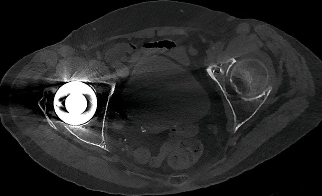
Işın sertleşmesi, polikromatik X-ışını demetindeki düşük enerjili fotonların kemik veya iyot gibi yoğun materyallerden geçerken daha fazla soğurulması ve geride daha yüksek ortalama enerjili bir demet kalmasıyla ortaya çıkar. Rekonstrüksiyon bu enerji değişimini tümüyle modelleyemediğinde kafa tabanında petroz kemikler arası koyu bantlar, yoğun kontrast çevresinde çizgilenme veya homojen yapıda merkezî atenüasyon düşüşü oluşabilir. Işın sertleşmesi artefaktı, özellikle kafa tabanı yakınındaki yapıların değerlendirilmesini zorlaştırabilir ve bazı durumlarda diğer patolojilerle karıştırılabilir. Işın sertleşmesi nedeniyle petroz kemikler arasında uzanan yapay koyu bantlar, komşu parankimi maskeleyerek tanıda zorluk çıkarabilir.
Faz ve pencere
- Kontrastsız tanısal
- Kontrastsız kafa BT'de petroz kemiklerden kaynaklanan ışın sertleşmesi, arka çukurda yapay atenüasyon bozukluğu ve artmış gürültüye yol açarak beyin sapı ve serebellumun görselleştirilmesini kısıtlayabilir ve doku tanısını zorlaştırabilir. Kontrast bu mekanizmanın şartı değildir; iyotlu kontrast ve yoğun metal de aynı fiziksel etkiyi artırabilir.
- Kemik rekonstrüksiyonu
- Petros kemikler arasındaki koyu bant, yumuşak doku penceresinde arka çukurun ve beyin sapının görselleştirilmesini kısıtlayan bir artefakt olarak görülür.
Önerilen pencereler: Beyin (G 80 / M 40), Kemik (G 1800 / M 400).
BT bulguları
- Petroz kemikler arası koyu bant — yüksek atenüasyonlu kafa tabanı kemikleri arasından geçen ışın yolunda düşük enerjili fotonların seçici kaybı posterior fossayı çizgileyebilir.
- Yoğun yapı çiftleri arasında çizgilenme — iki kemik, kontrastlı damar veya metal cisim arasında koyu ve parlak radyal şeritler gelişebilir.
- Cupping örüntüsünde, ışın sertleşmesi nedeniyle homojen yapının merkezi çevresine göre yapay olarak daha düşük atenüasyonlu görünür.
- Anatomik sınırları izlemeyen bant — yapay çizgi beyin sapı, serebellum ve BOS alanlarından geçerek kemik kaynaklı projeksiyon doğrultusunu sürdürür.
- Işın sertleşmesi artefaktı, yoğun petros kemiklerin bulunduğu kafa tabanı seviyesinde en belirgin şekilde izlenir.
- Komşu parankimde yalancı yoğunluk farkı — homojen dokunun içindeki merkez-çevre farkı, beklenen anatomik sınıra uymuyorsa cupping etkisini düşündürür.
- Yoğun kontrast yakınında koyu şerit — opak damar veya iyotlu materyalden geçen ışın doğrultusunda oluşan çizgi küçük damar ya da parankim bulgusunu örtebilir.
Normalde
Normal posterior fossada pons, serebellar hemisferler ve dördüncü ventrikül çevresindeki atenüasyonlar anatomik olarak tutarlı, beyin sapı konturu tek ve simetriktir. Petroz kemikler arasındaki şüpheli hipodens bandı serebellar hemisferler ve ponsun iki yanıyla kıyaslayın; lezyon anatomik sınırlara uymayıp kemiklerin arasından çizgi halinde geçiyorsa artefakt olasılığı artar.
Normal BT ile kıyasla
Aynı bölgenin, kaynak veri setinde patoloji içermediği belirtilen bir BT incelemesini kesit kesit kaydırın; organ sınırlarını açarak anatomiyi hasta görüntüsüyle karşılaştırın. Küçük rastlantısal bulgular tümüyle dışlanmamıştır.
Kaynak: TotalSegmentator v2.01 veri seti (Wasserthal ve ark., Radiology: Artificial Intelligence 2023), CC BY 4.0, vaka s0103; organ sınırları veri setinin otomatik segmentasyonundan, birleştirilerek sadeleştirildi. Görüntüler 2,5 mm kesit aralığına yeniden örneklendi.
Ayırıcı tanı
- Posterior fossa infarktı
- vasküler dağılıma uyan parankimal hipodensite ve gri-beyaz ayrımında silinme gösterir; kemikler arası doğrusal bant biçiminde olmaz.
- Akut intrakraniyal kanama
- beyin parankimi veya ekstraaksiyel kompartımanda anatomik sınırlı hiperdens odak oluşturur; ışın sertleşmesi ise parlak veya koyu şeritler oluşturarak komşu dokuyu maskeleyebilir.
- Serebellar kitle
- kitle etkisi, fokal doku yoğunluğu değişikliği veya çevresel ödemle anatomik bir lezyon oluştururken; ışın sertleşmesi serebellar görünürlüğü azaltarak gerçek lezyon değerlendirmesini zorlaştırır.
- Vazojenik ödem
- beyaz cevherde anatomik dağılımlı hipodensite ve bazen kitle etkisi görülür; petroz kemikler arasında doğrusal uzanmaz.
- Parsiyel hacim etkisi
- yoğun yapılar arasındaki zayıf kontrastlı geçişlerde rol oynar; ışın sertleşmesi ile birlikte zayıf atenüasyon gradyanlarına katkıda bulunabilir.
Tuzaklar
- Posterior fossa hipodensitesini tek kesitte infarkt kabul etmeyin; pons ve serebellumun komşu kesitlerdeki simetrisini, bandın petroz kemikler arasından geçişini kontrol edin.
- Işın sertleşmesi kaynaklı parlak veya koyu şeritler komşu dokuları maskeleyebilir veya sıvı boşluklarında yanılsama hiperdensiteyle karıştırılabilir.
- Beyin penceresinde görünmeyen kortikal detay için kemik serisini kullanın, fakat kemik penceresindeki doku kontrastıyla parankim tanısı koymayın.
- Kafa tabanı artefaktı içinde lezyon dışlanamayabilir; belirgin bant altında kalan yapıyı kesin normal saymak yerine değerlendirilebilirlik sınırını belirtin.
Kendini dene
Kontrastsız BT'de petroz kemikler arasında ponsu çaprazlayan koyu çizgi var; komşu kesitte silikleşiyor. En olası neden?
Cevabı göster
Işın sertleşmesi. Yoğun petroz kemikler arasında uzanan ve kafa tabanından uzaklaşınca azalan çizgisel hipodensite ışın sertleşmesiyle uyumludur. İnfarkt anatomik vasküler parankim alanına uyar; subdural kanama ekstraaksiyel hiperdens, kitle ise komşu kesitlerde süren yer kaplayan lezyondur.
Homojen serebellar parankimde merkezde yaygın atenüasyon düşüşü var; belirgin kitle etkisi yok. Hangi artefakt örüntüsü?
Cevabı göster
Cupping artefaktı. Homojen yapı merkezinin kenarlara göre yapay düşük atenüasyonlu görünmesi cupping örüntüsüdür. Ödem veya infarkt anatomik parankim dağılımı gösterir; subaraknoid kanama BOS aralıklarında hiperdens görünür.
İlgili konular
Kaynaklar
Bu sayfadaki 39 cümle ve 2 quiz sorusu aşağıdaki kaynaklardan alıntılarla tek tek karşılaştırıldı; 14 cümle bu karşılaştırmada düzeltildi (2026-10-08).
- Common computed tomography artifact: source and avoidancepmc.ncbi.nlm.nih.gov
- Reverse cupping–like appearance on CT: A beam hardening–related phenomenon with phantom correlationpmc.ncbi.nlm.nih.gov
- Optimization of Posterior Fossa Image Quality Using Virtual Monoenergetic Reconstructions from Dual-Layer Spectral CTpmc.ncbi.nlm.nih.gov
- A Beam Hardening Artifact Correction Method for CT Images Based on VGG Feature Extraction Networkspmc.ncbi.nlm.nih.gov
- Investigation of radiology professionals’ awareness of CT head artifactspmc.ncbi.nlm.nih.gov
- Imaging near orthopedic hardware.pubmed.ncbi.nlm.nih.gov
- Recognition, assessment, and treatment of non-union after surgical fixation of fractures: emphasis on 3D CT.pubmed.ncbi.nlm.nih.gov
- Applications of Dual-Energy Computed Tomography for Artifact Reduction in the Head, Neck, and Spine.pubmed.ncbi.nlm.nih.gov
- [Pitfall in the interpretation of head CT and CT angiography].pubmed.ncbi.nlm.nih.gov
- Current and Future Applications of Thoracic Dual-Energy CT in Children: Pearls and Pitfalls of Technique and Interpretation.pubmed.ncbi.nlm.nih.gov
- Quantitative myocardial CT perfusion: a pictorial review and the current state of technology development.pubmed.ncbi.nlm.nih.gov
- The Imaging Compass: Navigating Reperfusion in Acute Ischemic Stroke.pubmed.ncbi.nlm.nih.gov
- Overdiagnosis of Pulmonary Embolism by Pulmonary CT Angiography.pubmed.ncbi.nlm.nih.gov
- Direct quantitative material decomposition employing grating-based X-ray phase-contrast CT.pubmed.ncbi.nlm.nih.gov
- Assessing the Sensitivity of Dual-Energy Computed Tomography 3-Material Decomposition for the Detection of Gout.pubmed.ncbi.nlm.nih.gov
- Evaluation of raw-data-based and calculated electron density for contrast media with a dual-energy CT technique.pubmed.ncbi.nlm.nih.gov
- Impact of 64 multi-detector computed tomography for the evaluation of aortic paraprosthetic regurgitation.pubmed.ncbi.nlm.nih.gov
- Convolutional neural network based proton stopping-power-ratio estimation with dual-energy CT: a feasibility study.pubmed.ncbi.nlm.nih.gov
- Application of Different Levels of Advanced Modeling Iterative Reconstruction in Brain CT Scanning.pubmed.ncbi.nlm.nih.gov
- Reverse cupping-like appearance on CT: A beam hardening-related phenomenon with phantom correlation.pubmed.ncbi.nlm.nih.gov
- Introducing DIANA: dual-mode imaging analysis open-source simulation software.pubmed.ncbi.nlm.nih.gov
- Understanding Photon-Counting CT: Physics, Detector Technology, and Image Reconstructions.pubmed.ncbi.nlm.nih.gov
- Tradeoffs between Radiation Exposure to the Lens of the Eyes and Diagnostic Image Quality in Pediatric Brain Computed Tomography.pubmed.ncbi.nlm.nih.gov
- Dual-Energy CT in Head and Neck Imaging.pubmed.ncbi.nlm.nih.gov
- Pediatric Stroke Imaging.pubmed.ncbi.nlm.nih.gov
- Diagnostic accuracy of whole-brain computed tomography perfusion for detection of ischemic stroke in patients with mild neurological symptoms.pubmed.ncbi.nlm.nih.gov
- Artificial Intelligence and Digital Workflow in Craniofacial Bone Tissue Engineering: From Cone-Beam Computed Tomography (CBCT) to Personalized Bioceramic Implants.pubmed.ncbi.nlm.nih.gov
- Three-Dimensional Printing of a Spinal Interbody: Design Principles, Biomaterials, and Translational Considerations.pubmed.ncbi.nlm.nih.gov
- Clinical applications of portable CT scanners.pubmed.ncbi.nlm.nih.gov
- Imaging Evaluation of Acute Traumatic Brain Injury.pubmed.ncbi.nlm.nih.gov
- Spectral CT and Photon-counting Detector CT: Principles and Applications in Neuroradiology.pubmed.ncbi.nlm.nih.gov
- Novel High-Atomic-Number Element and Nanoparticle-Based CT Contrast Agents: Advanced Design Strategies and Clinical Translational Barriers.pubmed.ncbi.nlm.nih.gov
- Beyond the surface: A review of current and potential micro-CT applications in paleopathology.pubmed.ncbi.nlm.nih.gov
- Multimodal imaging in neuro-ophthalmology.pubmed.ncbi.nlm.nih.gov
- Non-contrast photon counting computed tomography of the head: optimized modeling of off-focal radiation to reduce calvarium-related tissue inhomogeneity.pubmed.ncbi.nlm.nih.gov
- A multi-stage deep learning framework for half-detector truncation and metal artifact reduction in CBCT.pubmed.ncbi.nlm.nih.gov
- Balancing radiopacity and artifact generation: a quantitative evaluation of CBCT artifacts induced by resin-based restorative materials.pubmed.ncbi.nlm.nih.gov
- Photon-Counting CT in Musculoskeletal Radiology: Technical Principles, Clinical Applications, and Future Directions.pubmed.ncbi.nlm.nih.gov
- Comparing energy-integrating detector and photon-counting detector-based breast cone beam CTs for microcalcification detection via Monte Carlo simulation.pubmed.ncbi.nlm.nih.gov
- Influence of reconstruction parameters of micro-computed tomography on the analysis of bone mineral density.pubmed.ncbi.nlm.nih.gov
- Commentary: an eye on PET quantification.pubmed.ncbi.nlm.nih.gov
- AAPM Task Group Report 238: 3D C-arms with volumetric imaging capability.pubmed.ncbi.nlm.nih.gov
- A mobile isocentric C-arm for intraoperative cone-beam CT: Technical assessment of dose and 3D imaging performance.pubmed.ncbi.nlm.nih.gov
- Acute Traumatic Aortic Injury: What the Radiologist Needs to Know.pubmed.ncbi.nlm.nih.gov
- Cardiac CT blooming artifacts: clinical significance, root causes and potential solutions.pubmed.ncbi.nlm.nih.gov
- Anatomically Precise Microsurgical Resection of a Posterior Fossa Cerebellar Metastasis in an Elderly Patient with Preservation of Venous Outflow, Dentate Nucleus, and Cerebrospinal Fluid Pathways.pubmed.ncbi.nlm.nih.gov
- Head of the Syrian Hamster (<i>Mesocricetus auratus</i>): Planar Anatomy with Correlative Micro-Computed Tomography and Magnetic Resonance Imaging.pubmed.ncbi.nlm.nih.gov
- Mapping Fetal Brain Development of 10 Weeks' Gestational Age with 9.4T Postmortem MRI and Histologic Sections.pubmed.ncbi.nlm.nih.gov
- Bilateral thalamic infarction in a young adult: The artery of Percheron conundrum.pubmed.ncbi.nlm.nih.gov
- Bilateral thalamic infarcts: Percheron territory.pubmed.ncbi.nlm.nih.gov
- CT perfusion imaging in aneurysmal subarachnoid hemorrhage. State of the art.pubmed.ncbi.nlm.nih.gov
- Low-flow vascular malformations without arteriovenous shunting of the central nervous system: a pictorial review.pubmed.ncbi.nlm.nih.gov
- Constrained Spherical Deconvolution White Matter Tractography in Neuro-Oncology and Deep Brain Stimulation: An Illustrative Case Series.pubmed.ncbi.nlm.nih.gov
- Posterior-to-anterior periventricular white matter hyperintensity area ratio: a supportive MRI feature for differentiating cerebral amyloid angiopathy in lobar intracerebral hemorrhage.pubmed.ncbi.nlm.nih.gov
- Consecutive PRES and diffuse alveolar hemorrhage in a patient with active lupus nephritis and antiphospholipid antibodies: a case report and systematic literature review.pubmed.ncbi.nlm.nih.gov
- Association of 24-Hour Computed Tomography Infarct Density on Functional Outcomes in Stroke: Secondary Analysis From the AcT Trial.pubmed.ncbi.nlm.nih.gov
- Amyloid-related imaging abnormalities (ARIA) in anti-amyloid therapies for Alzheimer's disease: An update from the Alzheimer's Association ARIA workgroup.pubmed.ncbi.nlm.nih.gov
- Review of image segmentation techniques for biomedical micro-CT: from laboratory absorption imaging to synchrotron phase-contrast.pubmed.ncbi.nlm.nih.gov
- Effect of Phantom Containing Implants Angulation on the Amount of Metal Artifacts Around the Implants in CBCT Images.pubmed.ncbi.nlm.nih.gov
- Developing a calibration method to utilize low-dose chest CT for assessment of coronary artery calcification score.pubmed.ncbi.nlm.nih.gov
- Robust slice-level stroke classification in non-contrast head CT via structural consistency regularization and counterfactual suppression.pubmed.ncbi.nlm.nih.gov
- Diagnostic Evaluation and Surgical Management of Self-Inflicted Trans-Frontal Sinus Penetrating Brain Injury in a Patient with Schizophrenia.pubmed.ncbi.nlm.nih.gov
- Validation of Synthetic Megavoltage Computed Tomography (MVCT) for Dose Calculation in Radiotherapy Treatment Planning.pubmed.ncbi.nlm.nih.gov
- Rapid progression of recurrent intracranial solitary fibrous tumor following a previously reported TERT-wild-type primary tumor: a case report.pubmed.ncbi.nlm.nih.gov
- Collaborative Diagnostic Pathways for Unilateral Orbital Disease: A Clinicopathological Series.pubmed.ncbi.nlm.nih.gov
- Orbital Anatomy: Anatomical Relationships of Surrounding Structures.pubmed.ncbi.nlm.nih.gov
- Clinical benefits and current challenges of photon-counting detector CT in vascular imaging.pubmed.ncbi.nlm.nih.gov
- Innovative approaches in precision radiation oncology: advanced imaging technologies and challenges which shape the future of radiation therapy.pubmed.ncbi.nlm.nih.gov
- Multifocal intracranial hemorrhage on emergency brain CT in a child with end-stage nephrotic syndrome.pubmed.ncbi.nlm.nih.gov
- Endoscopic transclival decompression for ventral brainstem tuberculous abscess: illustrative case.pubmed.ncbi.nlm.nih.gov
- Brain Shift Patterns: Upward, Lateral and Downward Herniation, Its Correlation with Clinical Patterns in Acute Intracranial Pathologies and Neurosurgical Management.pubmed.ncbi.nlm.nih.gov
- Association Between Pulse Wave Velocity and Cerebral Small Vessel Disease: A Scoping Review.pubmed.ncbi.nlm.nih.gov
- Posterior fossa extradural haematoma with cerebral venous sinus thrombosis precipitates haemophilia a diagnosis: a paediatric case report and literature review.pubmed.ncbi.nlm.nih.gov
- A diagnostic headache: A rare case of adult onset primary leptomeningeal medulloblastoma.pubmed.ncbi.nlm.nih.gov
- Non-contrast computed tomography at the end of the acute care phase as an additional prognostic tool after aneurysmal subarachnoid hemorrhage.pubmed.ncbi.nlm.nih.gov
- Contrast extravasation on Dyna-CT as a predictor of malignant brain edema after mechanical thrombectomy for acute anterior circulation large vessel occlusion.pubmed.ncbi.nlm.nih.gov
- Cytotoxic Edema and Intra-parenchymal Hemorrhage: A Mediated Pathway to Mortality and Functional Outcome in Cerebral Venous Sinus Thrombosis- A sub-analysis of the CLOT-VENUS Registry.pubmed.ncbi.nlm.nih.gov
- Optimization of Posterior Fossa Image Quality Using Virtual Monoenergetic Reconstructions from Dual-Layer Spectral CT.pubmed.ncbi.nlm.nih.gov
- Effects of recumbency and tube current on image quality of canine elbow computed tomography.pubmed.ncbi.nlm.nih.gov
- Precision imaging biomarkers for gout: artifact-specific spectral CT parameters for personalized differentiation of tophi in a prospective study.pubmed.ncbi.nlm.nih.gov
- Cardiac computed tomography with late contrast enhancement: A review.pubmed.ncbi.nlm.nih.gov
- Use of perfusion imaging and other imaging techniques to assess risks/benefits of acute stroke interventions.pubmed.ncbi.nlm.nih.gov
- Where Does the Signal Go? Technical Challenges of CT Perfusion in Lacunar and Infratentorial Stroke.pubmed.ncbi.nlm.nih.gov
- Methodological Limitations of CBCT-Derived Gray Values in Assessing Radiographic Attenuation Patterns After Peri-Implantitis Surgery: Secondary Analysis of a Prospective Clinical Cohort.pubmed.ncbi.nlm.nih.gov
- Assessment of dose distribution in pancreatic SABR in the presence of a metallic biliary stent using Monte Carlo modeling.pubmed.ncbi.nlm.nih.gov
- CT angiography: current technology and clinical use.pubmed.ncbi.nlm.nih.gov
- An Atlas of Neonatal Neurovascular Imaging Anatomy as Depicted with Microvascular Imaging: The Intracranial Arteries.pubmed.ncbi.nlm.nih.gov
- Imaging of Skull Base Tumors.pubmed.ncbi.nlm.nih.gov
- Imaging the facial nerve: a contemporary review.pubmed.ncbi.nlm.nih.gov
- Plaque-Adapted Virtual Monoenergetic Image Selection for Carotid Plaque Visualization Using Photon-Counting CT.pubmed.ncbi.nlm.nih.gov
- Hepatic portal venous gas in severe neurocritical illness after intracranial surgery: Two case reports and literature review.pubmed.ncbi.nlm.nih.gov
- Posterior fossa dynamic craniotomy utilizing NuCrani reversibly expandable bone flap fixation plates: patient series.pubmed.ncbi.nlm.nih.gov
- Trans-sylvian hematoma evacuation combined with lamina terminalis ventriculostomy for hypertensive basal ganglia hemorrhage: a retrospective cohort study.pubmed.ncbi.nlm.nih.gov
- CT-Guided Transthoracic Core-Needle Biopsy of Pulmonary Nodules: Current Practices, Efficacy, and Safety Considerations.pubmed.ncbi.nlm.nih.gov
- Diagnostic challenges of a giant cerebral cavernous malformation mimicking a glioma in a 16-year-old male.pubmed.ncbi.nlm.nih.gov
- Intentional Self-Inflicted Transcranial Screwdriver Injury: A Case Report.pubmed.ncbi.nlm.nih.gov
- Renal small cell neuroendocrine carcinoma: a case report and literature review.pubmed.ncbi.nlm.nih.gov
- Automated segmentation of ventricular volumes and subarachnoid hemorrhage from computed tomography images: Evaluation of a rule-based pipeline approach.pubmed.ncbi.nlm.nih.gov
- Time-resolved imaging of contrast kinetics magnetic resonance angiography for the assessment of vascular characteristics in intracranial and extracranial tumors in veterinary patients.pubmed.ncbi.nlm.nih.gov
- Dual energy imaging in cardiothoracic pathologies: A primer for radiologists and clinicians.pubmed.ncbi.nlm.nih.gov
- What scans we will read: imaging instrumentation trends in clinical oncology.pubmed.ncbi.nlm.nih.gov
- 128-slice acceletated-pitch dual energy CT angiography of the head and neck: comparison of different low contrast medium volumes.pubmed.ncbi.nlm.nih.gov
- CT Angiography of the Upper Extremities: Review of Acute Arterial Entities.pubmed.ncbi.nlm.nih.gov
- Patterns of injury and violence in Yaoundé Cameroon: an analysis of hospital data.pubmed.ncbi.nlm.nih.gov
Eğitim amaçlıdır; hasta başında tanı ve tedavi kararının yerine geçmez. İçerik yapay zekâ destekli bir süreçle güncel literatüre göre cümle cümle kaynaklarla karşılaştırıldı, ancak uzman radyolog incelemesinden geçmedi. Klinik kararlarda güncel kılavuzları, kurum protokollerini ve radyoloji raporunu esas alın.