Özet
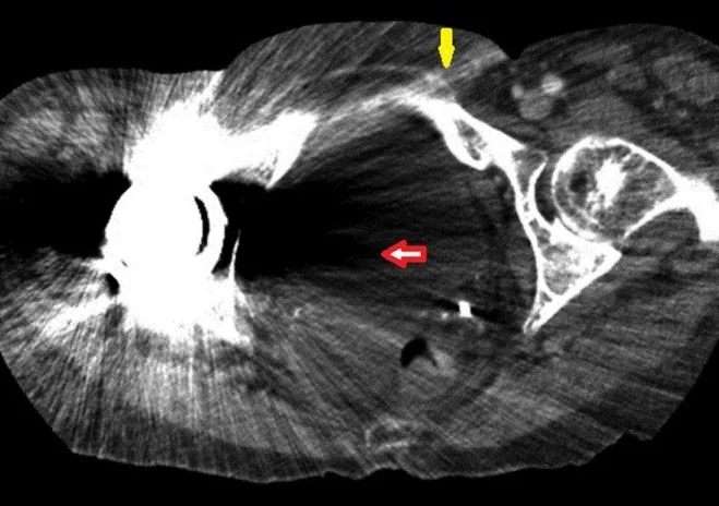
Metal implantlar, cerrahi klipsler, dental materyal ve metalik yabancı cisimler X-ışınını güçlü biçimde zayıflatarak çevre anatomiyi bozan çizgiler, bantlar ve kenar yapaylıkları üretir. Artefaktın şiddeti materyalin bileşimi ve geometrisinin yanı sıra tüp enerjisi, tüp akımı ve rekonstrüksiyon tekniğiyle değişir. Metalden dışarı ışınsal yayılan parlak-koyu çizgiler, foton açlığı ve ışın sertleşmesinin birlikte etkisini yansıtabilir; metal artefakt azaltma (MAR) serisi çevre dokuyu açığa çıkarırken yeni yalancı kenar veya doku da oluşturabilir. Bu nedenle implant çevresindeki kırık, gevşeme, apse veya kitle yalnız MAR görüntüsüne dayanılarak tanımlanmamalı; standart ve düzeltilmiş rekonstrüksiyonlar birlikte okunmalıdır.
Faz ve pencere
- Kontrastsız tanısal
- Metal implantın kendisi çok yüksek atenüasyonlu, çevresindeki yumuşak doku ise ışınsal parlak ve koyu çizgiler, koyu bantlar veya kayıp anatomik detayla bozulmuş görünür. Kontrastsız kemik incelemesi implant, korteks ve çevre kemik ilişkisini gösterir; kontrast fazı artefaktı ortadan kaldırmaz ve bu artefaktın değerlendirilmesi için gerekli değildir.
- Kemik algoritması tanısal
- Keskin kemik rekonstrüksiyonu kortikal sınırı ve implant-kemik arayüzünü gösterir; ancak metal yakınındaki çizgilenme bu algoritmada daha görünür olabilir. Aynı düzeyde standart ve varsa MAR rekonstrüksiyonunu karşılaştırmak yalancı periprostetik açıklığı gerçek gevşemeden ayırmaya yardım eder.
Önerilen pencereler: Kemik (G 1800 / M 400), Yumuşak doku (G 400 / M 40), Mediasten (G 350 / M 50).
BT bulguları
- Metal kaynaklı radyal çizgiler — yüksek atenüasyonlu implanttan komşu kesimlere doğru uzanan açık ve koyu bantlar kemik iliği ile yumuşak doku sınırlarını silebilir.
- Foton açlığı bandı — implantın karşılıklı yüzleri arasında kalan projeksiyonlarda dedektöre az foton ulaşması koyu ve kaba gürültülü alan yaratır.
- İmplant kenarında yalancı genişleme — kısmi hacim ve blooming artefaktları implant çevresindeki sınırların görünümünü bozabilir.
- Periprostetik kemik kaybı belirsizliği — vida veya protez çevresinde görülen koyu halka gerçek gevşemeyi taklit edebilir; standart seriyle doğrulanmalıdır.
- MAR ile açığa çıkan yumuşak doku — düzeltilmiş rekonstrüksiyonda implant çevresi daha okunur olabilir; düzeltme interpolasyonu yapay sınır ekleyebilir.
- MAR algoritmaları yeni artefaktlar oluşturabilir; omur kanalındaki hipodens araknoitise veya kemik çimentosu parçalanmasına benzetilebilir, bu nedenle artefakt düzeltilmemiş seri ile karşılaştırılmalıdır.
- Düzeltme serileri arası uyumsuzluk — yeni görünen çizgi veya odak yalnız MAR görüntüsünde yer alıyorsa gerçek patoloji kabul etmeden önce konvansiyonel görüntüde karşılığını arayın.
Normalde
İmplantlı görüntüde bu sınırların nerede gerçekten bozulduğunu, standart kemik/yumuşak doku rekonstrüksiyonu ile MAR serisinde aynı yerde bulunup bulunmadığını karşılaştırın.
Normal BT ile kıyasla
Aynı bölgenin, kaynak veri setinde patoloji içermediği belirtilen bir BT incelemesini kesit kesit kaydırın; organ sınırlarını açarak anatomiyi hasta görüntüsüyle karşılaştırın. Küçük rastlantısal bulgular tümüyle dışlanmamıştır.
Kaynak: TotalSegmentator v2.01 veri seti (Wasserthal ve ark., Radiology: Artificial Intelligence 2023), CC BY 4.0, vaka s1338; organ sınırları veri setinin otomatik segmentasyonundan, birleştirilerek sadeleştirildi. Görüntüler 2,5 mm kesit aralığına yeniden örneklendi.
Ayırıcı tanı
- Periprostetik kırık
- kortekste gerçek kırık hattı ve kesitler boyunca süren kemik basamağı aranır; tek seride metalden uzanan çizgi yeterli değildir.
- İmplant gevşemesi
- pedikül vidası çevresinde 2 mm'den geniş radyolusent alanlar gevşemeyi düşündürür; kesin tanı için seri görüntülerde implant konum değişikliği gösterilmelidir. MAR kaynaklı yalancı radyolusensi standart rekonstrüksiyonla dışlayın.
- Heterotopik ossifikasyon
- kas ve yumuşak doku içinde korteks/trabekül içeren gerçek kemikleşme oluşur; implanttan yayılan radyal çizgiler kemik matriksi göstermez.
- İntraosseöz litik lezyon
- medüller kemik içinde sınırları anatomik olarak izlenebilen destrüksiyon vardır; metal kaynaklı foton açlığı artefaktı ise düzensizdir ve projeksiyon yönüne bağımlıdır.
Tuzaklar
- MAR serisinde beliren vida çevresi radyolusent çizgi gevşemeyi taklit edebilir; şüpheli arayüzü standart rekonstrüksiyonla karşılaştırın.
- MAR algoritmaları patolojiyi taklit eden ikincil artefaktlar oluşturabilir; gevşeme değerlendirmesinde kortikal ve implant-kemik arayüzü bulgularını standart rekonstrüksiyonla karşılaştırın.
- Koyu çizgi veya bantları apse veya litik lezyon olarak atlayıcı teşhis etmeyin; metal kaynaklı foton açlığı ve ışın sertleşmesi nedeniyle oluşan radyal çizgileri ve koyu bantları tanıtın.
- Geniş görüntü penceresi implant çevresi yumuşak dokuyu görünür kılabilir, ancak kemik korteksindeki ince kırığı gizleyebilir; kemik ve yumuşak doku pencerelerini ayrı okuyun.
Kendini dene
Pedikül vidası çevresindeki radyolusent halka yalnız MAR serisinde görülüyor; standart görüntüde korteks sağlam. En doğru yorum?
Cevabı göster
MAR kaynaklı yalancı radyolusent görünüm. Yalnız MAR serisinde görülen ve standart rekonstrüksiyonda karşılığı olmayan radyolusent halo, düzeltme algoritmasına bağlı artefakttır. Gerçek gevşeme tanısı için standart görüntüde >2 mm radyolusensi ve seri incelemelerde pozisyon değişikliği aranmalıdır.
Kalça protezinden yayılan koyu bant asetabulumun bir bölümünü örtüyor. Bu görünümün temel nedeni hangisi?
Cevabı göster
Foton açlığı ve ışın sertleşmesi. Yoğun metal fotonları zayıflatır ve spektrumu sertleştirir; karşılıklı projeksiyonlarda sinyal yetersizliği rekonstrüksiyonda koyu bant ve çizgilenme oluşturur. Efüzyon eklem aralığındaki sıvıdır; kırık kortikal kesinti, heterotopik ossifikasyon ise yumuşak dokuda kemik matriksi gösterir.
Bu konunun yer aldığı turlar
İlgili konular
Kaynaklar
Bu sayfadaki 39 cümle ve 2 quiz sorusu aşağıdaki kaynaklardan alıntılarla tek tek karşılaştırıldı; 8 cümle bu karşılaştırmada düzeltildi (2026-10-08).
- Dual-Energy Computed Tomography, a New Metal Artifact Reduction Technique for Total Hip Arthroplasty: Is There a Light in the Darkness?pmc.ncbi.nlm.nih.gov
- Inflammatory Pseudotumor After Total Hip Arthroplasty‐An Increasingly Recognized Complicationpmc.ncbi.nlm.nih.gov
- CT Evaluation of Lumbar Interbody Fusion: A Comprehensive Review with an Integrated Framework for Principle-Based Interpretationpmc.ncbi.nlm.nih.gov
- Artificial Intelligence in Intraoperative Imaging and Navigation for Spine Surgery: A Narrative Reviewpmc.ncbi.nlm.nih.gov
- The effects of different implant crown restorative materials on the detection of proximal caries of adjacent teeth in cone-beam computed tomography (CBCT) scans using various machines, metal artifact-reduction (MAR) algorithm, and exposure protocolspmc.ncbi.nlm.nih.gov
- Deep Learning Image Reconstruction for CT: Technical Principles and Clinical Prospects.pubmed.ncbi.nlm.nih.gov
- Dual energy CT in radiotherapy: Current applications and future outlook.pubmed.ncbi.nlm.nih.gov
- Dual-Energy CT in Head and Neck Imaging.pubmed.ncbi.nlm.nih.gov
- Imaging of adverse reactions to metal debris.pubmed.ncbi.nlm.nih.gov
- Advanced MRI Techniques for the Ankle.pubmed.ncbi.nlm.nih.gov
- Imaging update in arthroplasty.pubmed.ncbi.nlm.nih.gov
- Postoperative MRI of the shoulder.pubmed.ncbi.nlm.nih.gov
- Postoperative Spinal CT: What the Radiologist Needs to Know.pubmed.ncbi.nlm.nih.gov
- MR Imaging of Knee Arthroplasty Implants.pubmed.ncbi.nlm.nih.gov
- Postoperative magnetic resonance imaging of the foot and ankle.pubmed.ncbi.nlm.nih.gov
- Metallic Implants in MRI - Hazards and Imaging Artifacts.pubmed.ncbi.nlm.nih.gov
- Dual-Energy Computed Tomography in Cardiothoracic Vascular Imaging.pubmed.ncbi.nlm.nih.gov
- Strategies to Improve Image Quality on Dual-Energy Computed Tomography.pubmed.ncbi.nlm.nih.gov
- Dual-Energy CT in the Acute Setting: Bowel Trauma.pubmed.ncbi.nlm.nih.gov
- Advanced Iterative Metal Artifact Reduction With Intrinsic Spectral Information From Photon-counting Detector Computed Tomography: Improving the Assessment of Metal-bone Interface.pubmed.ncbi.nlm.nih.gov
- Experimental investigation of metal artifact reduction using one-step spectral CT reconstruction.pubmed.ncbi.nlm.nih.gov
- Understanding Photon-Counting CT: Physics, Detector Technology, and Image Reconstructions.pubmed.ncbi.nlm.nih.gov
- Novel High-Atomic-Number Element and Nanoparticle-Based CT Contrast Agents: Advanced Design Strategies and Clinical Translational Barriers.pubmed.ncbi.nlm.nih.gov
- Metal artifact reduction and contrast agent performance as a function of source spectral shape.pubmed.ncbi.nlm.nih.gov
- Effects of recumbency and tube current on image quality of canine elbow computed tomography.pubmed.ncbi.nlm.nih.gov
- Application of streak metal artifact reduction technique in cone-beam computed tomography guided percutaneous transthoracic needle biopsy.pubmed.ncbi.nlm.nih.gov
- Assessment of metal artifacts from titanium wrist prostheses: photon-counting versus energy-integrating detector CT.pubmed.ncbi.nlm.nih.gov
- The Clinical Value of the MAR + Metal Artifact Reduction Algorithm for Postoperative Assessment of Lumbar Internal Fixation.pubmed.ncbi.nlm.nih.gov
- Dual-energy CT in musculoskeletal imaging: technical considerations and clinical applications.pubmed.ncbi.nlm.nih.gov
- Dosimetric impact of titanium, zirconia, and customized subperiosteal dental implants in head and neck radiotherapy: an <i>in vitro</i> study.pubmed.ncbi.nlm.nih.gov
- Nominal material density may not reflect TPS-relevant density for 3D-printed surgical reconstruction implants: An educational case study.pubmed.ncbi.nlm.nih.gov
- Metal Artifact Reduction in Oral Cavity MDCT Using Dental Overlays and the O-MAR Algorithm.pubmed.ncbi.nlm.nih.gov
- Deep learning-based metal artifact reduction in CT for total knee arthroplasty.pubmed.ncbi.nlm.nih.gov
- Photon-Counting Detector CT Applications in Musculoskeletal Radiology.pubmed.ncbi.nlm.nih.gov
- Photon-counting CT: image quality evaluation in patients with tibial plateau fracture treated with metallic osteosynthesis material.pubmed.ncbi.nlm.nih.gov
- Commissioning a single photon counting CT simulator for radiotherapy planning: Methods, challenges, and initial results.pubmed.ncbi.nlm.nih.gov
- Photon-Counting Detector CT-Based Virtual Monoenergetic Imaging With Metal Artifact Reduction for Endodontic Diagnostics.pubmed.ncbi.nlm.nih.gov
- <i>In vitro</i> assessment of CBCT metal artifact reduction algorithms in TMJ prosthetic components.pubmed.ncbi.nlm.nih.gov
- CT Evaluation of Lumbar Interbody Fusion: A Comprehensive Review with an Integrated Framework for Principle-Based Interpretation.pubmed.ncbi.nlm.nih.gov
- Photon-Counting CT in Musculoskeletal Radiology: Technical Principles, Clinical Applications, and Future Directions.pubmed.ncbi.nlm.nih.gov
- Integration of Photon-Counting CT into the Surgical Workflow of Complex Maxillofacial Reconstruction: A Pilot Feasibility Study.pubmed.ncbi.nlm.nih.gov
- Musculoskeletal CT Imaging: State-of-the-Art Advancements and Future Directions.pubmed.ncbi.nlm.nih.gov
- Metal artifact reduction and tumor detection using photon-counting multi-energy computed tomography.pubmed.ncbi.nlm.nih.gov
- AAPM Task Group Report 238: 3D C-arms with volumetric imaging capability.pubmed.ncbi.nlm.nih.gov
- Metal artifacts reduction in computed tomography: A phantom study to compare the effectiveness of metal artifact reduction algorithm, model-based iterative reconstruction, and virtual monochromatic imaging.pubmed.ncbi.nlm.nih.gov
- Optimizing Reconstruction Parameters for Detecting Peripheral In-Stent Restenosis with Photon-Counting Detector CT: A Phantom Study.pubmed.ncbi.nlm.nih.gov
- Clinical benefits and current challenges of photon-counting detector CT in vascular imaging.pubmed.ncbi.nlm.nih.gov
- Joint CT reconstruction of anatomy and implants using a mixed prior model.pubmed.ncbi.nlm.nih.gov
- Broadband and wide-temperature dissipation continuum in a supramolecular damping organohydrogel for motion-tolerant bioelectronics.pubmed.ncbi.nlm.nih.gov
- Biodegradable Magnesium-Based Implants in Sports Orthopedic Surgery: Advances in Alloy Design, Surface Engineering, and Translational Evidence.pubmed.ncbi.nlm.nih.gov
- Hibiscus derived nitrogen doped carbon nanochains for visual pH sensing and catalytic methyl orange degradation.pubmed.ncbi.nlm.nih.gov
- Effect of different artifact reduction methods for quantitative computed tomography measurements of bone mineral density: a pilot study.pubmed.ncbi.nlm.nih.gov
- Binary image acquisition and texture parameter calculation of asphalt pavement based on a U-Net model.pubmed.ncbi.nlm.nih.gov
- Mechanistic Insights into MoS<sub>2</sub> Functionalization via Thiol Groups of 4-Aminothiophenol.pubmed.ncbi.nlm.nih.gov
- Recurrent Giant Cell Tumor of the Lateral Condyle of the Distal Femur Managed by Repeated Extended Curettage and Bone Cementing: A Case Report.pubmed.ncbi.nlm.nih.gov
- Spatiotemporal mapping of osteomyelitis pathogenesis guides the design of functionally graded bone regeneration scaffolds.pubmed.ncbi.nlm.nih.gov
- Dissecting Hypoxia Inducible Factor-Driven High Bone Mass Phenotype in Mice Lacking Osteocytic Von Hippel-Lindau.pubmed.ncbi.nlm.nih.gov
- Use of PET-CT in diagnostic workup of periprosthetic infection of hip and knee joints: significance in detecting additional infectious focus.pubmed.ncbi.nlm.nih.gov
- Recent Advances in Musculoskeletal Radiology: Bridging Innovation and Clinical Application.pubmed.ncbi.nlm.nih.gov
- Case report: Focal heterotopic ossification in paravertebral muscles as a cause of neurogenic lameness in a dog.pubmed.ncbi.nlm.nih.gov
- Stepwise diagnostic algorithm for high-attenuation pulmonary abnormalities on CT.pubmed.ncbi.nlm.nih.gov
- Applications of spectral CT calcium suppression for percutaneous bone tumor ablation procedures.pubmed.ncbi.nlm.nih.gov
- Musculoskeletal infections through direct inoculation.pubmed.ncbi.nlm.nih.gov
- Bone and Joint Infections: The Role of Imaging in Tailoring Diagnosis to Improve Patients' Care.pubmed.ncbi.nlm.nih.gov
- The Role of CT and MR Imaging in Stereotactic Body Radiotherapy of the Spine: From Patient Selection and Treatment Planning to Post-Treatment Monitoring.pubmed.ncbi.nlm.nih.gov
- Multifunctional Ag-Poly( N -isopropylacrylamide/itaconic Acid) Hydrogel Nanocomposites Prepared by Gamma Irradiation for Potential Application as Topical Treatment Dressings.pubmed.ncbi.nlm.nih.gov
- Skin Relevant Biomaterials from Wound Healing, Medical Aesthetics, Flexible Electronics to Artificial Intelligence and Beyond.pubmed.ncbi.nlm.nih.gov
- Utility of Dual-Energy Computed Tomography in Clinical Conundra.pubmed.ncbi.nlm.nih.gov
- Noninvasive Imaging Prior to Biliary Interventions.pubmed.ncbi.nlm.nih.gov
- International Harmonization of Nomenclature and Diagnostic Criteria (INHAND): Non-proliferative and Proliferative Lesions of the Non-human Primate ( M. fascicularis ).pubmed.ncbi.nlm.nih.gov
- Strain-Insensitive Conductive Hydrogel Materials for Motion-Artifact-Free Flexible Bioelectronics.pubmed.ncbi.nlm.nih.gov
- Soft tissue recurrence in giant cell tumor of Bone: A comprehensive review of pathogenesis, imaging features, and clinical management.pubmed.ncbi.nlm.nih.gov
- Soft, Flexible, and Stretchable Platforms for Tissue-Interfaced Bioelectronics.pubmed.ncbi.nlm.nih.gov
- Congenital Scoliosis: A Comprehensive Review of Diagnosis, Management, and Surgical Decision-Making in Pediatric Spinal Deformity-An Expanded Narrative Review.pubmed.ncbi.nlm.nih.gov
- Patient-Specific 3-Dimensional-Printed Orthopedic Implants and Surgical Devices Are Potential Alternatives to Conventional Technology But Require Additional Characterization.pubmed.ncbi.nlm.nih.gov
- Application of 3D-printed porous prosthesis for the reconstruction of infectious bone defect with concomitant severe soft tissue lesion: a case series of 13 cases.pubmed.ncbi.nlm.nih.gov
- State-of-the-Art Imaging Techniques in Metastatic Spinal Cord Compression.pubmed.ncbi.nlm.nih.gov
- Metal artifact reduction of orthopedics metal artifact reduction algorithm in total hip and knee arthroplasty.pubmed.ncbi.nlm.nih.gov
- 3D-printed hip prostheses with regenerative integration: a state-of-the-art comprehensive review.pubmed.ncbi.nlm.nih.gov
- Transformer-based models in dentistry: a systematic review.pubmed.ncbi.nlm.nih.gov
- Power-drill fluoroscopy-controlled technique as an alternative to navigation-assisted pedicle screw placement: A propensity score-matched retrospective cohort study.pubmed.ncbi.nlm.nih.gov
- Occult cement plug causing femoral canal obstruction during nailing after TKA: a case report.pubmed.ncbi.nlm.nih.gov
- Implications of Size and Gender for Implant Fit and Coverage in Total Ankle Replacement.pubmed.ncbi.nlm.nih.gov
- Preoperative planning technique for anterior pelvis percutaneous intramedullary screw fixation.pubmed.ncbi.nlm.nih.gov
- The braincase anatomy of <i>Simosaurus gaillardoti</i> (Diapsida: Sauropterygia) revealed with X-ray micro-computed tomography.pubmed.ncbi.nlm.nih.gov
- Orthopaedic and non-orthopaedic applications of a single-energy iterative metal artefact reduction technique and other metal artefact reduction techniques explained.pubmed.ncbi.nlm.nih.gov
- Optimising complementary soft tissue synchrotron X-ray microtomography for reversibly-stained central nervous system samples.pubmed.ncbi.nlm.nih.gov
- Commentary: an eye on PET quantification.pubmed.ncbi.nlm.nih.gov
- High-affinity alphavbeta3 integrin targeted optical probe as a new imaging biomarker for early atherosclerosis: initial studies in Watanabe rabbits.pubmed.ncbi.nlm.nih.gov
- Patterns of injury and violence in Yaoundé Cameroon: an analysis of hospital data.pubmed.ncbi.nlm.nih.gov
- Postoperative Stiffness After Upper Limb Surgery: Prevention and Management.pubmed.ncbi.nlm.nih.gov
- Diagnostic and prognostic relevance of plain radiographs for periprosthetic joint infections of the hip: a literature review.pubmed.ncbi.nlm.nih.gov
- Advances in Bone Joint Imaging-Metal Artifact Reduction.pubmed.ncbi.nlm.nih.gov
- Novel marker-based cone-beam CT registration for enhanced accuracy in dental implant planning.pubmed.ncbi.nlm.nih.gov
- Applications of SPECT/CT in the Evaluation of Spinal Pathology: A Review.pubmed.ncbi.nlm.nih.gov
- Management of Bone Loss in Revision Total Knee Arthroplasty: An International Consensus Symposium.pubmed.ncbi.nlm.nih.gov
- Updates in postoperative imaging modalities following musculoskeletal surgery.pubmed.ncbi.nlm.nih.gov
- Dual-energy CT with metal artifact reduction for post-EVAR CTA surveillance: a prospective study with image quality analysis.pubmed.ncbi.nlm.nih.gov
- Innovative approaches in precision radiation oncology: advanced imaging technologies and challenges which shape the future of radiation therapy.pubmed.ncbi.nlm.nih.gov
- 2.5-mm articulated endoluminal forceps using a compliant mechanism.pubmed.ncbi.nlm.nih.gov
- The value of dual-energy CT virtual monoenergetic imaging and metal-artifacts reduction system after metal plate correction of congenital funnel chest in child: a single center retrospective study.pubmed.ncbi.nlm.nih.gov
- Photon-Counting Computed Tomography: Experience in Musculoskeletal Imaging.pubmed.ncbi.nlm.nih.gov
- Optimal combination periprosthetic vasculature visualization and metal artifact reduction by spectral computed tomography using virtual monoenergetic images in total hip arthroplasty.pubmed.ncbi.nlm.nih.gov
- Radon-Guided Wavelet-Domain Attention U-Net for Periodic Artifact Suppression in Brain MRI.pubmed.ncbi.nlm.nih.gov
- The role of imaging in extremity sarcoma surgery.pubmed.ncbi.nlm.nih.gov
- Osteoporosis Diagnostics in Dental Radiology (Panoramic Radiograph): The Mini Osteoporosis Pre-Screening (MOPS).pubmed.ncbi.nlm.nih.gov
- Murine model of high bone mass osteogenesis imperfecta exhibits bone matrix hyper-mineralization, misaligned mineral crystals, and altered osteoblast differentiation.pubmed.ncbi.nlm.nih.gov
- Imaging of pediatric musculoskeletal trauma: Age-specific considerations and challenges.pubmed.ncbi.nlm.nih.gov
- Local Regulatory T-cell Failure as a Potential Mechanism of the Halo Sign in Spinal Instrumentation: A Mechanistic Hypothesis.pubmed.ncbi.nlm.nih.gov
- Effect of blending Jersey and Holstein-Friesian milk on Cheddar cheese processing, composition, and quality.pubmed.ncbi.nlm.nih.gov
- Long-term safety and effectiveness of the "OptEase" vena cava filter.pubmed.ncbi.nlm.nih.gov
- Advancements in biomaterials and bioactive solutions for lumbar spine fusion cages: Current trends and future perspectives.pubmed.ncbi.nlm.nih.gov
- Advances in TCP-Modified PMMA Bone Cements: Relating Microstructure to Mechanical, Biological and Functional Performance-Systematic Review.pubmed.ncbi.nlm.nih.gov
- Transforming spinal surgery with innovations in biologics and additive manufacturing.pubmed.ncbi.nlm.nih.gov
- Postoperative MRI Artifact Burden After Carbon Fiber-Reinforced PEEK Versus Titanium Instrumentation in Metastatic Spinal Disease.pubmed.ncbi.nlm.nih.gov
Eğitim amaçlıdır; hasta başında tanı ve tedavi kararının yerine geçmez. İçerik yapay zekâ destekli bir süreçle güncel literatüre göre cümle cümle kaynaklarla karşılaştırıldı, ancak uzman radyolog incelemesinden geçmedi. Klinik kararlarda güncel kılavuzları, kurum protokollerini ve radyoloji raporunu esas alın.