Özet
Superior oftalmik ven trombozu, orbital venöz dönüşü bozarak ağrılı proptoz, kemozis ve göz hareket kısıtlılığına neden olabilen seyrek fakat görmeyi tehdit eden bir durumdur. Kontrastlı orbital BT venin genişlemesini ve lümen içi dolum defektini gösterirken kavernöz sinüs, sinüsler ve orbitadaki enfeksiyon odağını da araştırır. Tromboz septik veya aseptik olabilir ve karotis-kavernöz fistül ile kavernöz sinüs trombozu aynı klinik/görüntüleme alanında yer alır. Venöz tıkanıklığın ya da kaynağının atlanması görme ve intrakraniyal komplikasyonların fark edilmesini geciktirebilir. Superior oftalmik ven trombozu araştırılırken kavernöz sinüs tutulumu, karşı taraf ve enfeksiyon odağı birlikte değerlendirilmelidir.
Faz ve pencere
- Kontrastlı tanısal
- Kontrastlı görüntülemede superior oftalmik ven dilatasyonu ve intraluminal dolum defekti aranır; orbital yağdaki inflamatuvar değişiklikler, sino-orbital enfeksiyon bulguları ve kavernöz sinüs tutulumu da değerlendirilir. Damar opaklaşması çekim zamanlaması ve protokole bağlıdır; tromboz kuşkusunda BT anjiyografi veya MR venografi bulgularla birlikte değerlendirilir.
- Kontrastsız
- Kontrastsız görüntülerde genişlemiş ven veya hiperdens trombüs görülebilir ancak bulgu duyarlı değildir ve ven lümenindeki dolum defektini güvenilir biçimde dışlayamaz; kontrastsız seri eşlik eden kanama/kemiği değerlendirir, trombozu tek başına doğrulamak için yeterli değildir.
Önerilen pencereler: Yumuşak doku (G 400 / M 40), Kemik (G 1800 / M 400), Anjiyo (G 600 / M 150).
BT bulguları
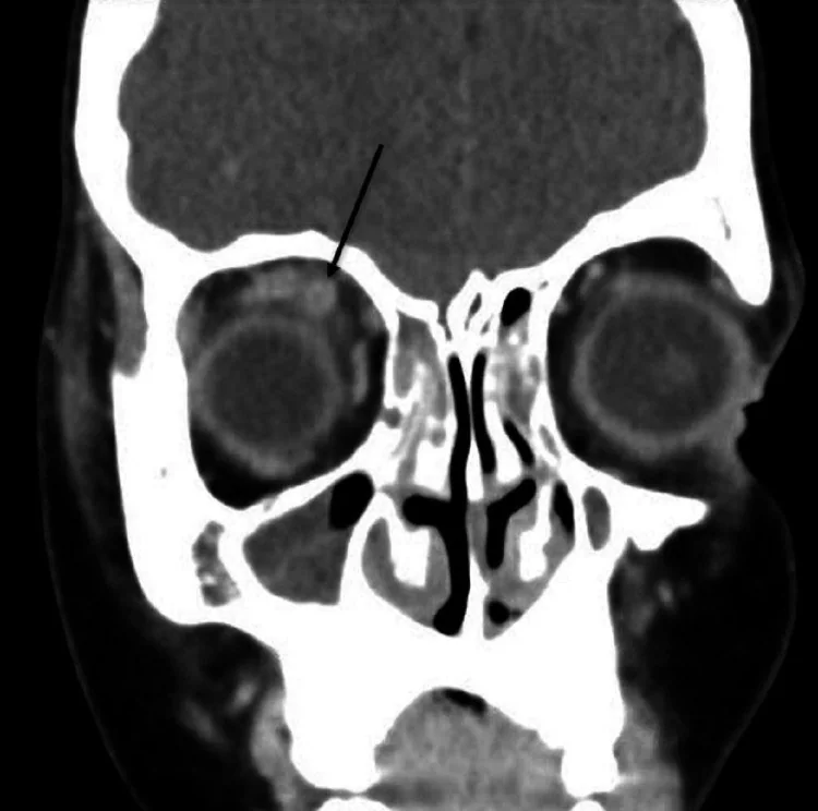
- Superior oftalmik ven, normalden geniş ve kıvrımlı tübüler yapı halinde optik sinir boyunca kavernöz sinüse uzanır.
- Kontrastlı incelemede venin opaklaşan lümeni içinde merkezî veya eksantrik dolum defekti trombüsü düşündürür.
- Ven çevresinde orbital yağ ödemi, rektus kaslarında dolgunluk ve proptoz venöz çıkış engelinin ikincil bulguları olabilir.
- Superior oftalmik ven trombozu kavernöz sinüs trombozuyla eşzamanlı bulunabilir; kavernöz sinüs tutulumu veya trombüs uzanımı ayrıca değerlendirilmelidir.
- Paranazal sinüslerde sinüzit veya opasifikasyon ve eşlik eden orbital yağ inflamasyonu septik kaynağı destekleyebilir.
- Arteriyel fazda erken venöz veya kavernöz sinüs dolumu izlenirse tromboz yerine akım şantı (karotis-kavernöz fistül) düşünülmeli ve damar yapısı buna göre yorumlanmalıdır.
- İzole trombozda diğer kavernöz sinüsün ve intrakraniyal venöz yapıların görünümü karşılaştırma için yararlıdır.
- Dolum defekti kuşkusunda kontrastlı görüntülerde superior oftalmik venin opaklaşmasını ve lümenindeki dolum defektini değerlendirin.
Normalde
Normal superior oftalmik ven ince, kontrastla homojen dolan tübüler damar olarak optik sinirin üst-medial komşuluğundan kavernöz sinüse ilerler; lümeninde kalıcı dolum kusuru bulunmaz. Aynı aksiyel/koronal düzlemde sağ ve sol venin kalibresini, lümen opaklaşmasını ve kavernöz sinüsle devamlılığını karşılaştır; damar dışı orbital ödemi ayrıca değerlendir.
Normal BT ile kıyasla
Aynı bölgenin, kaynak veri setinde patoloji içermediği belirtilen bir BT incelemesini kesit kesit kaydırın; organ sınırlarını açarak anatomiyi hasta görüntüsüyle karşılaştırın. Küçük rastlantısal bulgular tümüyle dışlanmamıştır.
Kaynak: TotalSegmentator v2.01 veri seti (Wasserthal ve ark., Radiology: Artificial Intelligence 2023), CC BY 4.0, vaka s0849; organ sınırları veri setinin otomatik segmentasyonundan, birleştirilerek sadeleştirildi. Görüntüler 2,5 mm kesit aralığına yeniden örneklendi.
Ayırıcı tanı
- Karotis-kavernöz fistül
- superior oftalmik ven geniş olabilir ancak arteriyel fazda erken venöz dolum ve kavernöz sinüs opaklaşması şantı destekler.
- Kavernöz sinüs trombozu
- sinüs içinde düzensiz dolum defekti, genişleme ve sinüzit/baş-boyun enfeksiyon odağı saptanabilir; trombüs oftalmik vene uzanabilir.
- Orbital selülit
- yağ dokusu ve ekstraoküler kaslarda inflamasyon görülür; belirgin ven içi dolum defekti olmadan da konjesyon yapabilir.
- Superior oftalmik ven variksi
- venöz yapı genişler fakat lümen genellikle açıktır; morfoloji ve dolum kusurunun yokluğu trombozdan ayırır.
- Orbital kitle
- komşu ven basıya uğrayabilir veya yer değiştirebilir; ven boyunca merkezî intraluminal trombüs görünümü beklenen bulgu değildir.
Tuzaklar
- Geniş superior oftalmik veni otomatik olarak tromboz sayma; fistül ve venöz variks de genişleme oluşturur, lümen dolumunu ve arteriyel zamanlamayı kontrol et.
- Tek kesitte kısmi opaklaşmama akım/zamanlama etkisi olabilir; çok düzlemli görüntülerde damarı baştan sona izle ve teknik yeterliliği değerlendir.
- Kontrastsız BT'de trombüsün görünmemesi trombozu dışlamaz; klinik şüphe sürüyorsa kontrastlı venöz görüntüleme veya MR venografi gerekir.
- Septik ven trombozu tek başına izole orbita bulgusu olmayabilir; paranazal sinüsleri ve kavernöz sinüsleri gözden kaçırma.
Kendini dene
Kontrast sonrası genişlemiş superior oftalmik ven içinde dolum defekti görülmesi en çok hangi tanıyı destekler?
Cevabı göster
Ven lümeninde tromboz. Kontrast sonrası genişlemiş superior oftalmik ven içindeki dolum defekti, tromboz tanımında kullanılan radyolojik bulgudur.
Kavernöz sinüste arteriyel fazda erken kontrastlanma ve genişlemiş superior oftalmik ven birlikte görülüyor. En olası tanı nedir?
Cevabı göster
Karotis-kavernöz fistül. Arteriyelize kavernöz sinüs ve genişlemiş superior oftalmik ven karotis-kavernöz fistülü düşündürür; serebral anjiyografi kesin tanı ve sınıflamada altın standarttır.
İlgili konular
Kaynaklar
Bu sayfadaki 37 cümle ve 2 quiz sorusu aşağıdaki kaynaklardan alıntılarla otomatik olarak karşılaştırıldı; 12 cümle bu karşılaştırmada düzeltildi (2026-10-09).
- Strep pyogenes infection causing rapid onset orbital cellulitis, superior ophthalmic vein thrombosis, and necrotizing scleritis with dehiscence of extraocular muscle following strabismus surgerypmc.ncbi.nlm.nih.gov
- Superior Ophthalmic Vein Thrombosis in Trinidad and Tobago: A Case Seriespmc.ncbi.nlm.nih.gov
- Superior Ophthalmic Vein Thrombosis Associated With Acute Suppurative Parotitis and Masticator Space Cellulitis: A Case Report and Review of the Literaturepmc.ncbi.nlm.nih.gov
- Pediatric Lemierre syndrome of rhinosinusogenic origin complicated by bilateral cavernous sinus thrombosis: A case reportpmc.ncbi.nlm.nih.gov
- Superior Ophthalmic Vein Thrombosis: A Case Report in a Previously Healthy Methicillin-Resistant Staphylococcus aureus -Positive Patientpmc.ncbi.nlm.nih.gov
- [Isolated superior ophthalmic vein thrombosis due to tamoxifen].pubmed.ncbi.nlm.nih.gov
- Impact of superior ophthalmic vein thrombosis: a case series and literature review.pubmed.ncbi.nlm.nih.gov
- Blindness caused by septic superior ophthalmic vein thrombosis in a Lemierre Syndrome variant.pubmed.ncbi.nlm.nih.gov
- Isolated Superior Ophthalmic Vein Thrombosis.pubmed.ncbi.nlm.nih.gov
- Combined central retinal vein, central retinal artery and cilioretinal artery occlusion with ischemic macular hole secondary to severe orbital cellulitis after black fly bite.pubmed.ncbi.nlm.nih.gov
- Oculomotor synkinesis: an uncommon sequela of paediatric cavernous sinus thrombosis.pubmed.ncbi.nlm.nih.gov
- Unilateral isolated superior ophthalmic vein thrombosis.pubmed.ncbi.nlm.nih.gov
- Superior ophthalmic vein thrombosis: What radiologist and clinician must know?pubmed.ncbi.nlm.nih.gov
- Right Superior Ophthalmic Vein Thrombosis Induced by Pansinusitis.pubmed.ncbi.nlm.nih.gov
- Superior ophthalmic vein thrombosis post manual carotid compression for indirect carotid-cavernous fistula.pubmed.ncbi.nlm.nih.gov
- Superior Ophthalmic Vein Thrombosis Induced by Orbital Cellulitis: An Ophthalmic Emergency.pubmed.ncbi.nlm.nih.gov
- Isolated Superior Ophthalmic Vein Thrombosis Associated with Orbital Cellulitis: Case Report.pubmed.ncbi.nlm.nih.gov
- Superior Ophthalmic Vein Thrombosis: The Role of Anticoagulation.pubmed.ncbi.nlm.nih.gov
- Superior ophthalmic vein thrombosis: A rare complication of Graves' orbitopathy.pubmed.ncbi.nlm.nih.gov
- Multisystem infection with cavernous sinus thrombosis in seropositive rheumatoid arthritis: a clinico-radiological case report.pubmed.ncbi.nlm.nih.gov
- Carotid Cavernous Fistula: A Case Report of a Rare Diagnosis and Presentation.pubmed.ncbi.nlm.nih.gov
- Spontaneous Resolution of a Bilateral Barrow Type D Indirect Carotid-Cavernous Fistula: A Rare Case Report and Literature Review.pubmed.ncbi.nlm.nih.gov
- Strep pyogenes infection causing rapid onset orbital cellulitis, superior ophthalmic vein thrombosis, and necrotizing scleritis with dehiscence of extraocular muscle following strabismus surgery.pubmed.ncbi.nlm.nih.gov
- Utility of High-Dose Steroids in Treatment-Resistant Carotid-Cavernous Fistulas: A Case Report.pubmed.ncbi.nlm.nih.gov
- Refractory cavernous sinus thrombophlebitis complicated by brain abscess and infectious hydrocephalus: a case report and literature review.pubmed.ncbi.nlm.nih.gov
- Glucocorticoids in the treatment of non-infectious superior ophthalmic vein thrombosis - Three cases and a review of the literature.pubmed.ncbi.nlm.nih.gov
- Intracranial venous thrombosis imaging by anatomical location: an educational review with mimics and diagnostic pitfalls.pubmed.ncbi.nlm.nih.gov
- Multimodality image fusion guidance in the microcatheter navigation to cerebrospinal arteriovenous fistula: Report of the two cases.pubmed.ncbi.nlm.nih.gov
- A Rare Case of Concurrent Carotid Cavernous Fistula and Orbital Cellulitis: Challenging Diagnosis and Divergent Management.pubmed.ncbi.nlm.nih.gov
- Magnetic resonance imaging of the orbit, Part 2: Characterization of orbital pathologies.pubmed.ncbi.nlm.nih.gov
- Imaging Evaluation of Acute Traumatic Brain Injury.pubmed.ncbi.nlm.nih.gov
- Multimodal imaging in neuro-ophthalmology.pubmed.ncbi.nlm.nih.gov
- Post-traumatic Carotid-Cavernous Fistula Following Penetrating Orbital Injury: A Case Report and Literature Review.pubmed.ncbi.nlm.nih.gov
- Subarachnoid haemorrhage associated with septic cavernous sinus thrombosis secondary to odontogenic maxillary sinusitis.pubmed.ncbi.nlm.nih.gov
- Imaging for Pediatric Head and Neck Emergencies: Critical Insights.pubmed.ncbi.nlm.nih.gov
- Cerebral venous disorders: clinical presentation, diagnostic strategy, and contemporary management.pubmed.ncbi.nlm.nih.gov
- The Developing Human Sphenoid Bone: Linking Embryological Development to Adult Morphology.pubmed.ncbi.nlm.nih.gov
- Ophthalmic Complications After Dental Procedures: Scoping Review.pubmed.ncbi.nlm.nih.gov
- Anatomical Variations of the Foramen Ovale and Trigeminal Neuralgia.pubmed.ncbi.nlm.nih.gov
- Superior Ophthalmic Vein Thrombosis Associated With Acute Suppurative Parotitis and Masticator Space Cellulitis: A Case Report and Review of the Literature.pubmed.ncbi.nlm.nih.gov
- Superior Ophthalmic Vein Thrombosis in Trinidad and Tobago: A Case Series.pubmed.ncbi.nlm.nih.gov
- Imaging of cerebrovascular complications from blunt skull base trauma.pubmed.ncbi.nlm.nih.gov
- A superior ophthalmic vein thrombosis following an acute pansinusitis: A case report.pubmed.ncbi.nlm.nih.gov
- Superior and inferior ophthalmic veins thrombosis with cavernous sinus meningioma.pubmed.ncbi.nlm.nih.gov
- Central nervous system vascular malformations: A clinical review.pubmed.ncbi.nlm.nih.gov
- Endovascular treatment of carotid cavernous sinus fistula: A systematic review.pubmed.ncbi.nlm.nih.gov
- Long-term safety and effectiveness of the "OptEase" vena cava filter.pubmed.ncbi.nlm.nih.gov
- A diagnostic dilemma in pediatric proptosis-from suspected malignancy to benign orbital cavernous venous malformation: a case report.pubmed.ncbi.nlm.nih.gov
- Ocular Misalignment and Motility Limitation in Orbital Tumors.pubmed.ncbi.nlm.nih.gov
- Traumatic Orbital Apex Syndrome With Acute Orbital Compartment Syndrome Secondary to Facial Fractures: A Case Report.pubmed.ncbi.nlm.nih.gov
- Systolic propulsion of the eyeballs in severe tricuspid regurgitation: a case series and review of the literature.pubmed.ncbi.nlm.nih.gov
- Guidelines for standard operation of imaging modalities in orbital diseases (2024).pubmed.ncbi.nlm.nih.gov
- Evaluation and management of cerebral venous thrombosis.pubmed.ncbi.nlm.nih.gov
- Pediatric Lemierre syndrome of rhinosinusogenic origin complicated by bilateral cavernous sinus thrombosis: A case report.pubmed.ncbi.nlm.nih.gov
- Split-bolus, single-phase contrast enhanced CT: a one-stop shop for invasive fungal sinusitis.pubmed.ncbi.nlm.nih.gov
- Ocular Complications Following Vaccination for COVID-19: A One-Year Retrospective.pubmed.ncbi.nlm.nih.gov
- Septic cerebral venous sinus thrombosis from maxillary sinusitis with concomitant right-sided sigmoid sinus hypoplasia: A case report and review of the literature.pubmed.ncbi.nlm.nih.gov
- Orbital apex syndrome in a man with rhino-ocular cerebral mucormycosis: a case report.pubmed.ncbi.nlm.nih.gov
- Lemierre-like syndrome presenting with extensive intracranial and central venous thrombosis without pulmonary embolism: illustrative case.pubmed.ncbi.nlm.nih.gov
- Orbital tuberculosis mimicking a vascular emergency: A case report of superior ophthalmic vein engorgement.pubmed.ncbi.nlm.nih.gov
- Clinical Outcomes of Severe Rhinosinusitis Complicated with Cavernous Sinus Syndrome.pubmed.ncbi.nlm.nih.gov
- Rhinosinusitis: Evidence and experience - 2024.pubmed.ncbi.nlm.nih.gov
- Critical infections in the head and neck: A pictorial review of acute presentations and complications.pubmed.ncbi.nlm.nih.gov
- Successful management of cavernous sinus thrombosis without anticoagulation: a neuro-ophthalmologic case report.pubmed.ncbi.nlm.nih.gov
- Unusual Presentation of Coronary Artery Fistula in Capillary Malformation Arteriovenous Malformation 2 Syndrome: A Case Report.pubmed.ncbi.nlm.nih.gov
- Low-flow carotid-cavernous fistula associated with cavernous sinus thrombosis: An ophthalmic presentation.pubmed.ncbi.nlm.nih.gov
- Technical Refinement of a Hybrid Surgical and Endovascular Approach for Cavernous Dural Arteriovenous Fistula with Leptomeningeal Venous Reflux: Direct Superficial Middle Cerebral Vein Exposure for Transvenous Embolization.pubmed.ncbi.nlm.nih.gov
- Rare sphenoid dural arteriovenous fistula treated with microsurgery: illustrative case.pubmed.ncbi.nlm.nih.gov
- The infraorbital vein: embryology, anatomy, histology, variations, and clinical relevance.pubmed.ncbi.nlm.nih.gov
- Non-Thrombotic Filling Defects in Cerebral Veins and Sinuses: When Normal Structures Mimic a Disease.pubmed.ncbi.nlm.nih.gov
- Direct superior ophthalmic vein puncture for cavernous sinus dural arteriovenous fistula with dual orbital venous outflow: illustrative case.pubmed.ncbi.nlm.nih.gov
- Ocular Color Doppler Ultrasound (OCDUS) in Diagnosis and Monitoring of Ophthalmological, Cerebrovascular and Systemic Diseases: A Narrative Review.pubmed.ncbi.nlm.nih.gov
- How I do it: Simpson grade I resection in a medial and inner ridge sphenoid wing meningioma.pubmed.ncbi.nlm.nih.gov
- Endovascular treatment of an intraosseous dural arteriovenous fistula of the greater wing of the sphenoid: A case report and literature review.pubmed.ncbi.nlm.nih.gov
- Co-occurring scalp arteriovenous malformation and dural arteriovenous fistula with diploic vein involvement: illustrative case.pubmed.ncbi.nlm.nih.gov
- Neuroimaging for neurovascular complications of traumatic brain injury.pubmed.ncbi.nlm.nih.gov
- Bilateral Carotid-Cavernous Fistula Following Traumatic Fall: A Case Report.pubmed.ncbi.nlm.nih.gov
- Optimizing imaging in orbital vascular anomalies: a review on matching modality to pathology for effective diagnosis and treatment planning.pubmed.ncbi.nlm.nih.gov
Eğitim amaçlıdır; hasta başında tanı ve tedavi kararının yerine geçmez, tıbbi tavsiye değildir. Metinler yapay zekâ desteğiyle yazıldı ve otomatik bir adımda kaynak alıntılarıyla karşılaştırıldı; bu bir tıbbi doğrulama değildir ve içerik uzman radyolog incelemesinden geçmedi. Klinik kararlarda güncel kılavuzları, kurum protokollerini ve radyoloji raporunu esas alın.