Özet
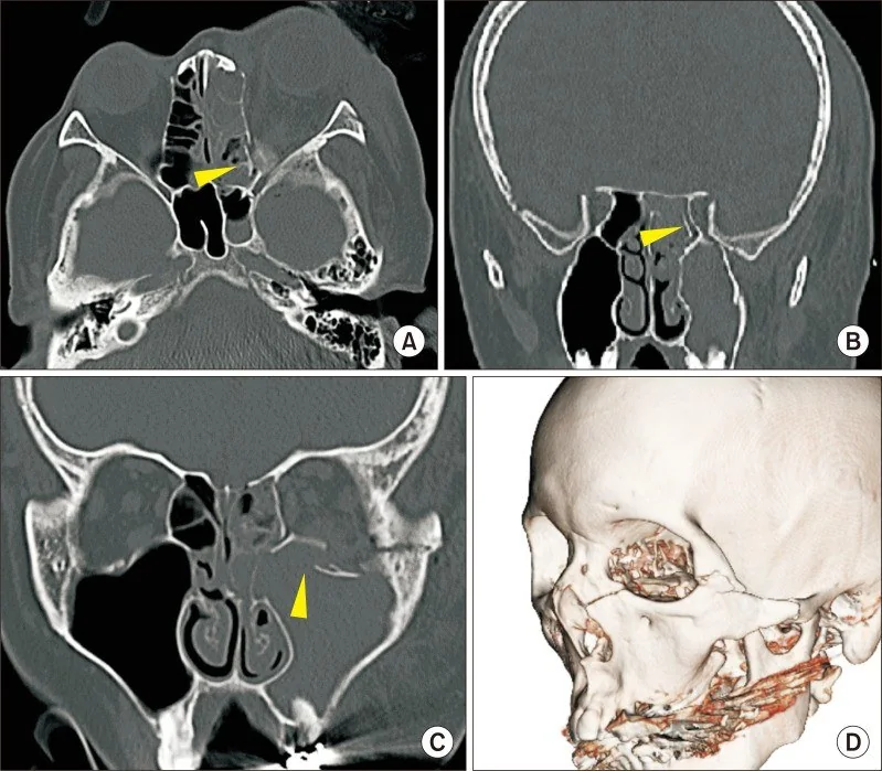
Künt kraniyofasiyal travma sonrası görme kaybı veya afferent pupilla kusuru olan hastada optik kanal kırığı, optik sinir sıkışmasıyla birlikte bulunabilir. İnce kesit kontrastsız BT kemik penceresinde kanal duvarındaki kırığı ve kanala uzanan kemik fragmanını gösterir; sinirin işlevsel hasarını BT tek başına dışlayamaz. Bulguların saptanması yaralanmanın anatomik düzeyini ve eşlik eden kafa tabanı travmasını ortaya koyar. Kanal içi bası veya eşlik eden hematom gözden kaçarsa görmeyi tehdit eden lezyonun ciddiyeti eksik raporlanabilir.
Faz ve pencere
- Kontrastsız tanısal
- Optik kanal korteksinde keskin kesinti, kanal lümenine uzanan kemik fragmanı ve komşu sfenoid/ön kafa tabanı kırığı aranır; kanal içi veya orbital apeks hematomu yumuşak doku dansitesinde olabilir. Optik kanalın kemik yapısı kontrastsız BT'de değerlendirilebilir.
- Kemik algoritması tanısal
- İnce kesit kemik rekonstrüksiyonlarında optik kanalın tüm çevresinde kortikal süreklilik, kanal çapındaki asimetri ve fragmanın optik sinir hattına yönelimi değerlendirilir.
Önerilen pencereler: Kemik (G 1800 / M 400), Yumuşak doku (G 400 / M 40), Beyin (G 80 / M 40).
BT bulguları
- Optik kanal korteksinde lineer kırık izlenir; kırık hattı sfenoid kemik yapısına veya komşu kafa tabanı bölgelerine uzanabilir.
- İntrakonal kemik fragmanı — kanal lümenine doğru yer değiştirmiş, optik sinir güzergâhına temas edebilen kemik parçası.
- Kanal kalibresinde daralma — kırık kenarlarının içe basısı veya fragman nedeniyle lümenin asimetrik küçülmesi.
- Orbital apeks hematomu — posterior intrakonal alanda sinir ve çevre yağ planlarını silen sınırlı yumuşak doku yoğunluğu.
- Optik sinir kılıfı çevresinde kan — sinir çevresindeki normal yağ halkasının kaybolması ve kılıf komşuluğunda yoğunlaşma.
- Kanal kırığının komşu sfenoidal yapılar (örneğin çatı, üst kenar veya foramen rotundum) ve kafa tabanı ile ilişkili olması.
- Pnömosefali veya intrakraniyal kanama — komşu kafa tabanı hasarını ve travmanın kapsamını gösteren eşlikçi bulgular.
Normalde
BT'de optik kanal kırığı ve kırık fragmanının optik sinire basısı değerlendirilebilir. Karşı tarafla eş düzeyde incelemede kanal duvarı süreksizliği, lümen çapı asimetrisi ve sinir çevresi yumuşak doku yoğunluk değişimleri değerlendirilir.
Normal BT ile kıyasla
Aynı bölgenin, kaynak veri setinde patoloji içermediği belirtilen bir BT incelemesini kesit kesit kaydırın; organ sınırlarını açarak anatomiyi hasta görüntüsüyle karşılaştırın. Küçük rastlantısal bulgular tümüyle dışlanmamıştır.
Kaynak: TotalSegmentator v2.01 veri seti (Wasserthal ve ark., Radiology: Artificial Intelligence 2023), CC BY 4.0, vaka s0849; organ sınırları veri setinin otomatik segmentasyonundan, birleştirilerek sadeleştirildi. Görüntüler 2,5 mm kesit aralığına yeniden örneklendi.
Ayırıcı tanı
- İzole indirekt travmatik optik nöropati
- klinik görme kusuru bulunabilir; BT'de kanal kırığı veya belirgin sinir basısı görülmeyebilir.
- Optik sinir kılıfı hematomu
- kanal kırığı olmadan sinir kılıfı çevresinde yumuşak doku yoğunluğu ve sinir çevresinde silinme yapar.
- Orbital apeks kontüzyonu
- apeks yağında düzensiz bulanıklık vardır, ancak kanal korteksinde kesinti şart değildir.
- Sfenoid sinüs duvarı kırığı
- sinüs çevresinde kırık izlenir; optik kanal korteksinin tutulumu ayrıca gösterilmelidir.
Tuzaklar
- Normal BT, indirekt travmatik optik nöropatiyi dışlamaz; mikrovasküler ve aksonal hasar BT çözünürlüğünün altında kalabilir.
- İnce kesitli BT ve çok düzlamlı yeniden yapılandırmalar (MPR), optik kanal ve komşu yapıların detaylı değerlendirilmesinde kullanılır.
- Kanal komşuluğundaki sfenoid kırığını optik kanal içine uzanmış kabul etmeyin; kanal korteksini aksiyel ve koronal planda ayrı izleyin.
Kendini dene
Yüz travması sonrası görme azalmış. İnce kesit BT'de kanal korteksi kesintili ve fragman lümene uzanıyor. En olası bulgu nedir?
Cevabı göster
Optik kanal kırığı. Kanal korteksindeki kesinti ve lümene uzanan fragman doğrudan optik kanal yaralanmasını gösterir. Diğer seçenekler farklı orbital duvar veya sinüs bölgelerindedir ve kanal lümenini bozmaz.
Travma sonrası akut görme kaybı ve mutlak afferent pupilla defekti (RAPD) gelişmiş; orbita ve kafa içi ince kesit BT normal bulunmuş. En uygun yaklaşım nedir?
Cevabı göster
İndirekt travmatik optik nöropati düşünülmeli ve yakın klinik/radyolojik takip planlanır. İndirekt travmatik optik nöropati (İTON), mekanik kopma veya kanal kırığı olmadan, iletilen kuvvetlere bağlı iskemi/strese bağlı gelişir ve akut BT'de yapısını bozan bir lezyon göstermeyebilir [S3, S7]. Optik sinirde görünür anatomik bozulma olmadan İTON gelişebileceğinden BT'de yapısal bozulma görülmemesi bu olasılığı dışlamaz; gözlem seçilirse seri klinik ve elektrofizyolojik izlem yapılmalı, görme kötüleşirse acil tekrar BT veya MR değerlendirilmelidir.
Kaynaklar
Bu sayfadaki 37 cümle ve 2 quiz sorusu aşağıdaki kaynaklardan alıntılarla tek tek karşılaştırıldı; 11 cümle bu karşılaştırmada düzeltildi (2026-10-08).
- A case report and literature review: Cerebral spinal fluid leak after COVID nasopharyngeal swab testingpmc.ncbi.nlm.nih.gov
- Minimally invasive resection of a spheno-orbital tumor: Review of surgical techniques and a case reportpmc.ncbi.nlm.nih.gov
- Indirect traumatic optic neuropathy in a high-risk diabetic adolescent: A rare clinical presentationpmc.ncbi.nlm.nih.gov
- Application of 3D Slicer-based three-dimensional visual surgical planning in endoscopic optic canal decompression surgery: a prospective randomised controlled studypmc.ncbi.nlm.nih.gov
- Anatomical variations of lamina papyracea, internal carotid artery, and optic canal dehiscence and protrusion: a computed tomography retrospective cohort studypmc.ncbi.nlm.nih.gov
- Optic nerve monitoring.pubmed.ncbi.nlm.nih.gov
- Traumatic optic neuropathy: a review.pubmed.ncbi.nlm.nih.gov
- Blowout fractures - clinic, imaging and applied anatomy of the orbit.pubmed.ncbi.nlm.nih.gov
- Radiological assessment of the lateral osteotomy line-lacrimal system distance on three-dimensional models.pubmed.ncbi.nlm.nih.gov
- [Traumatic optic neuropathy: a review of the literature in the light of personal experiences].pubmed.ncbi.nlm.nih.gov
- Radiological signs of periorbital trauma - the Singapore experience.pubmed.ncbi.nlm.nih.gov
- CT in the Evaluation of Acute Injuries of the Anterior Eye Segment.pubmed.ncbi.nlm.nih.gov
- Traumatic optic neuropathy: facial CT findings affecting visual acuity.pubmed.ncbi.nlm.nih.gov
- Post-traumatic optic neuropathy: our surgical and medical protocol.pubmed.ncbi.nlm.nih.gov
- A 16-Year Study on Incidence and Progression of Diseased Sphenoethmoidal (Onodi) Cell.pubmed.ncbi.nlm.nih.gov
- Optic Neuropathy with Delayed Onset After Trauma: Case Report and Review of the Literature.pubmed.ncbi.nlm.nih.gov
- Traumatic tympanic bulla fracture.pubmed.ncbi.nlm.nih.gov
- Mitochondrial Triglyceride Dysregulation in Optic Nerves Following Indirect Traumatic Optic Neuropathy.pubmed.ncbi.nlm.nih.gov
- Indirect traumatic optic neuropathy in a high-risk diabetic adolescent: A rare clinical presentation.pubmed.ncbi.nlm.nih.gov
- Application of 3D Slicer-based three-dimensional visual surgical planning in endoscopic optic canal decompression surgery: a prospective randomised controlled study.pubmed.ncbi.nlm.nih.gov
- Traumatic Orbital Apex Syndrome With Acute Orbital Compartment Syndrome Secondary to Facial Fractures: A Case Report.pubmed.ncbi.nlm.nih.gov
- Multimodal imaging in neuro-ophthalmology.pubmed.ncbi.nlm.nih.gov
- Neurolymphomatosis: A Comprehensive Review of Clinical and Imaging Features.pubmed.ncbi.nlm.nih.gov
- Guidelines for standard operation of imaging modalities in orbital diseases (2024).pubmed.ncbi.nlm.nih.gov
- A case report and literature review: Cerebral spinal fluid leak after COVID nasopharyngeal swab testing.pubmed.ncbi.nlm.nih.gov
- A CT radiomics nomogram predicts visual acuity improvement in patients with indirect traumatic optic neuropathy following optic canal decompression.pubmed.ncbi.nlm.nih.gov
- Neurosurgical robot-assisted transnasal endoscopic surgery in orbital-related diseases.pubmed.ncbi.nlm.nih.gov
- Conference Proceedings: Select Abstracts Presented at the 2025 Advocate Aurora Scientific Day.pubmed.ncbi.nlm.nih.gov
- Long-term safety and effectiveness of the "OptEase" vena cava filter.pubmed.ncbi.nlm.nih.gov
- Commentary: an eye on PET quantification.pubmed.ncbi.nlm.nih.gov
- Operative management of a nail wound projectile to the skull base with unusual trajectory: illustrative case.pubmed.ncbi.nlm.nih.gov
- Craniofacial speargun injuries: report of three cases, literature review and proposed management guidelines for maxillo-facial surgeons.pubmed.ncbi.nlm.nih.gov
- Critical anatomical variants in preoperative computed tomography of paranasal sinuses in a tertiary care center: a cross-sectional study.pubmed.ncbi.nlm.nih.gov
- Comparison of non-transport disc and conventional distraction osteogenesis for mandibular segmental defect reconstruction.pubmed.ncbi.nlm.nih.gov
- Characterization of human skeletal stem cells in closed and open tibia fractures: a single center pilot study.pubmed.ncbi.nlm.nih.gov
- Morphological and Epidemiological Analysis of the Sphenoid Sinus Based on Computed Tomography and Magnetic Resonance Imaging: A Narrative Review.pubmed.ncbi.nlm.nih.gov
- A refined radiological classification of anterior clinoid process pneumatization.pubmed.ncbi.nlm.nih.gov
- 3D-exoscopic extradural Hakuba-Dolenc approach with manipulation of pneumatized anterior clinoid process for the prevention of cerebrospinal fluid leakage: how I do it.pubmed.ncbi.nlm.nih.gov
- Endoscopic Endonasal Optic Nerve Decompression for Traumatic Optic Neuropathy: A Systematic Review.pubmed.ncbi.nlm.nih.gov
- Minimally Invasive Surgery for the Treatment of Lumbar Spinal Stenosis: A Comprehensive Review.pubmed.ncbi.nlm.nih.gov
- Unravelling the Unseen: A Case Series Exploring the Enigmas of Traumatic Optic Neuropathy.pubmed.ncbi.nlm.nih.gov
- The orthopaedic management of osteopetrosis in paediatric populations: a narrative review of the literature.pubmed.ncbi.nlm.nih.gov
- Totally endoscopic trans‑superior articular process lumbar interbody fusion: A case series on the development and preliminary evaluation of an innovative minimally invasive lumbar spine surgical technique.pubmed.ncbi.nlm.nih.gov
- Context-Aware Filtering of Unstructured Radiology Reports by Anatomical Region.pubmed.ncbi.nlm.nih.gov
- An oculoplastic perspective on Transorbital Neuroendoscopic Surgery: Anatomical corridors and clinical applications.pubmed.ncbi.nlm.nih.gov
- Technical Feasibility of the Endoscopic Endonasal Approach for Clipping an Ophthalmic Artery Aneurysm With an Origin Lateral to the Optic Nerve: A Case Report.pubmed.ncbi.nlm.nih.gov
- Response of the Rat Eye to Acute and Chronic Optic Nerve Pressure Modifications.pubmed.ncbi.nlm.nih.gov
- Profile and Outcomes of Neurofibromatosis Type 1 (NF1) Patients in a Tertiary Government Hospital in the Philippines: A Retrospective Descriptive Study.pubmed.ncbi.nlm.nih.gov
- A Case of Extensive Pneumocephalus Following an Electric Scooter Accident.pubmed.ncbi.nlm.nih.gov
- The Impact of a Horse Kick: A Systematic Review of Horse-Related Maxillofacial Trauma.pubmed.ncbi.nlm.nih.gov
- Sphenoid Temporal Meningioma With Granular Cell Features and Extracranial Infratemporal Extension.pubmed.ncbi.nlm.nih.gov
- Head of the Syrian Hamster (<i>Mesocricetus auratus</i>): Planar Anatomy with Correlative Micro-Computed Tomography and Magnetic Resonance Imaging.pubmed.ncbi.nlm.nih.gov
- Plastinated sectional anatomy applied in CT and MRI studies of the body of the crab-eating fox (Cerdocyon thous).pubmed.ncbi.nlm.nih.gov
- Impact of In-House 3D-Printed Models on Re-Operation Rates and Volumetric Precision in Orbital Floor Reconstruction: A Comparative Study.pubmed.ncbi.nlm.nih.gov
- Spontaneous Resolution of a Bilateral Barrow Type D Indirect Carotid-Cavernous Fistula: A Rare Case Report and Literature Review.pubmed.ncbi.nlm.nih.gov
- Visual function and cerebrospinal fluid volume along the optic nerve in patients with spontaneous cerebrospinal fluid rhinorrhea: a cross-sectional study.pubmed.ncbi.nlm.nih.gov
- A window into eye movement dysfunction following mTBI: A scoping review of magnetic resonance imaging and eye tracking findings.pubmed.ncbi.nlm.nih.gov
- Sphenoid sinus hyperpneumatization: anatomical variants, molecular blueprints, and AI-augmented roadmaps for skull base surgery.pubmed.ncbi.nlm.nih.gov
- Surgical Measures Improving Functional Limitations of the Masticatory System, Aesthetic Deficits, and Skeletal Malformations in Neurofibromatosis Type 1-associated Hemifacial Diffuse Plexiform Neurofibroma Complicated by Rapidly Growing Midfacial Peripheral Nerve Sheath Tumor.pubmed.ncbi.nlm.nih.gov
- Etiologic spectrum and predictors of visual acuity in non-glaucomatous optic atrophy.pubmed.ncbi.nlm.nih.gov
- The Dental Operating Microscope in Endodontics: A Narrative Review of Clinical Applications, Supporting Evidence, and Limitations.pubmed.ncbi.nlm.nih.gov
- The cavernous sinus: An anatomic study with clinical implication.pubmed.ncbi.nlm.nih.gov
- Sphenoid sinus anatomical variants and pathologies: pictorial essay.pubmed.ncbi.nlm.nih.gov
- Anatomical variations of lamina papyracea, internal carotid artery, and optic canal dehiscence and protrusion: a computed tomography retrospective cohort study.pubmed.ncbi.nlm.nih.gov
- Imaging of cerebrovascular complications from blunt skull base trauma.pubmed.ncbi.nlm.nih.gov
- Clinical analysis of 66 cases with orbital apex syndrome: a case series.pubmed.ncbi.nlm.nih.gov
- Malpositioned orbital implants: indications for implant removal and secondary reconstructive techniques.pubmed.ncbi.nlm.nih.gov
- Penetrating stab injury extending from the temporal region to the orbital apex and ethmoidal cells - A unique case report and review of the literature.pubmed.ncbi.nlm.nih.gov
- Trauma Unmasking Pituitary Apoplexy: Coexisting Epidural Hematoma and Sellar Mass - A Case Report with Selected Cases from the Literature.pubmed.ncbi.nlm.nih.gov
- Unfolding tale of post-traumatic globe luxation into the ethmoid sinus: an unusual encounter in ophthalmology.pubmed.ncbi.nlm.nih.gov
- Pulsatile Tinnitus: A Comprehensive Clinical Approach to Diagnosis and Management.pubmed.ncbi.nlm.nih.gov
- Magnetic resonance imaging of the cranial nerves in congenital, traumatic, and vascular diseases: a pictorial essay.pubmed.ncbi.nlm.nih.gov
- Neoplasms and tumor-like lesions of the sellar region: imaging findings with correlation to pathology and 2021 WHO classification.pubmed.ncbi.nlm.nih.gov
- Enhancement of Bond Strength, Degree of Conversion and Biocompatibility In Vitro Using a Hollow Glass Fiber Post With an Optical Illuminator in the Restoration of Endodontically Treated Teeth.pubmed.ncbi.nlm.nih.gov
- Comparison of the Efficacy and Prognosis of Different Treatment Strategies for Spinal Tuberculosis Patients Undergoing Vertebral Augmentation Surgery by Mistake.pubmed.ncbi.nlm.nih.gov
- Visual Loss Following Indirect Trauma: A Diagnostic Dilemma.pubmed.ncbi.nlm.nih.gov
- Mild Traumatic Brain Injury as a Heterogeneous Neurological Disorder: Definition, Epidemiology, Neurobiology, and Clinical Course.pubmed.ncbi.nlm.nih.gov
- 40 Years of diffusion MRI in the brain: From history to emerging frontiers.pubmed.ncbi.nlm.nih.gov
- Ultrastructural perspectives on the enteric nervous system in Parkinson's disease.pubmed.ncbi.nlm.nih.gov
- Presence of Antinuclear Antibodies in the Cerebrospinal Fluid and in the Serum in a Patient With Sjögren's Syndrome and Systemic Lupus: A Case Report.pubmed.ncbi.nlm.nih.gov
- Chronological Analysis of Visual Field and Optic Nerve Thickness for the Early Diagnosis of Bilateral Chronic Open-Angle Glaucoma With a Masquerading Growing Pituitary Tumor.pubmed.ncbi.nlm.nih.gov
- Pre-treatment and post-treatment nasopharyngeal carcinoma imaging: imaging updates, pearls and pitfalls.pubmed.ncbi.nlm.nih.gov
- Intracranial pressure, lateral sinus patency, and jugular ultrasound hemodynamics in patients with venous pulsatile tinnitus.pubmed.ncbi.nlm.nih.gov
- Brazilian Society of Otology task force - Otosclerosis: evaluation and treatment.pubmed.ncbi.nlm.nih.gov
- Penetrating Orbital and Intracranial Injury Due to a Ballpoint Pen: A Case Report.pubmed.ncbi.nlm.nih.gov
- Mycotic Sphenopalatine Sinusitis With Concurrent Compression of the Optic Nerves and Chiasm and Severe Visual Impairment in A Horse.pubmed.ncbi.nlm.nih.gov
- 3D Segmentation Algorithms for Computerized Tomographic Imaging: a Systematic Literature Review.pubmed.ncbi.nlm.nih.gov
- Anatomic, Radiographic, and Computed Tomographic Study of the Skull in the Persian Squirrel (Sciurus anomalus).pubmed.ncbi.nlm.nih.gov
- Primary Intraosseous Meningioma of the Sphenoid-Temporal Bone with Single-Stage, 3D-Planned Cranioplasty: Case Report.pubmed.ncbi.nlm.nih.gov
- Development of a novel ultraviolet-polymerized hydrogel as an orbital implant.pubmed.ncbi.nlm.nih.gov
- International Olympic Committee (IOC) consensus paper on sports-related ophthalmology issues in elite sports.pubmed.ncbi.nlm.nih.gov
- 2.5-mm articulated endoluminal forceps using a compliant mechanism.pubmed.ncbi.nlm.nih.gov
- What's that smell? A pictorial review of the olfactory pathways and imaging assessment of the myriad pathologies that can affect them.pubmed.ncbi.nlm.nih.gov
- Intraoperative CT evaluation of repositioning accuracy in zygomaticomaxillary complex fractures: 1-point versus multiple-point fixation.pubmed.ncbi.nlm.nih.gov
- A Reproducible 3D Classification of Orbital Morphology Derived from CBCT and FBCT Segmentation.pubmed.ncbi.nlm.nih.gov
- Efficacy of 3D-printed patient specific implant for orbital wall fracture repair in a series of 40 patients.pubmed.ncbi.nlm.nih.gov
- Structure-function correlates in anterior visual pathway lesions: a systematic review.pubmed.ncbi.nlm.nih.gov
- Surgical precision and clinical outcomes of 3D preformed titanium mesh in orbital blowout fracture reconstruction: a comparative study from Thailand.pubmed.ncbi.nlm.nih.gov
- Imaging the brain: diagnosis aided by structural features on neuroimaging studies.pubmed.ncbi.nlm.nih.gov
- Orbital apex disorders: Imaging findings and management.pubmed.ncbi.nlm.nih.gov
- Incidentally discovered asymptomatic extrasinusal mucocele of the sphenoid greater wing: A case report and review of differential diagnoses.pubmed.ncbi.nlm.nih.gov
- Evaluation of Ultra-High Molecular Weight Polyethylene (UHMWPE) Implants in Orbital Fracture Reconstruction: A Multicenter Cohort Study.pubmed.ncbi.nlm.nih.gov
- Effect of Taking a Break From Cochlear-Implant Use for Resolving Facial-Nerve Stimulation: A Case Series.pubmed.ncbi.nlm.nih.gov
- Advanced Digital Workflow for Lateral Orbitotomy in Orbital Dermoid Cysts: Integration of Point-of-Care Manufacturing and Intraoperative Navigation.pubmed.ncbi.nlm.nih.gov
- Feasibility of navigation-assisted endoscopic transnasal optic nerve decompression for the treatment of traumatic optic neuropathy in patients with midfacial fractures.pubmed.ncbi.nlm.nih.gov
- Endoscope-Assisted or Skin-Approach Osteosynthesis of Mandibular Condylar Fracture-A Comparison.pubmed.ncbi.nlm.nih.gov
- Radiologic Characterization of Invasive Fungal Infections of the Paranasal Sinuses and Skull Base: A Prospective Analysis.pubmed.ncbi.nlm.nih.gov
- Orbital floor repair and three-point fixation of zygomaticomaxillary complex fractures to restore orbital morphological features and ocular functions: a retrospective observational cohort study.pubmed.ncbi.nlm.nih.gov
- Guidelines for Orbital Defect Assessment and Patient-Specific Implant Design: Introducing OA<sup>2</sup> (Orbital Assessment Algorithm).pubmed.ncbi.nlm.nih.gov
- CT evaluation of the relationship between optic canal, anterior clinoid process, optic strut, caroticoclinoid foramen, and dimensions of sella turcica based on sphenoid sinus pneumatization patterns.pubmed.ncbi.nlm.nih.gov
- Surgical stabilization of rib fractures (SSRF): the WSES and CWIS position paper.pubmed.ncbi.nlm.nih.gov
- Minimally invasive resection of a spheno-orbital tumor: Review of surgical techniques and a case report.pubmed.ncbi.nlm.nih.gov
- Pediatric triad of craniofacial fibrous dysplasia, Chiari malformation type I and syringomyelia: a case report.pubmed.ncbi.nlm.nih.gov
- Brazilian Society of Otology task force - cochlear implant ‒ recommendations based on strength of evidence.pubmed.ncbi.nlm.nih.gov
- Clinician-Guided Deep Learning Segmentation of Skull Base Pneumatization on Computed Tomography Using 3D Slicer and MONAI Label.pubmed.ncbi.nlm.nih.gov
Eğitim amaçlıdır; hasta başında tanı ve tedavi kararının yerine geçmez. İçerik yapay zekâ destekli bir süreçle güncel literatüre göre cümle cümle kaynaklarla karşılaştırıldı, ancak uzman radyolog incelemesinden geçmedi. Klinik kararlarda güncel kılavuzları, kurum protokollerini ve radyoloji raporunu esas alın.