
Özet
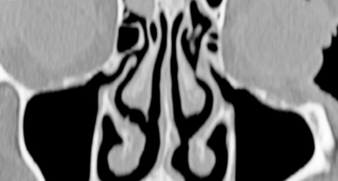
Agger nasi hücresi en anterior etmoid hava hücrelerinden biridir; frontal resesin ön sınırında, orta konkanın ön tutunma bölgesine komşu yer alır. Üstündeki ve çevresindeki frontal reses hücreleri frontal sinüs drenaj yolunun yönünü değiştirip daraltabilir. İnce kesit BT ve özellikle koronal-sagittal rekonstrüksiyonlar hücreleri, frontal ostiumu ve resesin çevre sınırlarını cerrahi öncesi gösterir. Frontal sinüs ve resese cerrahi erişim, hücrelerin isimlendirilmesinden ziyade agger nasi hücresinin pnömatizasyon derecesi ile belirlenir; bu nedenle çoklu düzlem BT'de kapsamlı değerlendirmenin yapılması isimlendirmeden daha kritiktir. Tek başına frontal reses hücresi varlığının frontal sinüzit gelişimi üzerinde anlamlı bir etkisi yoktur.
Okuma sırası
- Frontal sinüs ostiumu ve reses. Sagittal kesitte frontal sinüs ostiumundan aşağı inen hava yolunu bulun; çıkışın ön etmoiddeki devamını takip edin.
- Agger nasi hücresi. Koronal düzlemde orta konkanın ön tutunmasını belirleyip hemen önündeki kortike hücreyi agger nasi olarak tanımlayın.
- Frontal reses hücreleri. Agger nasi'nin üstünde ve arkasında kalan hücreleri tek tek izleyerek frontal reses hücresi olup olmadığını ve drenaj yolunu nereye ittiğini değerlendirin.
- Frontal drenaj yolu ve sinüs. Son olarak frontal ostiumda hava geçişini, sinüs mukozasını ve frontal sinüste eşlik eden opasifikasyonu inceleyin.
Faz ve pencere
- Kontrastsız tanısal
- Agger nasi hücresi frontal resesin ön sınırını oluşturan en anterior etmoid hücredir ve frontal beak ile birlikte resesi sınırlar; üstte supraorbital, suprabuller veya frontal bullar hücreler reses ve sinüs çıkışına komşu yer alır. Mukozal kalınlaşma veya sıvı-opasifikasyon drenaj yolunu daraltabilir; anatomi için kontrastsız ince kesit yeterlidir. Mukozal kalınlaşma ya da opasifikasyon hava kolonunu daraltabilir; kontrastlı faz gerekmez.
- Kontrastsız tanısal
- Kontrastsız ince kesit BT’nin koronal ve sagittal kemik rekonstrüksiyonlarında frontal beak, orta konka tutunması, etmoid bulla ve kafa tabanı sınırları izlenerek hücrenin frontal drenaj yoluna göre konumu belirlenir.
Önerilen pencereler: Kemik (G 1800 / M 400), Yumuşak doku (G 400 / M 40).
BT bulguları
- Agger nasi hücresi — orta konkanın ön tutunma bölgesinin önünde ve frontal resesin anteriorunda yer alan en ön etmoid hücre.
- Supra agger, suprabuller ve frontal septal hücreler gibi frontal reses varyantları, drenaj yolunu medial, posterior veya lateral yönde kaydırabilir; bu durum cerrahi planlamada kritik öneme sahiptir.
- Frontal çıkış daralması — hücre hacmi ya da eşlik eden mukozal kalınlaşma frontal ostium-reses hava kolonunu kısmen kapatabilir.
- Frontal sinüs ostiumu ve reses devamlılığı — sagittal planda sinüsten aşağıya doğru hava yolunun kesintisizliği değerlendirilir.
- Frontal sinüs mukozal hastalığı — reses daralmasının proksimalinde mukozal kalınlaşma veya sıvı-opasifikasyon eşlik edebilir; varyanttan ayrı tarif edilir.
Ölçütler ve sınıflamalar
- International Frontal Sinus Anatomy Classification (IFAC)
- Hücreleri frontal sinüs drenaj yoluna göre konumlandırır: anterior grup (agger nasi, supra agger, supra agger frontal), posterior grup (supra bulla, supra bulla frontal, supraorbital etmoid) ve medial grup (frontal septal) olarak adlandırılır. Agger nasi anterior gruptadır; eşlik eden hücreler konumlarına göre ayrıca isimlendirilir.
Normalde
Normal frontal reses hava kolonu frontal ostiumdan öne doğru uzanır; agger nasi hücresi ve diğer frontal reses hücreleri bu yolun sınırlarını belirleyen komşu ayrı kortike pnömatik boşluklar olarak izlenir. Karşı tarafı aynı koronal ve sagittal düzeyde karşılaştırıp orta konka ön tutunması, frontal beak, etmoid bulla ve drenaj yolunun açıklığına bakın.
Ayırıcı tanı
- Frontal sinüs hipoplazisi
- frontal sinüs hacmi küçüktür; reses önündeki ayrı kortike agger nasi hücresinden farklı olarak sinüsün kendisi gelişmemiş görünür.
- Frontal sinüs mukoseli
- frontal sinüste opak ekspansil boşluk ve düzgün kemik yeniden şekillenmesi yapar; agger nasi hücresi hava içeren ve reses önünde kalan bir hücredir.
- Etmoid bulla
- orta meatusun daha posteriorundaki belirgin ön etmoid hücredir; agger nasi'nin ön ve alt sınır komşuluğunda değildir.
- Frontal sinüs retansiyon kisti
- sinüs iç yüzünden kubbe biçimli yumuşak doku yoğunluğu olarak yükselir, kortike etmoid hücre oluşturmaz.
Tuzaklar
- Agger nasi hücresini frontal sinüsün alt bölümü sanmayın; orta konka ön tutunmasına ve frontal beak'e göre konumunu koronal-sagittal düzlemlerde doğrulayın.
- Frontal resesin karmaşık hücrelerini tek bir agger nasi etiketi altında toplamayın; ostium çevresindeki her ayrı hücrenin drenaj yoluna göre konumunu tarif edin.
- Hücre varlığını frontal sinüzit için yeterli kabul etmeyin; hava yolu daralması ve sinüs mukozal opasifikasyonunu ayrı ayrı gösterin.
Kendini dene
Orta konka ön tutunmasının önünde, frontal resesin anteriorunda ayrı kortike hava hücresi görülüyor. Adı nedir?
Cevabı göster
Agger nasi hücresi. Agger nasi, frontal resesin ön sınırında ve orta konkanın ön tutunmasına komşu en anterior etmoid hücredir. Etmoid bulla daha posteriordadır; Onodi posterior etmoid-sfenoid komşuluğunda, Haller ise orbita tabanının medialinde yer alır.
Agger nasi üstündeki hücre frontal drenaj yolunu posteriora itiyor. Bu ilişki hangi değerlendirme çerçevesine uyar?
Cevabı göster
IFAC hücre konumlandırması. IFAC frontal reses hücrelerini drenaj yoluna göre anterior, posterior ve medial gruplarda adlandırır. Keros olfaktör fossa derinliğini sınıflar; Lund-Mackay sinüs opasifikasyonunu puanlar; Onodi sfenoid üst-lateralindeki posterior etmoid hücredir.
Kaynaklar
Bu sayfadaki 39 cümle ve 2 quiz sorusu aşağıdaki kaynaklardan alıntılarla tek tek karşılaştırıldı; 2 cümle bu karşılaştırmada düzeltildi (2026-10-08).
- The Frontal Sinus and Frontal Recess: Anatomical, Radiological and Surgical Conceptspmc.ncbi.nlm.nih.gov
- Flying through congested airspaces: imaging of chronic rhinosinusitispmc.ncbi.nlm.nih.gov
- The Bifurcated Frontal Sinus.pubmed.ncbi.nlm.nih.gov
- Multidetector CT of the Nasal Cavity and Paranasal Sinuses Variations in 73 Patients.pubmed.ncbi.nlm.nih.gov
- Agger Nasi Cells Versus Lacrimal Cells and Uncinate Bullae in Cone-Beam Computed Tomography.pubmed.ncbi.nlm.nih.gov
- Anatomic variants of the paranasal sinuses in patients with recurrent acute rhinosinusitis.pubmed.ncbi.nlm.nih.gov
- Causes of failure in endoscopic frontal sinus surgery in chronic rhinosinusitis patients.pubmed.ncbi.nlm.nih.gov
- Sinonasal Anatomical Variants: CT and Endoscopy Study and Its Correlation with Extent of Disease.pubmed.ncbi.nlm.nih.gov
- Correlation between the extent of pneumatization of Agger agger Nasi nasi cells and the anterior-to-posterior length of the frontal recess: A a computer-assisted anatomical study.pubmed.ncbi.nlm.nih.gov
- Influence of opacification in the frontal recess on frontal sinusitis.pubmed.ncbi.nlm.nih.gov
- Mapping the frontal sinus ostia using virtual endoscopy.pubmed.ncbi.nlm.nih.gov
- Computed tomography analysis of frontal cell prevalence according to the International Frontal Sinus Anatomy Classification.pubmed.ncbi.nlm.nih.gov
- Is there any relationship between the frontal cell and the Agger nasi cell and the localization of the anterior ethmoid artery?pubmed.ncbi.nlm.nih.gov
- Impact of residual frontal recess cells on frontal sinusitis after endoscopic sinus surgery.pubmed.ncbi.nlm.nih.gov
- Osteomeatal Complex: A Study of Its Anatomical Variation Among Patients Attending North Bengal Medical College and Hospital.pubmed.ncbi.nlm.nih.gov
- Comparison Between Diagnostic Nasal Endoscopy and Plain Computed Tomography of the Paranasal Sinuses in Chronic Rhinosinusitis.pubmed.ncbi.nlm.nih.gov
- Clinical and Radiological Significance of Anatomical Variations in Paranasal Sinuses: A Retrospective CT-Based Study.pubmed.ncbi.nlm.nih.gov
- Multidimensional Morphology of the Ethmoid Roof and Anterior Ethmoidal Artery: A CT-Based Analysis and Proposal of the Akcan Classification.pubmed.ncbi.nlm.nih.gov
- Radiological Assessment of Frontal Recess Cells and Their Association with Frontal Sinusitis Among Omani Subjects: A Single-Center Computed Tomography-Based Study.pubmed.ncbi.nlm.nih.gov
- Episodic cluster headache with suspected giant cell arteritis, sinusitis-related headache, and dietary triggers: a case report.pubmed.ncbi.nlm.nih.gov
- Current Concepts in Frontal Sinus Fracture Management.pubmed.ncbi.nlm.nih.gov
- Sinus pneumatisation and Keros type as predictors of anterior ethmoidal artery configuration: a 3D-reconstructed computed tomography analysis.pubmed.ncbi.nlm.nih.gov
- Association of sphenoid sinus pneumatization patterns, Onodi cells, and rostrum pneumatization with natural ostium location: A cross-sectional CT study.pubmed.ncbi.nlm.nih.gov
- Assessment of Paranasal Sinus Growth with 3D Volumetric Measurements and the Effect of Anatomic Variations on Sinus Volume in a Pediatric Population.pubmed.ncbi.nlm.nih.gov
- CT Analysis of Variations in the Medial Maxillary Wall Relative to the Medial Orbital Wall: Implications for Surgical Risk Stratification from an Endoscopic Perspective.pubmed.ncbi.nlm.nih.gov
- Pre-FESS Imaging of Paranasal Sinuses and Nasal Cavity: Using Multi-detector Computed Tomography (MDCT) in Understanding Normal Anatomy and Anatomical Variations: Tips and Tricks.pubmed.ncbi.nlm.nih.gov
- Side-Specific Concordance Between Nasal Endoscopy and Computed Tomography Severity in Pediatric Chronic Rhinosinusitis.pubmed.ncbi.nlm.nih.gov
- Anatomical Variations Favouring Antrochoanal Polyp Genesis.pubmed.ncbi.nlm.nih.gov
- The maxillary sinus: physiology, development and imaging anatomy.pubmed.ncbi.nlm.nih.gov
- Frontal Sinus Irrigation After Draf 2a: Carolyn's Window Versus Angled Frontal Sinusotomy.pubmed.ncbi.nlm.nih.gov
- Tomographic assessment of anatomical boundaries in endoscopic frontal sinusotomy: retrospective radiologic analysis of Draf IIA and IIB approaches.pubmed.ncbi.nlm.nih.gov
- Physiology and pathophysiology of respiratory mucosa of the nose and the paranasal sinuses.pubmed.ncbi.nlm.nih.gov
- Long-term safety and effectiveness of the "OptEase" vena cava filter.pubmed.ncbi.nlm.nih.gov
- Morphological and Epidemiological Analysis of the Sphenoid Sinus Based on Computed Tomography and Magnetic Resonance Imaging: A Narrative Review.pubmed.ncbi.nlm.nih.gov
- Impact of RAMPA Therapy on Nasal Cavity Expansion and Paranasal Drainage: Fluid Mechanics Analysis, CAE Simulation, and a Case Study.pubmed.ncbi.nlm.nih.gov
- Rhinosinusitis: Evidence and experience - 2024.pubmed.ncbi.nlm.nih.gov
- Unusual Manifestations of Sinonasal Osteomas: A Narrative Review.pubmed.ncbi.nlm.nih.gov
- Anatomical Variations in Nasal Cavities and Paranasal Sinuses on Computed Tomography in Chronic Rhinosinusitis: Implications for Diagnosis and Treatment.pubmed.ncbi.nlm.nih.gov
- Clinical anatomy of the paranasal sinuses and its terminology.pubmed.ncbi.nlm.nih.gov
- Large-Scale Three-Dimensional CT Mapping of the Frontal Sinus Drainage Pathway: Anatomical Relationships and Surgical Validation.pubmed.ncbi.nlm.nih.gov
- Association of Frontal Recess Anatomy and Uncinate Process Variations With Frontal Sinusitis: A CT-Based Observational Study.pubmed.ncbi.nlm.nih.gov
- Analysis of the frontal recess pneumatization pattern in patients with chronic frontal sinusopathy.pubmed.ncbi.nlm.nih.gov
- Computed Tomographic Study of Frontal Sinus Cells and Opacification as Per the International Frontal Sinus Anatomy Classification.pubmed.ncbi.nlm.nih.gov
- Does Frontal Recess Cell Variation Associate with the Development of Frontal Sinusitis? A Narrative Review.pubmed.ncbi.nlm.nih.gov
- Evaluation of the Frequency of Anatomic Variations of the Paranasal Sinus Region by Using Multidetector Computed Tomography: A Hospital-Based Cross-Sectional Study.pubmed.ncbi.nlm.nih.gov
- Paranasal Sinus Hypoplasia and Sinonasal Anatomical Variants in Cystic Fibrosis Adult Patients: A Computed- Tomography-Based Volumetric Comparison with Healthy Controls.pubmed.ncbi.nlm.nih.gov
- Do Variations in Frontal Recess Anatomy Predispose to Mucocele Formation?pubmed.ncbi.nlm.nih.gov
- Ethmoid pneumatization and a large frontal-orbital-ethmoid mucocele.pubmed.ncbi.nlm.nih.gov
- Operative Corridors in Endoscopic Skull Base Tumor Surgery.pubmed.ncbi.nlm.nih.gov
- Magnetic resonance imaging of the orbit, Part 2: Characterization of orbital pathologies.pubmed.ncbi.nlm.nih.gov
- The Lamella Ostium Extent Mucosa (LOEM) system: a new classification and pilot study for endoscopic sinus surgery.pubmed.ncbi.nlm.nih.gov
- Inflammatory Modifications in Paranasal Sinuses and Ostiomeatal Complex Anatomical Variations in Jet Aircraft Pilots: A Computed Tomography Study.pubmed.ncbi.nlm.nih.gov
- Correlation between the Opacification Degree of Paranasal Sinuses on CT, Clinical Symptoms and Anatomical Variations of the Nose and Paranasal Sinuses in Patients with Chronic Rhinosinusitis.pubmed.ncbi.nlm.nih.gov
- Comprehensive review on endonasal endoscopic sinus surgery.pubmed.ncbi.nlm.nih.gov
- Anatomical Variations of the Nasal Cavities and Paranasal Sinuses: A Systematic Review.pubmed.ncbi.nlm.nih.gov
- Small ethmoid mucocele with intracranial extension associated with fibrous dysplasia: A rare case report.pubmed.ncbi.nlm.nih.gov
- Endoscopic visualization of CT-confirmed accessory maxillary ostium: a retrospective study.pubmed.ncbi.nlm.nih.gov
- Computed Tomographic Study of Remarkable Anatomic Variations in Paranasal Sinus Region and their Clinical Importance - A Retrospective Study.pubmed.ncbi.nlm.nih.gov
- Comparison of Low-Dose Non-contrast CT in Detecting Anatomical and Surgically Important Variants of Paranasal Sinuses to Standard Dose Non-contrast CT: Experience from a Tertiary Care Hospital in Sub-Himalayan Region of Northern India.pubmed.ncbi.nlm.nih.gov
- Surgical Instructions in Revision Endoscopic Sinus Surgery: Pearls and Pitfalls.pubmed.ncbi.nlm.nih.gov
- Craniofacial surgery for removing tumors in advanced stages that invade the anterior skull base.pubmed.ncbi.nlm.nih.gov
- Quantitative Assessment of Ethmoidal Air Cell Variations by Age and Gender Using Cone-Bone Computed Tomography.pubmed.ncbi.nlm.nih.gov
- Prevalence of Frontal Recess Cells and Their Effect on the Development of Frontal Sinusitis According to the International Frontal Sinus Anatomy Classification: A Retrospective Computed Tomography Analysis.pubmed.ncbi.nlm.nih.gov
- Retrograde Transillumination: A Reliable Guide for Frontal Sinus Opening in Endoscopic Sinus Surgery.pubmed.ncbi.nlm.nih.gov
- Critical anatomical variants in preoperative computed tomography of paranasal sinuses in a tertiary care center: a cross-sectional study.pubmed.ncbi.nlm.nih.gov
Eğitim amaçlıdır; hasta başında tanı ve tedavi kararının yerine geçmez. İçerik yapay zekâ destekli bir süreçle güncel literatüre göre cümle cümle kaynaklarla karşılaştırıldı, ancak uzman radyolog incelemesinden geçmedi. Klinik kararlarda güncel kılavuzları, kurum protokollerini ve radyoloji raporunu esas alın.