Özet
Haller hücresi, etmoid kökenli (tipik olarak ön etmoid) olup orbita tabanının medialinde ve maksiller sinüs tavanına komşuluğunda uzanan bir pnömatizasyon varyantıdır. Paranazal sinüs BT'sinde tanınması, hücrenin maksiller infundibulum ve ostiomeatal kompleksle ilişkisini cerrahi öncesi haritalamaya yardım eder. Geniş hücre veya içindeki mukozal hastalık drenaj yolunu daraltabilir; yalnızca hücrenin bulunması tek başına sinüzit nedeni olduğunu kanıtlamaz. Gözden kaçarsa maksiller sinüs çıkışındaki daralma açıklanmayabilir ve orbita tabanına komşu anatomide girişim sırasında yönelim zorlaşabilir.
Faz ve pencere
- Kontrastsız tanısal
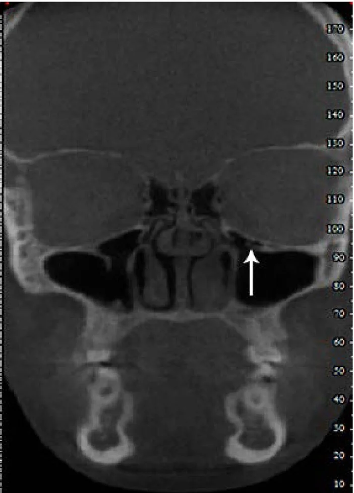
- Kortike ince kemik duvarlı, hava dansitesindeki hücre maksiller sinüs tavanı ile orbita tabanının medial kesimi arasında izlenir; yumuşak doku veya sıvı dansitesi varsa hücre içi opasifikasyon olarak değerlendirilir. Standart kontrastsız BT görüntülerinde kortike duvarlı hava hücresi maksiller sinüs tavanı ile orbita tabanı arasında net olarak izlenir.
- Kemik algoritması tanısal
- Koronal ve aksiyel kemik rekonstrüksiyonları hücrenin etmoid kökenli konumunu, maksiller infundibulumla komşuluğunu ve orbita tabanı korteksini gösterir; infundibulumun daralıp daralmadığı izlenir.
Önerilen pencereler: Kemik (G 1800 / M 400), Yumuşak doku (G 400 / M 40).
BT bulguları
- Infraorbital etmoid hücresi — maksiller sinüs tavanı boyunca, orbita tabanının medialinde uzanan kortike hava hücresi.
- Etmoid kapsülle devamlılık — hücre maksiller sinüs içindeki gerçek bir çıkıntıdan çok ön etmoid hava hücresi niteliğindedir.
- Maksiller infundibulum komşuluğu — hücrenin medial duvarı veya alt ucu infundibulumun dar bölümüne yaklaşabilir.
- İnfundibüler daralma — büyük hücre veya çevre mukozal kalınlaşma hava geçiş aralığını azaltabilir.
- Hücre içi opasifikasyon — hava yerine yumuşak doku/sıvı dansitesi görülmesi hücreye sınırlı inflamasyon ya da retansiyonu düşündürür.
- Orbita tabanı konturu — taban ince olabilir; eşlik eden kesinti veya aşağı deplasman varyant hücre tanımından ayrı raporlanır.
Normalde
Normal varyant anatomide maksiller sinüs tavanı ve orbita tabanının medial bölümü arasında ayrı bir infraorbital etmoid hücresi bulunmaz; maksiller infundibulum açık hava kolonu şeklinde izlenir. Karşı tarafın aynı koronal düzeyinde tavan-tabana mesafeyi, infundibulum lümenini ve mukozal örtüyü kıyaslayın.
Ayırıcı tanı
- Maksiller sinüs mukozal kisti
- sinüs içinde kubbe biçimli yumuşak doku yoğunluğu oluşturur.
- Maksiller sinüs retansiyon kisti
- sinüs içinde kubbe biçimli, düzgün sınırlı opasite olarak izlenir.
- Orbita tabanı blow-out kırığı
- kortikal süreksizlik, kemik fragmanı deplasmanı ve bazen orbital yağın sinüse herniasyonu vardır; düzenli kortike hücre duvarı beklenmez.
- Maksiller sinüs hipoplazisi
- sinüs genel olarak küçüktür ve duvarları yeniden şekillenmiştir; tek bir infraorbital hücre olarak sınırlı değildir.
Tuzaklar
- Hücreyi maksiller sinüs içine uzanan kemik çıkıntısı sanmayın; ardışık kesitlerde etmoid hava hücreleriyle devamlılığını izleyin.
- Haller hücresi varlığını tek başına maksiller sinüzitin nedeni olarak göstermeyin; lümen daralması ve eşlik eden mukozal hastalığı ayrıca belirtin.
- Opasifiye hücrede hava görünmemesi tanımı gizleyebilir; kortike duvarı ve orbita tabanı-maksiller tavan arasındaki konumu takip edin.
Kendini dene
Sinüs BT'sinde orbita tabanının medialinde, maksiller tavan boyunca kortike hava hücresi infundibuluma komşu. En olası varyant hangisidir?
Cevabı göster
Haller hücresi. Haller hücresi infraorbital ön etmoid hücredir ve maksiller sinüs tavanı-orbita tabanı komşuluğunda yer alır. Agger nasi frontal resesin önündedir; Onodi hücresi sfenoidin üst-lateralinde posterior etmoiddedir; supraorbital etmoid hücre orbita tavanına uzanır.
Geniş infraorbital etmoid hücre maksiller infundibuluma komşu, ancak sinüs mukozası normal. Bu bulgu en doğru nasıl yorumlanır?
Cevabı göster
Anatomik varyant. Hava içeren kortike hücre bir anatomik varyanttır; mukozal hastalık ve çıkış tıkanıklığı gösterilmeden enfeksiyon tanısı konmaz. Akut sinüzitte opasifikasyon, blow-out kırığında taban kesintisi/deplasman, mukoselde genişleme ve opasifikasyon beklenir.
Kaynaklar
Bu sayfadaki 35 cümle ve 2 quiz sorusu aşağıdaki kaynaklardan alıntılarla tek tek karşılaştırıldı; 6 cümle bu karşılaştırmada düzeltildi (2026-10-08).
- Deep learning classification of nasal anatomical variants on computed tomography for endoscopic sinus surgerypmc.ncbi.nlm.nih.gov
- Morphometric investigation of the infraorbital canal and determination of its “suspended” variant: a multidetector computed tomography studypmc.ncbi.nlm.nih.gov
- Infected Haller Cell Misdiagnosed as Invasive Fungal Sinusitis - A Parallaxpmc.ncbi.nlm.nih.gov
- Is There a Relationship Between Maxillary Sinus’s Inferior Pneumatisation and Sinonasal Variations? A Retrospective CBCT Studypmc.ncbi.nlm.nih.gov
- Anatomical variations of the ethmoid sinuses and their association with health or pathology of the ethmoid and maxillary sinuses in a Southern Chinese population: An analysis using cone-beam computed tomographypmc.ncbi.nlm.nih.gov
- Air spaces neighbouring the infraorbital canal.pubmed.ncbi.nlm.nih.gov
- [The CT image features of ethmomaxillary sinus and their significance in endoscopic surgery].pubmed.ncbi.nlm.nih.gov
- Radiological sinonasal anatomy. Exploring the Saudi population.pubmed.ncbi.nlm.nih.gov
- Evaluation of Infraorbital Canal in Cone Beam Computed Tomography of Maxillary Sinus.pubmed.ncbi.nlm.nih.gov
- CBCT analysis of haller cells: relationship with accessory maxillary ostium and maxillary sinus pathologies.pubmed.ncbi.nlm.nih.gov
- Anatomical Variations of the Nose and Paranasal Sinuses: A Computed Tomographic Study.pubmed.ncbi.nlm.nih.gov
- Clinically significant anatomical variants of the paranasal sinuses.pubmed.ncbi.nlm.nih.gov
- Sinonasal Anatomical Variants: CT and Endoscopy Study and Its Correlation with Extent of Disease.pubmed.ncbi.nlm.nih.gov
- Septal deviation is associated with maxillary sinus fungus ball in male patients.pubmed.ncbi.nlm.nih.gov
- Analysis of sinonasal anatomical variations associated with maxillary sinus fungal balls.pubmed.ncbi.nlm.nih.gov
- Medical therapy vs surgery for recurrent acute rhinosinusitis.pubmed.ncbi.nlm.nih.gov
- Computed Tomography Image Analysis and Clinical Correlations of Retromaxillary Cells.pubmed.ncbi.nlm.nih.gov
- Retromaxillary Pneumatization of Posterior Ethmoid Air Cells: Novel Description and Surgical Implications.pubmed.ncbi.nlm.nih.gov
- Correlations between anatomic variations of maxillary sinus ostium and postoperative complication after sinus lifting.pubmed.ncbi.nlm.nih.gov
- The effect of anatomic variations and maxillary sinus volume in antrochoanal polyp formation.pubmed.ncbi.nlm.nih.gov
- A juxta-ethmoidal course of the infraorbital canal in cases with large ethmomaxillary sinuses or giant Haller cells.pubmed.ncbi.nlm.nih.gov
- Middle meatal nasal recesses of the maxillary sinuses and dangerously modified nasal anatomy.pubmed.ncbi.nlm.nih.gov
- Anatomical variations and abnormalities of the maxillary region and clinical implications: A systematic review and metaanalysis.pubmed.ncbi.nlm.nih.gov
- Deep learning classification of nasal anatomical variants on computed tomography for endoscopic sinus surgery.pubmed.ncbi.nlm.nih.gov
- Anatomical Variations in Nasal Cavities and Paranasal Sinuses on Computed Tomography in Chronic Rhinosinusitis: Implications for Diagnosis and Treatment.pubmed.ncbi.nlm.nih.gov
- The Associations between the Maxillary Sinus Volume, Infraorbital Ethmoid Cells, and the Infraorbital Canal: A CT-Based Study.pubmed.ncbi.nlm.nih.gov
- Side-Specific Concordance Between Nasal Endoscopy and Computed Tomography Severity in Pediatric Chronic Rhinosinusitis.pubmed.ncbi.nlm.nih.gov
- Assessment of the Relationship Between Haller Cells, Accessory Maxillary Ostium, and Maxillary Sinus Pathologies: A Cross-Sectional CBCT Study.pubmed.ncbi.nlm.nih.gov
- Comparison Between Diagnostic Nasal Endoscopy and Plain Computed Tomography of the Paranasal Sinuses in Chronic Rhinosinusitis.pubmed.ncbi.nlm.nih.gov
- Cone Beam CT Analysis of Haller Cells: Prevalence and Relationship with Orbital Floor Dehiscence.pubmed.ncbi.nlm.nih.gov
- Inter Observer Agreement Among Radiologist and Otorhinolaryngologists on Paranasal Sinus Computed Tomography Scans in Chronic Rhinosinusitis.pubmed.ncbi.nlm.nih.gov
- Prospective Study to Evaluate the Role of Multidetector Computed Tomography in Evaluation of Paranasal Sinus Pathologies.pubmed.ncbi.nlm.nih.gov
- The maxillary sinus: physiology, development and imaging anatomy.pubmed.ncbi.nlm.nih.gov
- Evaluation of the Relationship of Dimensions of Maxillary Sinus Drainage System with Anatomical Variations and Sinusopathy: Cone-Beam Computed Tomography Findings.pubmed.ncbi.nlm.nih.gov
- Ossifying Fibroma of the Frontal Sinus: A Case Report and Literature Review.pubmed.ncbi.nlm.nih.gov
- Management of Maxillary Sinus Inverted Papilloma Using Endoscopic Modified Medial Maxillectomy (EMMM) and Direct Approach to the Anterior and Lateral Maxillary Sinus (DALMA) With Nasolacrimal Duct Preservation: A Case Report.pubmed.ncbi.nlm.nih.gov
- Diagnostic challenges in hepatic metastatic olfactory neuroblastoma: a case report and literature review.pubmed.ncbi.nlm.nih.gov
- Description of the Uncinate Process: A Computed Tomography Cross-Sectional Study.pubmed.ncbi.nlm.nih.gov
- Inflammatory Modifications in Paranasal Sinuses and Ostiomeatal Complex Anatomical Variations in Jet Aircraft Pilots: A Computed Tomography Study.pubmed.ncbi.nlm.nih.gov
- Systematic anatomical validation of the endoscopic mononostril transethmoid-paraseptal approach to the central skull base.pubmed.ncbi.nlm.nih.gov
- Establishment of AI-assisted diagnosis of the infraorbital posterior ethmoid cells based on deep learning.pubmed.ncbi.nlm.nih.gov
- Anatomical variations of the ethmoid sinuses and their association with health or pathology of the ethmoid and maxillary sinuses in a Southern Chinese population: An analysis using cone-beam computed tomography.pubmed.ncbi.nlm.nih.gov
- Imaging anatomy of the vidian canal and its clinical significance.pubmed.ncbi.nlm.nih.gov
- Volumetric analysis of maxillary sinus and assessment of various sinonasal anatomic variants on multi-detector computed tomography (MDCT) and their association with chronic rhinosinusitis.pubmed.ncbi.nlm.nih.gov
- Anatomical Variations of the Sinonasal Area and Their Clinical Impact on Sinus Pathology: A Systematic Review.pubmed.ncbi.nlm.nih.gov
- Evaluation of osteomeatal complex by cone-beam computed tomography in patients with maxillary sinus pathology and nasal septum deviation.pubmed.ncbi.nlm.nih.gov
- Rhinosinusitis: Evidence and experience - 2024.pubmed.ncbi.nlm.nih.gov
- Computed tomography scans of paranasal sinuses before functional endoscopic sinus surgery.pubmed.ncbi.nlm.nih.gov
- The Infra-Bullar Groove: Assessing a Novel Surgical Landmark for Identifying the Natural Maxillary Ostium.pubmed.ncbi.nlm.nih.gov
- Incidental Detection of a Posterior Inferior Cerebellar Artery (PICA)-Terminating Vertebral Artery Variant During CT Evaluation of Venous Sinus Thrombosis: A Case Report.pubmed.ncbi.nlm.nih.gov
- Foramen rotundum versus canal of the maxillary verve.pubmed.ncbi.nlm.nih.gov
- Silent Sinus Syndrome in a Paediatric Patient: An Incidental Diagnosis Following Facial Trauma.pubmed.ncbi.nlm.nih.gov
- Facial pain as a presenting symptom of a left maxillary sinus cyst: Case report and literature review.pubmed.ncbi.nlm.nih.gov
- Sinus Lift With Simultaneous Mucous Retention Cyst Drainage for Implant Rehabilitation: Clinical, Tomographic, and Histologic Assessments in a Six-Year Follow-Up Case Report.pubmed.ncbi.nlm.nih.gov
- Severe facial pain and removal of maxillary sinus mucous retention cyst.pubmed.ncbi.nlm.nih.gov
- Ectopic maxillary third molar associated with an odontogenic keratocyst in the maxillary Sinus presenting as refractory unilateral sinonasal symptoms: a case report with implications for early dental diagnosis.pubmed.ncbi.nlm.nih.gov
- Chronic Sinonasal Mucocele Manifesting as a Distinctive Collapsed Fibrous Hyalinized Cyst Wall.pubmed.ncbi.nlm.nih.gov
- Endoscopic Management of a Right Maxillary Sinus Mucocele With Palatal and Medial Wall Erosion.pubmed.ncbi.nlm.nih.gov
- Spontaneous Orbital Wall Fracture Secondary to Neglected Graves' Orbitopathy: A Case Report and Review of Current Literature.pubmed.ncbi.nlm.nih.gov
- The Dilemma of Reconstructive Material Choice for Orbital Floor Fracture: A Narrative Review.pubmed.ncbi.nlm.nih.gov
- Radiologic Evaluation of Odontogenic Sinusitis and Its Etiologic Factors: Lessons Learned from a Retrospective Study with a Proposed Imaging-Guided Management Pathway.pubmed.ncbi.nlm.nih.gov
- Full Clinical Pathological and Bacteriological Investigation of a Giant Nasal Polyp Associated With the Developing Apex of the Permanent Fourth Premolar (Triadan 108) in a 3-year-old Connemara Filly: A Case Report.pubmed.ncbi.nlm.nih.gov
- Radioanatomy of the Silent Sinus Syndrome.pubmed.ncbi.nlm.nih.gov
- The Big Nose Pattern at the Second Upper Molar-A Retrospective CBCT Study.pubmed.ncbi.nlm.nih.gov
- Multidetector CT of the Nasal Cavity and Paranasal Sinuses Variations in 73 Patients.pubmed.ncbi.nlm.nih.gov
- Osteomeatal Complex: A Study of Its Anatomical Variation Among Patients Attending North Bengal Medical College and Hospital.pubmed.ncbi.nlm.nih.gov
- Genera of phytopathogenic fungi known from culture: 1-379.pubmed.ncbi.nlm.nih.gov
- Flying through congested airspaces: imaging of chronic rhinosinusitis.pubmed.ncbi.nlm.nih.gov
- Pediatric Congenital Tracheobronchial Variants and Their Clinical Significance for Endotracheal Intubation and Airway Management: A Scoping Review of Fetal, Neonatal, and Pediatric Cases.pubmed.ncbi.nlm.nih.gov
- Heart Disease and Stroke Statistics-2023 Update: A Report From the American Heart Association.pubmed.ncbi.nlm.nih.gov
- Congenital Chest Wall Deformities in Children: A Narrative Review.pubmed.ncbi.nlm.nih.gov
- Comprehensive review on endonasal endoscopic sinus surgery.pubmed.ncbi.nlm.nih.gov
- Association Between DMFT Index and Sinonasal Anatomical Variations in Children: A Retrospective Cone-Beam Computed Tomography Study.pubmed.ncbi.nlm.nih.gov
- Assessment of Paranasal Sinus Growth with 3D Volumetric Measurements and the Effect of Anatomic Variations on Sinus Volume in a Pediatric Population.pubmed.ncbi.nlm.nih.gov
- Anatomical Variations Favouring Antrochoanal Polyp Genesis.pubmed.ncbi.nlm.nih.gov
- ESR Essentials: imaging in nasal obstruction and epistaxis-practice recommendations by the European Society of Head and Neck Radiology.pubmed.ncbi.nlm.nih.gov
- Critical anatomical variants in preoperative computed tomography of paranasal sinuses in a tertiary care center: a cross-sectional study.pubmed.ncbi.nlm.nih.gov
- The ethmomaxillary sinus: A rare anatomical variant.pubmed.ncbi.nlm.nih.gov
- Superior Mesenteric Artery Syndrome After Previous Nuss Procedure: A Case Report of Anatomical Realignment With Unintended Consequences.pubmed.ncbi.nlm.nih.gov
Eğitim amaçlıdır; hasta başında tanı ve tedavi kararının yerine geçmez. İçerik yapay zekâ destekli bir süreçle güncel literatüre göre cümle cümle kaynaklarla karşılaştırıldı, ancak uzman radyolog incelemesinden geçmedi. Klinik kararlarda güncel kılavuzları, kurum protokollerini ve radyoloji raporunu esas alın.