Özet
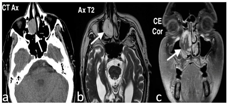
Lobüler kapiller hemanjiom, septum veya konka mukozasında gelişebilen benign vasküler nodüldür ve tekrarlayan burun kanaması ya da tıkanıklıkla başvurabilir. Kontrastsız BT lezyonun yerini ve kemikle ilişkisini gösterir; kontrastlı BT'de yoğun boyanma vasküler niteliği destekler. Küçük lezyonda kemik değişikliği olmayabilir, kemikte yer değiştirme, remodelasyon veya erozyon görülebilir. Hipervasküler kitleyi polip olarak adlandırmak ve çevre kemik değişikliğini incelememek tanısal ayrımı geciktirebilir.
Faz ve pencere
- Kontrastsız tanısal
- Septum veya konka mukozasından çıkan yumuşak doku nodülü ve komşu kemik korteksinin yer değiştirmesi izlenebilir; küçük lobüler kapiller hemanjiomda kemik değişikliği bulunmayabilir.
- Kontrastlı tanısal
- Kapiller ve lobüler kapiller hemanjiomlarda BT’de erken fazda orta veya belirgin kontrastlanma görülebilir ve bu kontrastlanma gecikmiş fazda azalabilir. Kapiller ve lobüler kapiller hemanjiomlarda BT’de erken fazda orta veya belirgin kontrastlanma görülebilir ve bu kontrastlanma gecikmiş fazda azalabilir.
Önerilen pencereler G genişlik, M merkez; değerler Hounsfield birimi (HU)
- Yumuşak dokuG 400 / M +40
- KemikG 1800 / M +400
BT bulguları
- Nazal septum ön kısmı veya alt konkada sık görülen, iyi sınırlı lobüleli yumuşak doku nodülü.
- Kontrastlı görüntüde lezyonda yoğun veya lobüler kontrast tutulumu.
- Küçük kitlede komşu kemik korteksinin korunması.
- Komşu kemikte yer değiştirme veya erozyon görülebilir; erozyon tek başına malignite kanıtı değildir.
- Nazal kavite hava yolunda lokal daralma ve posterior koanaya doğru uzanım.
- Kapiller ve lobüler kapiller hemanjiomlarda BT’de erken fazda orta veya belirgin kontrastlanma görülebilir ve bu kontrastlanma gecikmiş fazda azalabilir; kontrastlanma özellikleri histolojik tiplemeye katkı sağlayabilir.
- Maksiller sinüs ve etmoid hücrelerde eşlik eden sekresyon veya mukoza kalınlaşması.
Normalde
Normal nazal kavitede septum orta hattadır; konkaların kemik çekirdekleri ince, düzgün ve üzerindeki mukoza simetriktir. Karşı taraf septum ve konka konturlarını kıyaslayarak nodülün yapışma yerini, kontrastlanmasını ve komşu kemikte bası olup olmadığını ayırt et.
Normal BT ile kıyasla
Aynı bölgenin, kaynak veri setinde patoloji içermediği belirtilen bir BT incelemesini kesit kesit kaydırın; organ sınırlarını açarak anatomiyi hasta görüntüsüyle karşılaştırın. Küçük rastlantısal bulgular tümüyle dışlanmamıştır.
Kaynak: TotalSegmentator v2.01 veri seti (Wasserthal ve ark., Radiology: Artificial Intelligence 2023), CC BY 4.0, vaka s0849; organ sınırları veri setinin otomatik segmentasyonundan, birleştirilerek sadeleştirildi. Görüntüler 2,5 mm kesit aralığına yeniden örneklendi.
Ayırıcı tanı
- Juvenil nazofaringeal anjiofibrom
- posterior nazal/nazofarenks merkezli, pterigopalatin fossa genişleten vasküler kitle.
- Devrik papillom
- BT ve MR’da karakteristiki konvolüze serebriform deseni gösterir; kemik yıkımı ve lokal agresyon ile ayrılır.
- İnflamatuvar polip
- tek taraflı nazal kitlelerin ayırıcı tanısında değerlendirilir.
- Sinonazal melanom
- ilerleyici tek taraflı burun tıkanıklığı, tekrarlayan epistaksi ve kemik invazyonu ile seyreden agresif bir mukozal tümördür.
- Sinonazal malignite
- düzensiz kemik yıkımı ve komşu doku infiltrasyonunun kuşkulanıldığı agresif bir süreçtir.
Tuzaklar
- Belirgin kontrastlanma tek başına hemanjiom tanısı değildir; tümörün septum/konka kökenini ve posterior pterigopalatin alanı incele.
- Lobüler kapiller hemanjiomda komşu kemikte yeniden şekillenme veya erozyon olmayabilir; kemik değişikliklerinin yokluğu tanının dışlanması için yeterli değildir.
- Kontrastsız incelemede yumuşak doku diğer poliplerle benzer görünebilir; damar niteliği için kontrastlı inceleme ve klinik kanama öyküsü önemlidir.
- Belirgin vasküler nazal kitlelerde görüntüleme lezyonun vasküler özelliklerini ve yayılımını değerlendirmeye yardımcı olur; yüksek vaskülarite biyopsi sırasında kanama riski taşıyabilir ve juvenil nazofaringeal anjiofibrom ayırıcı tanısında sfenopalatin foramen ile pterigopalatin fossa ilişkisi incelenmelidir.
Kendini dene
Ön nazal septumda polilobüle ve temasla kanayan bir lezyon görülüyor; BT bulguları lobüler kapiller hemanjiomu düşündürmektedir. En olası tanı hangisidir?
Cevabı göster
Lobüler kapiller hemanjiom. Ön septumdaki polilobüle, temasla kanayan lezyon ve BT bulguları lobüler kapiller hemanjiomu akla getirir; kesin tanı histopatolojik inceleme ile konur.
Posterior nazofarenks kitlesi pterigopalatin fossayı genişletip maksiller arka duvarı öne itiyor. Hangi lezyon öne çıkar?
Cevabı göster
Juvenil anjiofibrom. Posterior nazal köken ve pterigopalatin fossa genişlemesi juvenil anjiofibrom için tipiktir; lobüler kapiller hemanjiom çoğunlukla septum veya konka kaynaklıdır.
İlgili konular
Kaynaklar
Bu sayfadaki 36 cümle ve 2 quiz sorusu aşağıdaki kaynaklardan alıntılarla otomatik olarak karşılaştırıldı; 13 cümle bu karşılaştırmada düzeltildi (2026-10-09).
- Pyogenic Granuloma in the Sinonasal Cavity: A Potential 38-Year Latency Following Foreign Body Entrapmentpmc.ncbi.nlm.nih.gov
- A silent vascular mimicker of Sinonasal disease: Nasal cavity angioma presenting with progressive epistaxis and obstructionpmc.ncbi.nlm.nih.gov
- Benign Sinonasal Tumors: A 17-Year Single Center Experiencepmc.ncbi.nlm.nih.gov
- Cavernous venous malformation of the frontal sinus: CT and MRI imaging findingspmc.ncbi.nlm.nih.gov
- Post‐Operative Surveillance After Resection of Uncommon Benign Sinonasal Tumors: A Multi‐Institutional Reviewpmc.ncbi.nlm.nih.gov
- Computed tomography and magnetic resonance imaging findings of nasal cavity hemangiomas according to histological type.pubmed.ncbi.nlm.nih.gov
- Uncommon Nasal Mass Presentation: A Radiological Case Series.pubmed.ncbi.nlm.nih.gov
- Sinonasal Lobular Capillary Hemangioma of the Nasal Cavity: A Rare Giant Vascular Tumor Managed With Preoperative Embolization and Medial Maxillectomy.pubmed.ncbi.nlm.nih.gov
- Pyogenic Granuloma in the Sinonasal Cavity: A Potential 38-Year Latency Following Foreign Body Entrapment.pubmed.ncbi.nlm.nih.gov
- A silent vascular mimicker of Sinonasal disease: Nasal cavity angioma presenting with progressive epistaxis and obstruction.pubmed.ncbi.nlm.nih.gov
- Endoscopic Classification of Severe Spontaneous Epistaxis: A Case-Series of 445 Patients.pubmed.ncbi.nlm.nih.gov
- A Huge Intraosseous Hemangioma in the Maxillary Sinus Misdiagnosed as Osteosarcoma: A Case Report.pubmed.ncbi.nlm.nih.gov
- Nasal kaposiform hemangioendothelioma: a rare disease in a rare location-a review article with a case report.pubmed.ncbi.nlm.nih.gov
- The application of ultrasonography in the differential diagnosis of orbital lesions.pubmed.ncbi.nlm.nih.gov
- Bilateral rhinoliths causing long-standing nasal obstruction: a case report and review of the literature.pubmed.ncbi.nlm.nih.gov
- Sinonasal schwannoma masquerading as an IgG4-related nasal mass.pubmed.ncbi.nlm.nih.gov
- Imaging features of rhinosporidiosis on contrast CT.pubmed.ncbi.nlm.nih.gov
- Use of the Term "Atypical Hepatic Hemangioma" in Radiology Reports: An Analysis of Diagnostic Precision.pubmed.ncbi.nlm.nih.gov
- A case of incidental adrenal capillary hemangioma after a partial nephrectomy for renal clear cell carcinoma.pubmed.ncbi.nlm.nih.gov
- A case report: Masson's tumor (intravascular papillary endothelial hyperplasia) of the abdominal cavity.pubmed.ncbi.nlm.nih.gov
- Primary splenic angiosarcoma presenting with multiple bone destruction as the initial manifestation: a case report and narrative review.pubmed.ncbi.nlm.nih.gov
- Case Report: Gastric glomus tumor with long-term dynamic multimodal imaging findings.pubmed.ncbi.nlm.nih.gov
- Bleeding polyp of the nasal septum: A rare lesion in adults.pubmed.ncbi.nlm.nih.gov
- Cavernous venous malformation of the frontal sinus: CT and MRI imaging findings.pubmed.ncbi.nlm.nih.gov
- Pediatric Central Giant Cell Granuloma in the Nasal Cavity: A Case Report and Literature Review.pubmed.ncbi.nlm.nih.gov
- Imaging for Pediatric Head and Neck Emergencies: Critical Insights.pubmed.ncbi.nlm.nih.gov
- Craniofacial surgery for removing tumors in advanced stages that invade the anterior skull base.pubmed.ncbi.nlm.nih.gov
- Ancient Diseases in Vertebrates: Tumours through the Ages.pubmed.ncbi.nlm.nih.gov
- Recurrent Epistaxis as a Rare Presentation of Extramedullary Multiple Myeloma.pubmed.ncbi.nlm.nih.gov
- Leiomyosarcoma of the maxillary sinus presenting with epiphora: a case report.pubmed.ncbi.nlm.nih.gov
- A Rare Nasopharyngeal Hemangioma Presenting as Recurrent Cyanotic Apnea in a Neonate: A Case Report and Literature Review.pubmed.ncbi.nlm.nih.gov
- Congenital Anomalies of the Nose: From Embryologic to Surgical Treatment.pubmed.ncbi.nlm.nih.gov
- Decoding calvarial lesions: Contemporary neurosurgical management integrating radiological and histopathological features.pubmed.ncbi.nlm.nih.gov
- Cardiac Hemangiomas: A Five-Year Systematic Review of Diagnosis, Treatment, and Outcomes.pubmed.ncbi.nlm.nih.gov
- Developmental venous anomalies and cerebral cavernous malformations: Partners in crime.pubmed.ncbi.nlm.nih.gov
- Combined corticosteroid therapy enhances outcomes of endoscopic sinus surgery in chronic rhinosinusitis with nasal polyps: a prospective cohort study.pubmed.ncbi.nlm.nih.gov
- Algorithmic Approach to Fibroinflammatory Sinonasal Tract Lesions.pubmed.ncbi.nlm.nih.gov
- Neuroendoscopic single-nostril nasoseptal approach for tuberculum sellae meningiomas: technical description and preliminary clinical experience.pubmed.ncbi.nlm.nih.gov
- Radiologic and cadaveric spatial relation between nasopalatine canal and the middle turbinate axilla.pubmed.ncbi.nlm.nih.gov
- Nasal Cavity Dimensions and Pharyngeal Airway Space in Simple Snoring: A Cross-Sectional CT-Based Study.pubmed.ncbi.nlm.nih.gov
- Diagnostic Performance of Infrared Nasal Thermography for the Detection of Enzootic Nasal Adenocarcinoma in Goats.pubmed.ncbi.nlm.nih.gov
- A nasolabial cyst is It a rare cyst or not? case series of bilateral and unilateral patterns.pubmed.ncbi.nlm.nih.gov
- Evaluation of treatment outcomes of zygomaticomaxillary complex (ZMC) fractures using Foley catheter ballooning technique.pubmed.ncbi.nlm.nih.gov
- Recurrent Juvenile Nasopharyngeal Angiofibroma: Two Cases Emphasizing the Role of Follow-Up and Staging in Surgical Planning.pubmed.ncbi.nlm.nih.gov
- Juvenile Nasopharyngeal Angiofibroma in an Adult Patient: A Rare Presentation with Fahr Syndrome and Multiple Comorbidities-A Case Report and Literature Review.pubmed.ncbi.nlm.nih.gov
- Adult-onset juvenile nasopharyngeal angiofibroma with delayed adult recurrence managed by revision endoscopic resection and external carotid branch ligation.pubmed.ncbi.nlm.nih.gov
- Prespecified Structured CT and MRI Scores for Sinonasal Inverted Papilloma: A Dual-reader Diagnostic Accuracy Study.pubmed.ncbi.nlm.nih.gov
- Epithelial Markers in Sinonasal Inverted Papilloma with Malignant Transformation: A Scoping Review and Proposal for an Integrative Model of Malignant Risk Stratification.pubmed.ncbi.nlm.nih.gov
- Management of Maxillary Sinus Inverted Papilloma Using Endoscopic Modified Medial Maxillectomy (EMMM) and Direct Approach to the Anterior and Lateral Maxillary Sinus (DALMA) With Nasolacrimal Duct Preservation: A Case Report.pubmed.ncbi.nlm.nih.gov
- Extranasopharyngeal angiofibroma of the sinonasal tract: A systematic review.pubmed.ncbi.nlm.nih.gov
- Pharmacological, Technological, and Digital Innovative Aspects in Rhinology.pubmed.ncbi.nlm.nih.gov
- STAT6 Blockade Abrogates Aspergillus -Induced Eosinophilic Chronic Rhinosinusitis and Asthma, A Model of Unified Airway Disease.pubmed.ncbi.nlm.nih.gov
- Imaging More than Skin-Deep: Radiologic and Dermatologic Presentations of Systemic Disorders.pubmed.ncbi.nlm.nih.gov
- Rare Presentation of Sinonasal Synovial Sarcoma With Frontal Sinus and Limited Intracranial Extension Associated With Probable Paraneoplastic Vasculitis: A Case Report.pubmed.ncbi.nlm.nih.gov
- Sinonasal Mucosal Epithelioid Melanoma with Rapid Skull-Base and Orbital Progression.pubmed.ncbi.nlm.nih.gov
- Leptomeningeal carcinomatosis in sinonasal NUT carcinoma: a case report and review of intracranial involvement.pubmed.ncbi.nlm.nih.gov
- Clinicopathologic Characteristics of Intraglomerular Malignancy in Kidney Biopsies.pubmed.ncbi.nlm.nih.gov
- Update from the 5th Edition of the World Health Organization Classification of Head and Neck Tumors: Soft Tissue Tumors.pubmed.ncbi.nlm.nih.gov
- Soft Tissue Special Issue: Perivascular and Vascular Tumors of the Head and Neck.pubmed.ncbi.nlm.nih.gov
- Oral Hobnail Hemangioma of the Maxillary Labial Mucosa: Clinicopathologic Findings of a Rare Case.pubmed.ncbi.nlm.nih.gov
- Rapidly progressive angiomatous epulis: How to differentiate a benign gingival lesion from malignancy?pubmed.ncbi.nlm.nih.gov
- Comparison of Imaging Findings between Granulomatosis with Polyangiitis and Eosinophilic Granulomatosis with Polyangiitis on Sinus CT: Importance of High-Density Opacification of the Paranasal Sinuses.pubmed.ncbi.nlm.nih.gov
- The radiological perspective of juvenile nasopharyngeal angiofibroma: A case report.pubmed.ncbi.nlm.nih.gov
- Sinonasal Inflammation or Neoplasm: Raise the Red Flagsǃ-A Pictorial Review.pubmed.ncbi.nlm.nih.gov
- Orbital tuberculosis mimicking a vascular emergency: A case report of superior ophthalmic vein engorgement.pubmed.ncbi.nlm.nih.gov
- Surviving childhood cancer: the hidden threat of second malignancies.pubmed.ncbi.nlm.nih.gov
- Clinical and imaging manifestations of metastases from rare sites.pubmed.ncbi.nlm.nih.gov
- Endoscopic Endonasal Resection of Fibro-Osseous Lesions Involving the Nasal Turbinates: A Report of Two Cases and a Literature Review.pubmed.ncbi.nlm.nih.gov
- Orbital Extension of Sinonasal Mucocele Presenting as Proptosis: A Report of Two Cases.pubmed.ncbi.nlm.nih.gov
- Sinonasal extramedullary plasmacytoma: A case report of unilateral nasal obstruction with epistaxis.pubmed.ncbi.nlm.nih.gov
- Small ethmoid mucocele with intracranial extension associated with fibrous dysplasia: A rare case report.pubmed.ncbi.nlm.nih.gov
- The management of extensive juvenile nasopharyngeal angiofibroma involving the carotid artery: An illustrative case series and literature review original.pubmed.ncbi.nlm.nih.gov
- Diagnostic Insights and Surgical Outcomes of Glomangiopericytoma: A Tertiary Center Case Series.pubmed.ncbi.nlm.nih.gov
- Clinical Presentation, Diagnostic Challenges, and Management Strategies for Asymptomatic Advanced Stage 4B Juvenile Nasal Angiofibroma: A Rare Pediatric Case Report and Literature Review.pubmed.ncbi.nlm.nih.gov
- Epistaxis and Destructive Computed Tomographic Features in Canine Nasal Transitional Carcinoma: Frequent Cribriform Plate Destruction but Uncommon CT-Detected Brain Parenchymal Involvement.pubmed.ncbi.nlm.nih.gov
- Kiesselbach's area nasal septal gouty tophus with an integrated "3 + 2" diagnostic algorithm: a case report.pubmed.ncbi.nlm.nih.gov
- Advanced tumour-induced osteomalacia secondary to sinonasal phosphaturic mesenchymal tumour.pubmed.ncbi.nlm.nih.gov
- Benign Sinonasal Tumors: A 17-Year Single Center Experience.pubmed.ncbi.nlm.nih.gov
- Sinonasal tract angiosarcoma: a clinicopathologic and immunophenotypic study of 10 cases with a review of the literature.pubmed.ncbi.nlm.nih.gov
- Pre-surgical Embolization for Nasal Lobular Capillary Hemangioma: A Case Report.pubmed.ncbi.nlm.nih.gov
- Rare case of littoral cell angioma recognised on CT simulation for adjuvant radiation treatment for early stage breast cancer.pubmed.ncbi.nlm.nih.gov
- Imaging in Rare and Atypical Sinonasal Masses: An Interesting Case Series.pubmed.ncbi.nlm.nih.gov
- Solitary Fibrous Tumor of the Nasal Vestibule: A Case Report.pubmed.ncbi.nlm.nih.gov
- Rare Isolated Sphenoid Sinus Lesions: A Multi-case Study.pubmed.ncbi.nlm.nih.gov
- Radiologic Features for Differentiating Sinonasal Inverted Papilloma and Squamous Cell Carcinoma Arising From Inverted Papilloma Without Bone Destruction.pubmed.ncbi.nlm.nih.gov
- Differences in Imaging and Histology Between Sinonasal Inverted Papilloma with and Without Squamous Cell Carcinoma.pubmed.ncbi.nlm.nih.gov
- Role of radiological corroboration in a locally aggressive inverted papilloma: a case report.pubmed.ncbi.nlm.nih.gov
- Chinese expert consensus on imaging examination and diagnosis of nasal cavity and paranasal sinus tumors.pubmed.ncbi.nlm.nih.gov
- Rhinosinusitis: Evidence and experience - 2024.pubmed.ncbi.nlm.nih.gov
- Tumors of the Lacrimal Drainage System: Diagnosis and Management.pubmed.ncbi.nlm.nih.gov
- Radiologic overview of sinonasal lesions.pubmed.ncbi.nlm.nih.gov
- Unusual progression of Rosai-Dorfman disease in a pediatric patient.pubmed.ncbi.nlm.nih.gov
- CT Findings of Feline Nasal Hamartomas Mimicking Malignant Growth.pubmed.ncbi.nlm.nih.gov
- Vasculitis syndromes: large vessel vasculitides and ANCA-associated vasculitides from a neuroradiologist's perspective.pubmed.ncbi.nlm.nih.gov
- Comparative Imaging of Allergic Fungal Sinusitis With Orbital and Intracranial Extension: A Case Report.pubmed.ncbi.nlm.nih.gov
- Craniofacial Ossifying Fibromas in Children: Clinical Variability and Surgical Outcomes in a Case Series.pubmed.ncbi.nlm.nih.gov
- Rare Diseases of the Orbit.pubmed.ncbi.nlm.nih.gov
- Dermatologic Manifestations of Monogenic Autoinflammatory Diseases.pubmed.ncbi.nlm.nih.gov
- Nasal lobular capillary hemangioma: Report of a case managed by endoscopic excision and pre-operative angio-embolization.pubmed.ncbi.nlm.nih.gov
- Respiratory Epithelial Adenomatoid Hamartoma in a Young Female: A Diagnostic Dilemma.pubmed.ncbi.nlm.nih.gov
- Sinonasal hemangiopericytoma: A rare sinonasal tumor.pubmed.ncbi.nlm.nih.gov
- The Endovascular Treatment of Epistaxis.pubmed.ncbi.nlm.nih.gov
- Evaluating the Effectiveness of Yttrium-90 Glass Microspheres in the Treatment of Hepatocellular Carcinoma, Intrahepatic Cholangiocarcinoma, and Metastatic Colorectal Cancer in Practice: Protocol for the Prospective PROACTIF Phase IV Registry Study in France.pubmed.ncbi.nlm.nih.gov
- Extra-Axial Cavernous Angioma: A Case Report and Review of the Literature.pubmed.ncbi.nlm.nih.gov
- Unique case of a post-traumatic nasoseptal arteriovenous malformation.pubmed.ncbi.nlm.nih.gov
- CT features of lobular capillary hemangioma of the nasal cavity.pubmed.ncbi.nlm.nih.gov
Eğitim amaçlıdır; hasta başında tanı ve tedavi kararının yerine geçmez, tıbbi tavsiye değildir. Metinler yapay zekâ desteğiyle yazıldı ve otomatik bir adımda kaynak alıntılarıyla karşılaştırıldı; bu bir tıbbi doğrulama değildir ve içerik uzman radyolog incelemesinden geçmedi. Klinik kararlarda güncel kılavuzları, kurum protokollerini ve radyoloji raporunu esas alın.