Özet
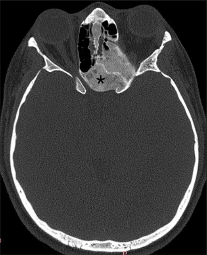
Kraniofasiyal fibroz displazi, normal kemik dokusunun fibro-osseöz matriksle yer değiştirdiği benign bir kemik hastalığıdır ve sinüs çevresindeki kemikleri genişleterek hava boşluklarını daraltabilir. Kontrastsız ince kesit BT, buzlu cam veya karma matriksi, kemik genişlemesini ve sinüs anatomisine etkisini gösterir. Fibroz displazi, radyografik özellikleri diğer fibro-osseöz lezyonlarla örtüşebildiğinden kesin tanı için histopatoloji gerekli olup genişleyici ve destrüktif olmayan seyir gösterir. Optik kanal, orbital duvar veya kafa tabanı komşuluğundaki daralma atlanırsa işlevsel etkilenim ve hastalık yayılımı eksik tarif edilebilir.
Faz ve pencere
- Kontrastsız tanısal
- Kemik algoritmalı ince kesitlerde genişlemiş kemik içinde buzlu cam, sklerotik ya da karma fibro-osseöz matriks ve sinüs lümeninde biçim değişikliği görülür.
Önerilen pencereler: Kemik (G 1800 / M 400).
BT bulguları
- Maksilla veya başka kraniofasiyal kemik segmentinde düzgün ekspansiyon ve kortikal kabukta korunma ya da incelme.
- Medüller boşluğun homojen buzlu cam görünümünde yoğun matriksle silinmesi.
- Olgunlaşma ve kemik içeriğine bağlı sklerotik veya yamalı karma yoğunluk alanları.
- Lezyon, kaynaklarda belirtildiği üzere genişleyici ve non-destrüktif seyir izler; radyografik özellikler lezyonun boyutuna ve derecesine göre değişkenlik gösterir.
- Maksiller sinüs tabanının veya yan duvarının içe itilmesiyle sinüs hacminde azalma ve asimetri.
- Fibroz displaziye bağlı kemik genişlemesi, etmoid hücrelerin, sfenoid sinüs duvarlarının veya frontal resesin daralmasına ve sinüs ostiumlarının kemiksel deformasyona uğramasına yol açabilir.
- Orbita tabanı, optik kanal veya kafa tabanı açıklıklarında komşuluk nedeniyle oluşan daralma.
Normalde
BT’de lezyonun yerleşimini ve matriksini tanımlayın; uzanımını ve çevre yapılarla ilişkisini değerlendirin.
Normal BT ile kıyasla
Aynı bölgenin, kaynak veri setinde patoloji içermediği belirtilen bir BT incelemesini kesit kesit kaydırın; organ sınırlarını açarak anatomiyi hasta görüntüsüyle karşılaştırın. Küçük rastlantısal bulgular tümüyle dışlanmamıştır.
Kaynak: TotalSegmentator v2.01 veri seti (Wasserthal ve ark., Radiology: Artificial Intelligence 2023), CC BY 4.0, vaka s0849; organ sınırları veri setinin otomatik segmentasyonundan, birleştirilerek sadeleştirildi. Görüntüler 2,5 mm kesit aralığına yeniden örneklendi.
Ayırıcı tanı
- Ossifiye fibrom
- keskin sınırlı, çoğu kez kapsüllü ve kemiği dışa doğru iten ekspansil odak oluşturur.
- Osteoma
- sınırlı, çok yoğun kemik odağı olarak sinüs lümenine projekte olur; kemiğin geniş bir segmentini tutmaz.
- Düşük dereceli osteosarkom
- korteks yıkımı, düzensiz periostal reaksiyon veya ekstraosseöz yumuşak doku bileşeni varsa benign displaziden uzaklaşılır.
Tuzaklar
- Buzlu cam görünümünü tek başına tanı kabul etmeyin; lezyonun kemiği genişletme biçimi ve sınırının medullaya geçişi ossifiye fibromdan ayrımda önemlidir.
- Sinüs lümenindeki opasifikasyon yalnızca mukozal hastalık olarak yorumlanmamalı; fibroz displazide karakteristik kemik genişlemesi ve duvarların itilmesi ayrıntılı incelenmelidir.
- Belirgin asimetriyi agresif destrüksiyonla eşitlemeyin; düzgün konturlu genişleme fibroz displazide görülebilir, korteks dışına çıkan yumuşak doku kitlesi ise atipiktir.
Kendini dene
Genç erişkinin sinüs BT’sinde maksiller kemik düzgün genişlemiş, buzlu cam matriksli ve sinüs tabanı içe itilmiş. En olası tanı nedir?
Cevabı göster
Fibroz displazi. Kemiğin segmental ve düzgün genişlemesiyle buzlu cam matriks fibroz displaziyi destekler. Ossifiye fibrom daha keskin sınırlı bir ekspansil odak oluşturur; osteit duvar kalınlaşmasıdır, osteom ise fokal yoğun kemiktir.
Buzlu cam matriksli kraniofasiyal kemik lezyonunda sinüs anatomisinin etkisini belirlemek için hangi kıyas en yararlıdır?
Cevabı göster
Karşı maksiller sinüs hacmi. Karşı maksiller sinüs aynı anatomik düzeyde kontrol sağlar; taban ve duvarların yer değiştirmesi ile lümen daralmasını görünür kılar.
İlgili konular
Kaynaklar
Bu sayfadaki 30 cümle ve 2 quiz sorusu aşağıdaki kaynaklardan alıntılarla tek tek karşılaştırıldı; 7 cümle bu karşılaştırmada düzeltildi (2026-10-08).
- Small ethmoid mucocele with intracranial extension associated with fibrous dysplasia: A rare case reportpmc.ncbi.nlm.nih.gov
- A Huge Intraosseous Hemangioma in the Maxillary Sinus Misdiagnosed as Osteosarcoma: A Case Reportpmc.ncbi.nlm.nih.gov
- Endoscopic management of uncommon benign maxillary sinus lesions: a case seriespmc.ncbi.nlm.nih.gov
- Benign Sinonasal Tumors: A 17-Year Single Center Experiencepmc.ncbi.nlm.nih.gov
- Intracranial aneurysmal bone cysts in association with fibrous dysplasia: Case report and scoping review of case reportspmc.ncbi.nlm.nih.gov
- Characteristics of sinonasal fibrous dysplasia: experience from a single department.pubmed.ncbi.nlm.nih.gov
- Ossifying fibromas of the paranasal sinuses: diagnosis and management.pubmed.ncbi.nlm.nih.gov
- Orbital Involvement In Sinonasal Diseases.pubmed.ncbi.nlm.nih.gov
- [Application of 3D computer-assisted printing technique combined with plastic titanium mesh in the reconstruction of maxillary defect].pubmed.ncbi.nlm.nih.gov
- An alternative management approach to paranasal sinus fibro-osseous lesions.pubmed.ncbi.nlm.nih.gov
- A Clinical and Radiological Approach to the Management of Benign Mesenchymal Sinonasal Tumors.pubmed.ncbi.nlm.nih.gov
- Sinonasal disease in polyostotic fibrous dysplasia and McCune-Albright Syndrome.pubmed.ncbi.nlm.nih.gov
- [Surgical management of sinonasal and adjacent skull base benign fibro-osseous lesions].pubmed.ncbi.nlm.nih.gov
- Role of intraoperative CT-updates during image-guided endoscopic sinus surgery for sinonasal fibro-osseous lesions.pubmed.ncbi.nlm.nih.gov
- Hyperostotic esthesioneuroblastoma: rare variant and fibrous dysplasia mimicker.pubmed.ncbi.nlm.nih.gov
- Small ethmoid mucocele with intracranial extension associated with fibrous dysplasia: A rare case report.pubmed.ncbi.nlm.nih.gov
- Intracranial aneurysmal bone cysts in association with fibrous dysplasia: Case report and scoping review of case reports.pubmed.ncbi.nlm.nih.gov
- Endoscopic Endonasal Resection of Fibro-Osseous Lesions Involving the Nasal Turbinates: A Report of Two Cases and a Literature Review.pubmed.ncbi.nlm.nih.gov
- Endoscopic management of uncommon benign maxillary sinus lesions: a case series.pubmed.ncbi.nlm.nih.gov
- Cavernous venous malformation of the frontal sinus: CT and MRI imaging findings.pubmed.ncbi.nlm.nih.gov
- Sculpting Solutions: A Case Report of Resection and Reconstruction in an Aggressive Mandibular Juvenile Ossifying Fibroma and Review of Literature.pubmed.ncbi.nlm.nih.gov
- Benign incidental do-not-touch bone lesions.pubmed.ncbi.nlm.nih.gov
- Nonproliferative and Proliferative Lesions of the Rat and Mouse Skeletal Tissues (Bones, Joints, and Teeth).pubmed.ncbi.nlm.nih.gov
- Unusual Manifestations of Sinonasal Osteomas: A Narrative Review.pubmed.ncbi.nlm.nih.gov
- Polyostotic fibrous dysplasia: Unveiling the complexity of a rare bone disorder through a case report.pubmed.ncbi.nlm.nih.gov
- Imaging of the skull base and orbital tumors.pubmed.ncbi.nlm.nih.gov
- When plasmacytoma defies expectations: An unusual maxillary presentation with mixed imaging features in a young patient.pubmed.ncbi.nlm.nih.gov
- Ossifying fibroma of the maxilla: A case report with literature review.pubmed.ncbi.nlm.nih.gov
- Imaging of Skull Base Tumors.pubmed.ncbi.nlm.nih.gov
- Aesthetic and Functional Rehabilitation in Juvenile Ossifying Fibroma: A Case Report.pubmed.ncbi.nlm.nih.gov
- "Maxillary juvenile ossifying fibroma: A case study of severe tooth displacement and orbital involvement in a pediatric patient".pubmed.ncbi.nlm.nih.gov
- Cone-beam computed tomography imaging of benign odontogenic lesions in the maxilla: A report of 2 cases.pubmed.ncbi.nlm.nih.gov
- The GNAS Gene: Fibrous Dysplasia, McCune-Albright Syndrome, and Skeletal Structure and Function.pubmed.ncbi.nlm.nih.gov
- Malignant Transformation of Musculoskeletal Lesions with Imaging-Pathology Correlation-Part 1: Bone Lesions.pubmed.ncbi.nlm.nih.gov
- Unveiling Primary Bone Tumors of the Spine: A Review of Essential Imaging Clues.pubmed.ncbi.nlm.nih.gov
- Musculoskeletal tumors and tumor-like lesions with "dark" signal intensity on T2-weighted MR images: A pictorial review.pubmed.ncbi.nlm.nih.gov
- Soft Tissue Special Issue: Gnathic Fibro-Osseous Lesions and Osteosarcoma.pubmed.ncbi.nlm.nih.gov
- EWSR1 -The Most Common Rearranged Gene in Soft Tissue Lesions, Which Also Occurs in Different Bone Lesions: An Updated Review.pubmed.ncbi.nlm.nih.gov
- Conventional radiography for the assessment of focal bone lesions of the appendicular skeleton: fundamental concepts in the modern imaging era.pubmed.ncbi.nlm.nih.gov
- International Harmonization of Nomenclature and Diagnostic Criteria (INHAND): Non-proliferative and Proliferative Lesions of the Non-human Primate ( M. fascicularis ).pubmed.ncbi.nlm.nih.gov
- Nonproliferative and Proliferative Lesions of the Rat and Mouse Endocrine System.pubmed.ncbi.nlm.nih.gov
- The maxillary sinus: physiology, development and imaging anatomy.pubmed.ncbi.nlm.nih.gov
- Computed Tomography in Craniofacial Fibrous Dysplasia: A Case Series with Review of Literature and Classification Update.pubmed.ncbi.nlm.nih.gov
- Masses of developmental and genetic origin affecting the paediatric craniofacial skeleton.pubmed.ncbi.nlm.nih.gov
- A practical overview of CT and MRI features of developmental, inflammatory, and neoplastic lesions of the sphenoid body and clivus.pubmed.ncbi.nlm.nih.gov
- Ocular findings in Jansen metaphyseal chondrodysplasia.pubmed.ncbi.nlm.nih.gov
- 23-year review of spheno-orbital meningioma: clinical, radiological, and pathological insights from 100 cases.pubmed.ncbi.nlm.nih.gov
- Relationship between maxillary sinus volume and alveolar trabeculation at orthodontic mini-implant sites across vertical skeletal patterns: a cross-sectional CBCT analysis.pubmed.ncbi.nlm.nih.gov
- Dental Radiographic/Digital Radiography Technology along with Biological Agents in Human Identification.pubmed.ncbi.nlm.nih.gov
- Neurofibromatosis Type 1 With Cherubism-like Phenotype, Multiple Osteolytic Bone Lesions of Lower Extremities, and Alagille-syndrome: Case Report With Literature Survey.pubmed.ncbi.nlm.nih.gov
- Imaging-Guided Surgical Decision-Making and Bone Healing in Mandibular Cystic and Tumor-like Lesions: A Case-Based Radiologic Observation.pubmed.ncbi.nlm.nih.gov
- Jansen metaphyseal chondrodysplasia: analysis of craniofacial manifestations.pubmed.ncbi.nlm.nih.gov
- Craniovertebral Junction Instability after Oncological Resection: A Narrative Review.pubmed.ncbi.nlm.nih.gov
- Bone-Preserving Management of Maxillary Cemento-Ossifying Fibroma With Orbital Floor Thinning: A Case Report.pubmed.ncbi.nlm.nih.gov
- Aggressive Juvenile Trabecular Ossifying Fibroma in a 4-Year-Old: Case Report Bringing Diagnostic Challenges and Surgical Management.pubmed.ncbi.nlm.nih.gov
- Extensive ossifying fibroma of the mandible: segmental resection and free fibula flap reconstruction.pubmed.ncbi.nlm.nih.gov
- Review of imaging modalities and radiological findings of calvarial lesions.pubmed.ncbi.nlm.nih.gov
- Benign and malignant lesions of the mandible and maxilla: imaging findings and systematic approach.pubmed.ncbi.nlm.nih.gov
- Osteomyelitis of the Jaw Bones and Its Mimics: Resolving the Diagnostic Enigma.pubmed.ncbi.nlm.nih.gov
- Imaging Features of Mandibular Ramus Osteoma on Computed Tomography: A Case Report With Literature Analysis.pubmed.ncbi.nlm.nih.gov
- Fibrous dysplasia of the jaws: four cases with conservative and resective surgical management.pubmed.ncbi.nlm.nih.gov
- Giant Peripheral Osteoma of the Mandibular Antegonial Notch in an Elderly Patient: A Case Report.pubmed.ncbi.nlm.nih.gov
- Cystic bone Lesions: Diagnostic pitfalls and therapeutic considerations.pubmed.ncbi.nlm.nih.gov
- Long-term safety and effectiveness of the "OptEase" vena cava filter.pubmed.ncbi.nlm.nih.gov
- Fibrous Dysplasia Presenting as an Exophytic Gingival Mass: A Rare Clinical Presentation.pubmed.ncbi.nlm.nih.gov
- Desmoplastic Fibroma of the Mandible in a 10-Year-Old Female Patient: A Case Report.pubmed.ncbi.nlm.nih.gov
- Prevalence of Incidental Maxillary Sinus Anomalies on CBCT Scans: A Radiographic Study.pubmed.ncbi.nlm.nih.gov
- Craniofacial Osteomas: From Diagnosis to Therapy.pubmed.ncbi.nlm.nih.gov
- Bone in Parathyroid Diseases Revisited: Evidence From Epidemiological, Surgical and New Drug Outcomes.pubmed.ncbi.nlm.nih.gov
- Radiological review of skull lesions.pubmed.ncbi.nlm.nih.gov
- Imaging approach for jaw and maxillofacial bone tumors with updates from the 2022 World Health Organization classification.pubmed.ncbi.nlm.nih.gov
- A Review of Posteromedial Lesions of the Chest Wall: What Should a Chest Radiologist Know?pubmed.ncbi.nlm.nih.gov
- Bone-related disorders of the jaw: A clinico-radiological diagnostic algorithm.pubmed.ncbi.nlm.nih.gov
- Salvaging Vision: A Study of Non-Traumatic Optic Neuropathies.pubmed.ncbi.nlm.nih.gov
- Sphenoclival fibrous dysplasia with cyst formation causing abducens nerve palsy in an older patient: illustrative case.pubmed.ncbi.nlm.nih.gov
- Special Considerations in Pediatric Endoscopic Skull Base Surgery.pubmed.ncbi.nlm.nih.gov
- Congenital and Breed-Associated Vertebral Anomalies in Dogs and Cats: Developmental Anatomy, Genetic Determinants, and Biological Significance.pubmed.ncbi.nlm.nih.gov
- Augmented Reality Navigation in Craniomaxillofacial/Head and Neck Surgery.pubmed.ncbi.nlm.nih.gov
- MRI Investigation of Kidneys, Ureters and Urinary Bladder in Rabbits.pubmed.ncbi.nlm.nih.gov
- A case of craniofacial polyostotic fibrous dysplasia.pubmed.ncbi.nlm.nih.gov
- A review of the oral health of individuals with disabilities in Puerto Rico and among U.S. Hispanics.pubmed.ncbi.nlm.nih.gov
- A Huge Intraosseous Hemangioma in the Maxillary Sinus Misdiagnosed as Osteosarcoma: A Case Report.pubmed.ncbi.nlm.nih.gov
- Histological Diversity, Diagnostic Challenges, and Surgical Treatment Strategies of Fibrous Dysplasia of Upper and Mid-Thirds of the Craniomaxillofacial Complex.pubmed.ncbi.nlm.nih.gov
- Clival and Paraclival Lesions: A Pictorial Review.pubmed.ncbi.nlm.nih.gov
- Rare Diseases of the Middle Ear and Lateral Skull Base.pubmed.ncbi.nlm.nih.gov
- Giant Oncocytic Sinonasal Papilloma: A Rare Case.pubmed.ncbi.nlm.nih.gov
- Adjacent Maxillary Bone Microarchitecture in Patients with Asymmetric Bilateral Lund-Mackay Scores: A Pilot Retrospective Within-Subject Analysis Using CBCT.pubmed.ncbi.nlm.nih.gov
- Uremic leontiasis ossea: distinctive imaging features allow differentiation from other clinical causes of leontiasis ossea.pubmed.ncbi.nlm.nih.gov
- Efficiency and Safety of Dental Implantation in the Area of Hyperdense Jaw Lesions: A Narrative Review.pubmed.ncbi.nlm.nih.gov
- Surgical Management of a Case of Frontoparietal Fibrous Dysplasia.pubmed.ncbi.nlm.nih.gov
- Surgically Treated Benign Bone Tumors and Tumor-like Conditions in the Pediatric Population-A 10-Year Institutional Experience.pubmed.ncbi.nlm.nih.gov
- Craniofacial Fibrous Dysplasia to Affect or Not the Optic Nerve in Long-Term Follow-Up of Three Cases.pubmed.ncbi.nlm.nih.gov
- Bone Regeneration After Maxillary Sinus Augmentation with Allogeneic and Xenogeneic Biomaterials with Adjunctive Photobiomodulation: Histological and Radiological Secondary Outcomes of a Randomized Clinical Trial.pubmed.ncbi.nlm.nih.gov
- From Structure to Aesthetics: The Importance of Nasal Bone Thickness in Rhinoplasty Planning.pubmed.ncbi.nlm.nih.gov
- Craniofacial Ossifying Fibromas in Children: Clinical Variability and Surgical Outcomes in a Case Series.pubmed.ncbi.nlm.nih.gov
- Emerging and re-emerging infectious disease in otorhinolaryngology.pubmed.ncbi.nlm.nih.gov
- Endoscopic endonasal transsphenoidal surgery for unusual sellar lesions: eight cases and review of the literature.pubmed.ncbi.nlm.nih.gov
- A Rare and Atypical Manifestation of Intraosseous Hemangioma in the Zygomatic Bone.pubmed.ncbi.nlm.nih.gov
- Biomechanical Effects of Horizontal, Vertical, and Combined Misfits in Full-Arch Implant-Supported Titanium Frameworks: A Three-Dimensional Finite Element Analysis.pubmed.ncbi.nlm.nih.gov
- Rare Diseases of the Orbit.pubmed.ncbi.nlm.nih.gov
- Comprehensive review on endonasal endoscopic sinus surgery.pubmed.ncbi.nlm.nih.gov
- Osseous Hemangioma of the Inferior Turbinate: A Case Report and Review of Literature.pubmed.ncbi.nlm.nih.gov
- Ectopic ACTH-Secreting Pituitary Adenoma of the Maxillary Sinus: A Rare Cause of Cushing's Syndrome.pubmed.ncbi.nlm.nih.gov
- Ossifying Fibroma of the Frontal Sinus: A Case Report and Literature Review.pubmed.ncbi.nlm.nih.gov
- Craniofacial fibrous dysplasia: Molecular insights, long-term institutional experience, and evolving surgical strategies.pubmed.ncbi.nlm.nih.gov
- Modern technology complements classic technique: 3D-printed inter-locking cutting guides for frontal sinus osteoma resection and reconstruction with split-calvarial bone graft.pubmed.ncbi.nlm.nih.gov
- Cone-Beam Computed Tomography Imaging of Primary Non-Hodgkin Lymphoma of the Jaws: A 6-Case Series.pubmed.ncbi.nlm.nih.gov
- Radiographic Assessment of Normal Periapical Bone Density: A Descriptive Analysis Based on Digital Imaging.pubmed.ncbi.nlm.nih.gov
- Non-Ossifying Fibromas: A 2025 Review.pubmed.ncbi.nlm.nih.gov
- Radiolucent Lesions of the Jaws: An Attempted Demonstration of the Use of Co-Word Analysis to List Main Similar Pathologies.pubmed.ncbi.nlm.nih.gov
- CT Anatomy of the Skull in Lamb and Adult Romane Sheep.pubmed.ncbi.nlm.nih.gov
- Cadaveric Study of the Juvenile Grey Heron's (Ardea cinerea) Nasal Cavity and Associated Structures Through Computed Tomography and Anatomical Sections.pubmed.ncbi.nlm.nih.gov
- Ectopic maxillary third molar associated with an odontogenic keratocyst in the maxillary Sinus presenting as refractory unilateral sinonasal symptoms: a case report with implications for early dental diagnosis.pubmed.ncbi.nlm.nih.gov
- How Clinical and Radiological Findings in Chronic Mandibular Osteomyelitis Do Not Always Correlate: Diagnostic Dilemmas in Dental-Related Bone Inflammations.pubmed.ncbi.nlm.nih.gov
- Pediatric Desmoplastic Fibroma of the Jaws: A Comprehensive Review of Clinical Presentation, Management, and Outcomes.pubmed.ncbi.nlm.nih.gov
Eğitim amaçlıdır; hasta başında tanı ve tedavi kararının yerine geçmez. İçerik yapay zekâ destekli bir süreçle güncel literatüre göre cümle cümle kaynaklarla karşılaştırıldı, ancak uzman radyolog incelemesinden geçmedi. Klinik kararlarda güncel kılavuzları, kurum protokollerini ve radyoloji raporunu esas alın.