Özet
Sinüslerdeki sıvı birikimi ve yumuşak doku değişiklikleri görüntülerde opasite oluşturabilir; ince kesitli çok düzlemli BT, sinonazal anatominin ve normal varyasyonlarının değerlendirilmesini sağlar. Bu ayrım — görünümün yalancı opasite mi gerçek lezyon mu olduğunun ayırt edilmesi — özellikle tek bir oblik kesitte ostium çevresinde beliren opasitede önemlidir; tanı için görünümün komşu kesitlerde ve ortogonal düzlemlerde devam edip etmediğine bakılır. Sinüs BT'si hava, sıvı, mukozal kalınlaşma ve kemik sınırlarını gösterir; sinüs anatomisinin ve varyasyonlarının anlaşılmasına yardımcı olur. Sinüs lezyonlarının benign inflamatuar süreçler ile malignant tümörler arasındaki ayrımı yapmak erken tanı ve iyi prognoz açısından kritiktir; kırmızı bayrak bulgular gözden kaçırıldığında hastalarda gecikmiş tanı ve kötü sonuçlar görülebilir.
Okuma sırası
- Maksiller sinüsler. Koronal bilgisayarlı tomografi görüntülerinde maksiller sinüslerin hava dolu alanlarını değerlendirmeli ve şüpheli opasitenin hangi sinüs boşluğunda veya ostium kenarında ortaya çıktığını belirtmelisiniz.
- Maksiller ostium. Aksiyel komşu kesitleri sırayla inceleyerek opasitenin aynı anatomik bölgede süreklilik gösteren bir doku yapısı olup olmadığını veya kesit düzlemi değiştiğinde kaybolan bir kenar izlenimini verip vermediğini karşılaştırmalısın.
- Maksiller sinüs lümeni. BT görüntülerinde sinüs opasifikasyonları arasında kısmi veya total opasite ile hava-sıvı seviyesi sınıflandırmaları yapılırken, patolojinin yayılımı ve özellikleri detaylıca raporlanmalıdır.
- Ostiomeatal kompleks ve sinüs duvarları. Kemik penceresinde ostiumun bony sınırlarını ve kontralateral açık drenaj yolunu gözden geçirmeli; gerçek bir kitle varlığında komşu yumuşak doku süregidenliği veya kemikte yıkım gibi değişikliklere yönelik arama yapmalısın.
Faz ve pencere
- Kontrastsız tanısal
- Sinüs lezyonlarının değerlendirmesinde BT kullanılır: lümendeki sıvı/sekresyon, hava-sıvı ara yüzü, ostium açıklığı ve kemik kenarlar değerlendirilir.
Önerilen pencereler: Kemik (G 1800 / M 400), Yumuşak doku (G 400 / M 40).
BT bulguları
- Bağımlı sinüs bölümünde yerçekimine göre tabakalanan sıvı ve üzerinde hava, düzgün bir hava-sıvı seviyesi oluşturabilir.
- Sekresyon veya mukozal kalınlaşma sinüs lümeninde kısmi veya total opasifikasyona neden olabilir ve bu durum CBCT veya multidedektörlü BT görüntülerinde sınıflandırılır.
- Mukoza kalınlaşması sinüs patolojileri arasında ayrı bir alt grup olarak sınıflandırılır ve genellikle duvarı izleyen yumuşak doku yoğunluğunda artış olarak görülür.
- Ostium hizasındaki küçük opasite, sinonazal anatomik varyasyonları değerlendirmek için komşu kesitlerde ve çok düzlemli rekonstrüksiyonlarda incelenmelidir.
- Oblik ostium düzlemi, komşu konkayı veya sinüs duvarını lümen içine taşmış yumuşak doku gibi gösterebilir.
- Gerçek polipoid kitle ardışık kesitlerde aynı anatomik yerde devam eder ve sinüs lümeninde hacim kaplayan yumuşak doku olarak izlenir.
- Kemik penceresinde ostium kenarının devamlılığı, yalancı lümen opasitesini kemik çıkıntı ya da komşu hücreyle karıştırmayı önler.
Normalde
Maksiller sinüs ve sinonazal bölgenin normal anatomisi değişkenlik gösterebilir; bu yapılar BT'de çok düzlemli rekonstrüksiyonlarla değerlendirilmelidir. Aynı tarafta komşu kesitleri ve karşı maksiller ostiumu kıyaslayarak opasitenin lümende kalıcı bir kitle mi, duvara bağımlı mukozal çizgi mi, yoksa oblik kesitte kaybolan anatomik kenar mı olduğunu belirle.
Normal BT ile kıyasla
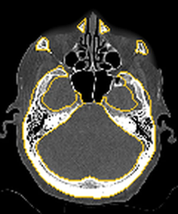
Aynı bölgenin, kaynak veri setinde patoloji içermediği belirtilen bir BT incelemesini kesit kesit kaydırın; organ sınırlarını açarak anatomiyi hasta görüntüsüyle karşılaştırın. Küçük rastlantısal bulgular tümüyle dışlanmamıştır.
Kaynak: TotalSegmentator v2.01 veri seti (Wasserthal ve ark., Radiology: Artificial Intelligence 2023), CC BY 4.0, vaka s0849; organ sınırları veri setinin otomatik segmentasyonundan, birleştirilerek sadeleştirildi. Görüntüler 2,5 mm kesit aralığına yeniden örneklendi.
Ayırıcı tanı
- Retansiyon kisti
- sinüs duvarına geniş tabanla oturan kubbe biçimli opasite yapar, ostiumu dolduran devamlı kitle gibi davranmaz.
- Mukozal kalınlaşma ve inflamatuar polipler, sinüs patolojileri arasında kısmi veya total opasifikasyon ile birlikte sınıflandırılan yaygın inflamatuvar bulgulardır.
- Mukosel
- sinüsü genişleten homojen içerik ve düzgün kemik incelmesi/remodeling ile seyredebilir.
- Fungal enfeksiyonlar (mikotik top veya alerjik fungal rinosinüzit), sinüs lümeninde yüksek yoğunluklu odaklar ve çevresinde mukozal kalınlaşma şeklinde izlenebilir; bu radyoopak yapılar konvolütör BT veya multidedektörlü BT ile net olarak saptanır.
- Sinonazal tümör
- kalıcı solid doku, komşu boşluklara uzanım veya kemikte fokal yıkım gösterebilir.
Tuzaklar
- Tek bir kesitte görünen opasitelerin anatomik varyasyon mu yoksa patoloji mi olduğundan emin olmak için altmilimetrik dilim kalınlığında ve çoklu düzlem rekonstrüksiyonlarında üç boyutlu veri setinin analizi yapılmalıdır.
- Hava-sıvı seviyesi görüldüğünde sıvı dansitesini solid doku olarak adlandırma; yatay ara yüzü ve bağımlı dağılımı diğer düzlemlerde doğrula.
- Tam opasifikasyon bulunan sinüste yumuşak doku veya intrakraniyal uzanımdan şüphelenildiğinde BT'yi MRG ile tamamlayın.
Kendini dene
Sinonasal bölgede kontrastlı BT'de heterojen enfekte, lokal invaziv yumuşak doku lezyonu ve kemikte erozyon görülüyor. En olası tanı nedir?
Cevabı göster
Sinonazal malign tümör. S50 ve S49'a göre, lokal invazyon, heterojen enfekte yapı ve kemik yıkımı sinonazal maligniteyi işaret eden kırmızı bayraklardır; kist ve mukoseller genelde ekspansil ama yıkıcı değildir.
BT görüntülerinde sinüs lümeninde bağımlı bölgede sıvı seviyesi ve üzerindeki hava kesiti görülürken sinüs duvarlarında belirgin bir deformasyon saptanmıyor. Bu bulgu en çok aşağıdakilerden hangisini düşündürür?
Cevabı göster
Tabakalanan sekresyon veya mukopiyosel. S80'a göre hava-sıvı seviyesi sinüs patolojileri arasında ayrı bir alt gruptur. Mukoseller, sinüsü genişletebilen ve ilerleyici büyümeleri kemikte yeniden şekillenmeye yol açabilen benign, lokal agresif lezyonlardır.
Kaynaklar
Bu sayfadaki 34 cümle ve 2 quiz sorusu aşağıdaki kaynaklardan alıntılarla tek tek karşılaştırıldı; 13 cümle bu karşılaştırmada düzeltildi (2026-10-08).
- Ultra-low 70-kV cardiac CT angiography in neonates: a comparative dose-optimized approach with preserved image quality.pubmed.ncbi.nlm.nih.gov
- CT Imaging for Valvular Interventions.pubmed.ncbi.nlm.nih.gov
- Branch-Critical Clipping of a Ruptured Carotid-Posterior Communicating Aneurysm with Fetal PCA Configuration.pubmed.ncbi.nlm.nih.gov
- Sinonasal Inflammation or Neoplasm: Raise the Red Flagsǃ-A Pictorial Review.pubmed.ncbi.nlm.nih.gov
- Imaging of Skull Base Tumors.pubmed.ncbi.nlm.nih.gov
- Chronic sphenoid rhinosinusitis: management challenge.pubmed.ncbi.nlm.nih.gov
- Computed Tomographic and Magnetic Resonance Imaging Diagnosis of Concurrent Sinonasal Aspergillosis and Meningoencephalocele in a Dog.pubmed.ncbi.nlm.nih.gov
- Computed tomographic features of severe horn infection in a male Scimitar-horned oryx (Oryx dammah).pubmed.ncbi.nlm.nih.gov
- Cardiac lead perforation: Mechanisms, detection, and therapeutic approaches.pubmed.ncbi.nlm.nih.gov
- Antimicrobial prescribing guidelines for horses in Australia.pubmed.ncbi.nlm.nih.gov
- Neuroimaging Patterns of Intracranial Infections: Meningitis, Cerebritis, and Their Complications.pubmed.ncbi.nlm.nih.gov
- A comprehensive framework for navigating patient care in systemic sclerosis: A global response to the need for improving the practice of diagnostic and preventive strategies in SSc.pubmed.ncbi.nlm.nih.gov
- Spontaneous rupture of a giant mature teratoma in the lateral ventricle: a case report.pubmed.ncbi.nlm.nih.gov
- Endoscopic Management of Post-Bariatric Surgery Complications: Diagnostic Work-Up and Innovative Approaches for Leak, Fistula, and Stricture Management.pubmed.ncbi.nlm.nih.gov
- Panoramic description of ROS-based nanotechnology for osteomyelitis therapy: Challenges, opportunities, and prospects.pubmed.ncbi.nlm.nih.gov
- Cavernous venous malformation of the frontal sinus: CT and MRI imaging findings.pubmed.ncbi.nlm.nih.gov
- Role of magnetic resonance in bowel pathologies: Beyond inflammatory bowel disease.pubmed.ncbi.nlm.nih.gov
- Giant Oncocytic Sinonasal Papilloma: A Rare Case.pubmed.ncbi.nlm.nih.gov
- The Initial Diagnostic Approach to Adult Congenital Heart Disease: A Practical Imaging Pathway for General Cardiologists and Cardiac Imagers.pubmed.ncbi.nlm.nih.gov
- Chinese expert consensus on imaging examination and diagnosis of nasal cavity and paranasal sinus tumors.pubmed.ncbi.nlm.nih.gov
- Society for Cardiovascular Magnetic Resonance 2024 Cases of SCMR case series.pubmed.ncbi.nlm.nih.gov
- Cardiac Computed Tomography in Structural Heart Interventions: From Preprocedural Planning to Procedural Strategy.pubmed.ncbi.nlm.nih.gov
- Management of patients with atrial tachycardia: a clinical consensus statement of the European Heart Rhythm Association (EHRA) of the ESC, endorsed by the Heart Rhythm Society (HRS), the Asia Pacific Heart Rhythm Society (APHRS), the Latin American Heart Rhythm Society (LAHRS), and the Association for European Paediatric and Congenital Cardiology (AEPC).pubmed.ncbi.nlm.nih.gov
- Native aorto-ostial coronary lesions on CT coronary angiogram.pubmed.ncbi.nlm.nih.gov
- Commentary: an eye on PET quantification.pubmed.ncbi.nlm.nih.gov
- Long-term safety and effectiveness of the "OptEase" vena cava filter.pubmed.ncbi.nlm.nih.gov
- Intensive care course after stage 1 Norwood procedure: are there early predictors of failure?pubmed.ncbi.nlm.nih.gov
- Endoscopic visualization of CT-confirmed accessory maxillary ostium: a retrospective study.pubmed.ncbi.nlm.nih.gov
- Association of sphenoid sinus pneumatization patterns, Onodi cells, and rostrum pneumatization with natural ostium location: A cross-sectional CT study.pubmed.ncbi.nlm.nih.gov
- Posterior superior alveolar artery in severely atrophic maxillae prior to lateral sinus augmentation: a retrospective cross-sectional CBCT imaging study.pubmed.ncbi.nlm.nih.gov
- Morphological and Epidemiological Analysis of the Sphenoid Sinus Based on Computed Tomography and Magnetic Resonance Imaging: A Narrative Review.pubmed.ncbi.nlm.nih.gov
- Assessment of the impact of nasal septum deviation types on sinonasal variations and infundibulum morphometry in relation to gender and age using CT images.pubmed.ncbi.nlm.nih.gov
- Association Between DMFT Index and Sinonasal Anatomical Variations in Children: A Retrospective Cone-Beam Computed Tomography Study.pubmed.ncbi.nlm.nih.gov
- Ostiomeatal complex inflammation with a rare ethmoid sinolith utilizing cone-beam computed tomography: A clinical and radiological approach to diagnosis.pubmed.ncbi.nlm.nih.gov
- Comprehensive Management of Cholesteatoma in Otitis Media: Diagnostic Challenges, Imaging Advances, and Surgical Outcome.pubmed.ncbi.nlm.nih.gov
- Management of Adult Bronchiectasis: Consensus-Based Guidelines of the German Respiratory Society.pubmed.ncbi.nlm.nih.gov
- International Harmonization of Nomenclature and Diagnostic Criteria (INHAND): Nonproliferative and Proliferative Lesions of the Rabbit.pubmed.ncbi.nlm.nih.gov
- International Society of Paediatric Surgical Oncology (IPSO) Surgical Practice Guidelines.pubmed.ncbi.nlm.nih.gov
- Coexistence of Autoantibodies against the Golgi Complex and Ro52 Antigen in a Patient with Nonspecific Interstitial Pneumonia.pubmed.ncbi.nlm.nih.gov
- MR Imaging of the Upper Urinary Tract: Techniques, Diagnostic Performance, and Imaging Biomarkers.pubmed.ncbi.nlm.nih.gov
- Frontal Mucopyocoele Presenting With Recurrent Periorbital Cellulitis: A Delayed Diagnosis.pubmed.ncbi.nlm.nih.gov
- EACR 2025 Congress: Innovative Cancer Science, 16-19 June 2025.pubmed.ncbi.nlm.nih.gov
- Small ethmoid mucocele with intracranial extension associated with fibrous dysplasia: A rare case report.pubmed.ncbi.nlm.nih.gov
- Recurrent Post-traumatic Frontal Mucocele Following Previous Endoscopic Management: Alternative Surgical Reconstruction With Partial Capsule Resection.pubmed.ncbi.nlm.nih.gov
- Persistent presumed allergic fungal rhinosinusitis during tezepelumab therapy with improvement after revision surgery: a case report.pubmed.ncbi.nlm.nih.gov
- Sphenoid sinus anatomical variants and pathologies: pictorial essay.pubmed.ncbi.nlm.nih.gov
- Imaging of thoracic tuberculosis: pulmonary and extrapulmonary.pubmed.ncbi.nlm.nih.gov
- A practical overview of CT and MRI features of developmental, inflammatory, and neoplastic lesions of the sphenoid body and clivus.pubmed.ncbi.nlm.nih.gov
- SMARCB1-deficient sinonasal carcinoma in a pediatric patient: Imaging findings and differential diagnosis of a rare entity.pubmed.ncbi.nlm.nih.gov
- Case Report: Sinonasal plasmablastic lymphoma in a human immunodeficiency virus-negative elderly woman diagnosed by immunohistochemistry.pubmed.ncbi.nlm.nih.gov
- Delayed Diagnosis of Primary Sinonasal Mucosal Melanoma.pubmed.ncbi.nlm.nih.gov
- The Role of Computed Tomography in Imaging Non-neurologic Disorders of the Head in Equine Patients.pubmed.ncbi.nlm.nih.gov
- The Frontal Sinus and Frontal Recess: Anatomical, Radiological and Surgical Concepts.pubmed.ncbi.nlm.nih.gov
- Uncommon Urinary Actinomycosis Mimicking Upper Urinary Tract Urothelial Tumor: Case Report and Literature Review.pubmed.ncbi.nlm.nih.gov
- Narrative review of chest wall ultrasound: a practical approach.pubmed.ncbi.nlm.nih.gov
- Biomechanical and Aerodynamic Modulation for Sinonasal Homeostasis in Craniofacial Orthopedics: A Comprehensive Review of RAMPA Therapy.pubmed.ncbi.nlm.nih.gov
- Connecting the Airways: Current Trends in United Airway Diseases.pubmed.ncbi.nlm.nih.gov
- From inflammatory initiation to fibrotic remodeling: mechanisms and precision therapeutic strategies in otorhinolaryngologic involvement of IgG4-related disease.pubmed.ncbi.nlm.nih.gov
- Post-Traumatic Orbital Reconstruction Using Titanium Patient-Specific Implants: A Clinical and Radiological Cohort Study Focusing on Paranasal Sinuses Physiology.pubmed.ncbi.nlm.nih.gov
- Intravascular papillary endothelial hyperplasia of the maxillary sinus extending into the contralateral nasal cavity.pubmed.ncbi.nlm.nih.gov
- Juvenile psammomatoid ossifying fibroma of the ethmoid sinus successfully treated by endoscopic sinus surgery: a case report.pubmed.ncbi.nlm.nih.gov
- Mechanotherapeutic Modulation of the Nasal Microenvironment: RAMPA-Induced Maxillofacial Remodeling and Its Pathophysiological Impact on Mucus Rheology and Chronic Inflammation.pubmed.ncbi.nlm.nih.gov
- Reassessment of two finite-element studies of infant head trauma: limits of the modelling evidence for shaking-induced bridging-vein rupture and protection by enlarged subarachnoid spaces.pubmed.ncbi.nlm.nih.gov
- Impact of RAMPA Therapy on Nasal Cavity Expansion and Paranasal Drainage: Fluid Mechanics Analysis, CAE Simulation, and a Case Study.pubmed.ncbi.nlm.nih.gov
- Odontogenic Sinusitis: The Selective Role of a Modified Caldwell-Luc Approach in Two CT/CBCT-Documented Cases.pubmed.ncbi.nlm.nih.gov
- Mucosal Grafts and Flaps in Draf IIb and Draf III: A Systematic Review and Meta-analysis.pubmed.ncbi.nlm.nih.gov
- Generalized Tetanus in an Unvaccinated 52-Year-Old Male Gardener: Diagnostic and Therapeutic Challenges in a Low-Resource Setting.pubmed.ncbi.nlm.nih.gov
- ESR Essentials: imaging in nasal obstruction and epistaxis-practice recommendations by the European Society of Head and Neck Radiology.pubmed.ncbi.nlm.nih.gov
- Contiguous orbital inflammation from paranasal sinus abnormalities in etiology of thyroid-associated ophthalmopathy.pubmed.ncbi.nlm.nih.gov
- Head of the Syrian Hamster (<i>Mesocricetus auratus</i>): Planar Anatomy with Correlative Micro-Computed Tomography and Magnetic Resonance Imaging.pubmed.ncbi.nlm.nih.gov
- Role of Imaging Spectrum Along With Other Diagnostic Modalities in Rhino-Orbital-Cerebral Mucormycosis (ROCM).pubmed.ncbi.nlm.nih.gov
- International Harmonization of Nomenclature and Diagnostic Criteria (INHAND): Non-proliferative and Proliferative Lesions of the Non-human Primate ( M. fascicularis ).pubmed.ncbi.nlm.nih.gov
- Turkish Guideline for Diagnosis and Treatment of Allergic Rhinitis (ART).pubmed.ncbi.nlm.nih.gov
- New advances in jellyfish anatomy: the benefits of endocasts and X-ray microtomography in the investigation of the gastrovascular system of Cotylorhiza tuberculata (Scyphozoa; Rhizostomeae; Cepheidae).pubmed.ncbi.nlm.nih.gov
- Standards for quantitative assessments by coronary computed tomography angiography (CCTA): An expert consensus document of the society of cardiovascular computed tomography (SCCT).pubmed.ncbi.nlm.nih.gov
- 3D-Printing to Plan Complex Transcatheter Paravalvular Leaks Closure.pubmed.ncbi.nlm.nih.gov
- Head and neck manifestations of extramedullary plasmacytomas and their differential diagnoses: a pictorial review.pubmed.ncbi.nlm.nih.gov
- Pre-FESS Imaging of Paranasal Sinuses and Nasal Cavity: Using Multi-detector Computed Tomography (MDCT) in Understanding Normal Anatomy and Anatomical Variations: Tips and Tricks.pubmed.ncbi.nlm.nih.gov
- The maxillary sinus: physiology, development and imaging anatomy.pubmed.ncbi.nlm.nih.gov
- Relationships Between Radiomorphometric Parameters of the Maxillary Sinus and Development of Sinusitis: A Cone Beam Computed Tomography Study.pubmed.ncbi.nlm.nih.gov
- Operative Corridors in Endoscopic Skull Base Tumor Surgery.pubmed.ncbi.nlm.nih.gov
- Best Practice Recommendations for Infertility Management.pubmed.ncbi.nlm.nih.gov
- 2.5-mm articulated endoluminal forceps using a compliant mechanism.pubmed.ncbi.nlm.nih.gov
- Inflammatory Modifications in Paranasal Sinuses and Ostiomeatal Complex Anatomical Variations in Jet Aircraft Pilots: A Computed Tomography Study.pubmed.ncbi.nlm.nih.gov
- Uncommon Nasal Mass Presentation: A Radiological Case Series.pubmed.ncbi.nlm.nih.gov
- Foreign body-associated maxillary sinus aspergillosis caused by Endomethasone extrusion: diagnostic challenges, biological mechanisms, and surgical management.pubmed.ncbi.nlm.nih.gov
- Radiologic Characterization of Invasive Fungal Infections of the Paranasal Sinuses and Skull Base: A Prospective Analysis.pubmed.ncbi.nlm.nih.gov
- Diagnosis of Mucormycosis: Current Situation, Challenges and Future Prospects.pubmed.ncbi.nlm.nih.gov
- High-affinity alphavbeta3 integrin targeted optical probe as a new imaging biomarker for early atherosclerosis: initial studies in Watanabe rabbits.pubmed.ncbi.nlm.nih.gov
- Rhinosinusitis: Evidence and experience - 2024.pubmed.ncbi.nlm.nih.gov
- Danger points, complications and medico-legal aspects in endoscopic sinus surgery.pubmed.ncbi.nlm.nih.gov
- Emerging and re-emerging infectious disease in otorhinolaryngology.pubmed.ncbi.nlm.nih.gov
- Intracranial venous thrombosis imaging by anatomical location: an educational review with mimics and diagnostic pitfalls.pubmed.ncbi.nlm.nih.gov
- Imaging and physiology across the high-low cerebrospinal fluid pressure spectrum: Navigating diagnostic uncertainty in headache practice.pubmed.ncbi.nlm.nih.gov
- Neurolymphomatosis: A Comprehensive Review of Clinical and Imaging Features.pubmed.ncbi.nlm.nih.gov
- Imaging of congenital lung diseases presenting in the adulthood: a pictorial review.pubmed.ncbi.nlm.nih.gov
- Complex adult congenital heart disease on cross-sectional imaging: an introductory overview.pubmed.ncbi.nlm.nih.gov
- Intracardiac eustachian valve cyst in an adult detected with other cardiac anomalies: usefulness of multidetector CT in diagnosis.pubmed.ncbi.nlm.nih.gov
- Morphometric Analysis of the Maxillary Sinus Using CT-Based Air-Cavity Reconstruction: A Retrospective Study.pubmed.ncbi.nlm.nih.gov
- A case of respiratory epithelial adenomatoid hamartoma in a patient suffering from non-steroidal anti-inflammatory drug - exacerbated respiratory disease.pubmed.ncbi.nlm.nih.gov
- Occult recurrence of sinonasal inverted papilloma masked by a mucocele: a case report.pubmed.ncbi.nlm.nih.gov
- Beyond Angiography: Cardiac CT for Planning Complex PCI in Calcified Coronary Lesions.pubmed.ncbi.nlm.nih.gov
- Trauma of the midface.pubmed.ncbi.nlm.nih.gov
- Paranasal sinus occupancy assessed from magnetic resonance images-associations with clinical indicators in patients with systemic lupus erythematosus.pubmed.ncbi.nlm.nih.gov
- Bilateral Sinonasal Inverted Papilloma: Report of an Uncommon Case Involving Sinuses of the Face and Orbital Cavity.pubmed.ncbi.nlm.nih.gov
- Hemodynamic Significance of APEXCT Coronary Angiography Combined With Dynamic CT Myocardial Perfusion Imaging in Assessing Restenosis After Coronary Stenting.pubmed.ncbi.nlm.nih.gov
- High CT Attenuation Values Relative to the Brainstem Predict Fungal Hyphae Within the Sinus.pubmed.ncbi.nlm.nih.gov
- Society for Cardiovascular Magnetic Resonance/European Society of Cardiovascular Imaging/American Society of Echocardiography/Society for Pediatric Radiology/North American Society for Cardiovascular Imaging Guidelines for the Use of Cardiac Magnetic Resonance in Pediatric Congenital and Acquired Heart Disease: Endorsed by The American Heart Association.pubmed.ncbi.nlm.nih.gov
- Severe facial pain and removal of maxillary sinus mucous retention cyst.pubmed.ncbi.nlm.nih.gov
- Clinician-Guided Deep Learning Segmentation of Skull Base Pneumatization on Computed Tomography Using 3D Slicer and MONAI Label.pubmed.ncbi.nlm.nih.gov
Eğitim amaçlıdır; hasta başında tanı ve tedavi kararının yerine geçmez. İçerik yapay zekâ destekli bir süreçle güncel literatüre göre cümle cümle kaynaklarla karşılaştırıldı, ancak uzman radyolog incelemesinden geçmedi. Klinik kararlarda güncel kılavuzları, kurum protokollerini ve radyoloji raporunu esas alın.