Özet
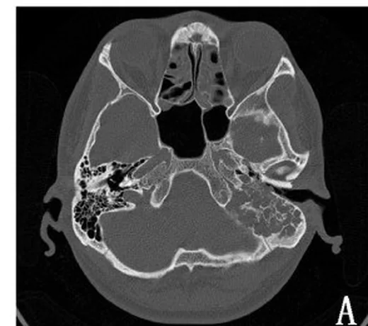
Endolenfatik kese tümörü, posterior petroz temporal kemikte endolenfatik duktus-kese kompleksinden gelişen nadir, lokal invaziv bir neoplazmdır ve von Hippel–Lindau hastalığıyla ilişkili olabilir. İnce kesit kemik algoritmalı temporal kemik BT, posterior petroz kemikteki yıkımı ve lezyon içindeki spiküle, noktalı veya retiküler yüksek dansiteli odakları gösterebilir. Kitle labirente, mastoide, orta kulağa veya kafa içi kompartımanlara uzanabilir; MR kitlenin özelliklerini değerlendirirken anjiyografi tümörün damar yapısı ve beslenmesini göstermeye yardımcı olur. ELST'nin merkezi tipik olarak retrolabirentin alanda, vestibüler akuadukt düzeyinde izlenirken, ayırıcı tanıda yer alan paragangliomlar juguler foramen veya promontoryum civarında merkezlenir; bu anatomik ayrım lezyonun doğru merkezini belirlemek için kritiktir.
Faz ve pencere
- Kontrastsız tanısal
- İnce kesit kemik algoritmalı temporal kemik BT'de vestibüler akuadukt çevresinde retrolabirentin litik-destrüktif değişiklik, genişlemiş veya silinmiş akuadukt konturu, düzensiz reaktif kemik ve tümör içinde kemiksi spiküller aranır. Kemik merkezini saptamak için kontrastsız BT tanısaldır; yumuşak doku kontrastlanmasını değerlendirmek bu BT fazının amacı değildir.
Önerilen pencereler: Kemik (G 1800 / M 400), Yumuşak doku (G 400 / M 40).
BT bulguları
- İnce kesit kemik algoritmalı BT'de posterior petroz kemikte genişleyici litik yıkım ve lezyon içinde spiküle, noktalı veya retiküler yüksek dansiteli odaklar görülebilir; bu odaklar kalıntı kemik spiküllerini yansıtabilir.
- Akuadukt kanalının çevre kemik erozyonu içinde genişlemiş görünmesi.
- Destrüktif lezyon içinde ince, düzensiz kemiksi spiküller ya da reaktif kemik adacıkları
- Posterior petroz kemik konturunda ekspansiyon ve dış kortekste incelme
- Mastoide, posterior timpanik kaviteye, juguler fossa kenarına veya posterior fossa dura komşuluğuna yumuşak doku uzanımı
- İleri olguda semisirküler kanal, vestibül veya koklea çevresindeki otik kapsül sınırında bozulma
- Kontrastlı MR'da ELST heterojen görünümlü ve kontrastlanan bir kitle olarak izlenebilir; kontrastsız T1 ağırlıklı görüntülerdeki hiperintens odaklar kan ürünleriyle ilişkili olabilir.
Normalde
Vestibüler akuadukt, endolenfatik duktusun içinden geçtiği kemik kanaldır. Lezyonun vestibüler akuadukt ve retrolabirent kemikte mi, yoksa juguler foramen veya promontoryumda mı merkezlendiğini karşılaştır; ELST kökeni genellikle retrolabirentin alanda izlenirken temporal kemik paragangliomları sıklıkla juguler foramen bölgesinde veya promontoryumda yer alır.
Ayırıcı tanı
- Jugulotimpanik paragangliom
- kemik değişikliği juguler foramen çevresinde veya promontoryumda merkezlenir; endolenfatik kese bölgesi başlangıç odağı değildir.
- Orta kulak adenomu/nöroendokrin tümörü
- orta kulak ve mastoidle sınırlıdır; kemik yıkımı nadirdir.
- Temporal kemik metastazı
- ayırıcı tanıda özellikle tiroid veya böbrek kaynaklı metastatik adenokarsinomlar yer alır.
- Geniş vestibüler akuadukt
- ELST erken dönemde BT’de yalnız vestibüler akuadukt genişlemesiyle görülebilir.
- Kolesteatom
- orta kulak yumuşak doku dansitesi skutum erozyonuyla birlikte görülebilir.
- Özellikle hipervasküler renal hücreli karsinom veya tiroid kaynaklı metastazlar, retrolabirentin kemik yıkımı yapan kitlelerde ayırıcı tanıda düşünülmelidir.
Tuzaklar
- Orta kulak ya da mastoidde görülen yumuşak doku tümörün başlangıç yeri olmayabilir; kemik penceresinde geriye doğru iz sürerek retrolabirentin odağı saptamadan merkez tanımlama.
- Lezyon içindeki retiküler, spiküle veya noktalı yüksek dansiteli odaklar doğrudan kalsifikasyon olarak adlandırılmamalıdır; bu görünümler kalıntı kemik spiküllerinden kaynaklanabilir.
- Vestibüler akuaduktun geniş görünmesi tek başına tümörü dışlamaz; erken ELST’de BT’de yalnız genişleme görülebilir.
- BT'de dura veya labirentle temas invazyonun derecesini göstermez; yumuşak doku ve intrakraniyal yayılım kuşkusunu MR ile tamamla.
Kendini dene
Retrolabirentin bölgede, vestibüler akuadukt ve endolenfatik kese çevresinde yerleşen; içinde spiküle veya retiküler yüksek dansite odakları bulunan kemik yıkımı görülüyor. En olası tanı hangisidir?
Cevabı göster
Endolenfatik kese tümörü. Retrolabirentin yerleşim, kemik erozyonu ve tümör içindeki spiküle ya da retiküler yüksek dansite odakları endolenfatik kese tümörünü destekler. Temporal kemik paragangliomları sıklıkla juguler foramen bölgesinde veya promontoryumda yer alır; orta kulak adenomu orta kulak ve mastoidle sınırlı olabilir ve kemik yıkımı nadirdir.
Erken ELST'de temporal kemik BT'sinde görülebilecek bulgu hangisidir?
Cevabı göster
Yalnız kemik erozyonu veya vestibüler akuadukt genişlemesi. Erken ELST'de BT'de yalnız kemik erozyonu veya vestibüler akuadukt genişlemesi görülebilir.
Kaynaklar
Bu sayfadaki 36 cümle ve 2 quiz sorusu aşağıdaki kaynaklardan alıntılarla otomatik olarak karşılaştırıldı; 21 cümle bu karşılaştırmada düzeltildi (2026-10-09).
- Surgical Management of Endolymphatic Sac Tumor: A Systematic Review and Meta-Analysispmc.ncbi.nlm.nih.gov
- Current Treatment of Endolymphatic Sac Tumor of the Temporal Bonepmc.ncbi.nlm.nih.gov
- Endolymphatic sac tumor: case report and review of the literaturepmc.ncbi.nlm.nih.gov
- What the neurosurgeon should know about hemangioblastoma, both sporadic and in Von Hippel-Lindau disease: A literature reviewpmc.ncbi.nlm.nih.gov
- Case report: endolymphatic sac tumor masquerading as Meniere’s diseasepmc.ncbi.nlm.nih.gov
- What the neurosurgeon should know about hemangioblastoma, both sporadic and in Von Hippel-Lindau disease: A literature review.pubmed.ncbi.nlm.nih.gov
- Endolymphatic sac tumor: case report and review of the literature.pubmed.ncbi.nlm.nih.gov
- [Endolymphatic sac tumor with symptoms of endolymphatic hydrops: a case report and reviews].pubmed.ncbi.nlm.nih.gov
- Plasmacytoma of the Temporal Bone, a Great Imitator: Report of Seven Cases and Comprehensive Review of the Literature.pubmed.ncbi.nlm.nih.gov
- Endolymphatic sac tumor with von Hippel-Lindau disease: report of a case with analysis of von Hippel-Lindau gene and review.pubmed.ncbi.nlm.nih.gov
- Gamma knife treatment of an endolymphatic sac tumor: unique features of a case and review of the literature.pubmed.ncbi.nlm.nih.gov
- Von Hippel-Lindau disease.pubmed.ncbi.nlm.nih.gov
- Intralabyrinthine Cholesterol Granuloma.pubmed.ncbi.nlm.nih.gov
- Endolymphatic sac tumor.pubmed.ncbi.nlm.nih.gov
- Clinicoradiologic characteristics of endolymphatic sac tumors.pubmed.ncbi.nlm.nih.gov
- [Typical tumors of the petrous bone].pubmed.ncbi.nlm.nih.gov
- 68Ga-DOTATATE Uptake in an Endolymphatic Sac Tumor: Radiologic-Pathologic Correlation.pubmed.ncbi.nlm.nih.gov
- Endolymphatic Sac Tumor in Two Dogs.pubmed.ncbi.nlm.nih.gov
- Gamma Knife Radiosurgery for Endolymphatic Sac Tumors: A Single-Institution Experience.pubmed.ncbi.nlm.nih.gov
- Von Hippel-Lindau syndrome presenting as a lateral ventricular space-occupying lesion: a case report.pubmed.ncbi.nlm.nih.gov
- Von Hippel-Lindau Syndrome: An Updated Narrative Review for the First-Contact Clinician.pubmed.ncbi.nlm.nih.gov
- Case report: endolymphatic sac tumor masquerading as Meniere's disease.pubmed.ncbi.nlm.nih.gov
- Von Hippel-Lindau Disease-Associated Endolymphatic Sac Tumours: Seven Cases and Genotype-Phenotype Features.pubmed.ncbi.nlm.nih.gov
- Case report: Cochlear implantation for deafness caused by endolymphatic sac tumors in patients with von Hippel-Lindau syndrome.pubmed.ncbi.nlm.nih.gov
- Case report: Endolymphatic sac tumor with blurred vision.pubmed.ncbi.nlm.nih.gov
- Auriculotemporal Carcinoma - A Retrospective Case Series.pubmed.ncbi.nlm.nih.gov
- CAIX and pax-8 Commonly Immunoreactive in Endolymphatic Sac Tumors: A Clinicopathologic Study of 26 Cases with Differential Considerations for Metastatic Renal Cell Carcinoma in von Hippel-Lindau Patients.pubmed.ncbi.nlm.nih.gov
- Imaging in Pulsatile Tinnitus: Case Based Review.pubmed.ncbi.nlm.nih.gov
- Cross Sectional Imaging of the Ear and Temporal Bone.pubmed.ncbi.nlm.nih.gov
- Bilateral intradiploic dermoid cysts with unilateral rupture causing hearing loss: illustrative case.pubmed.ncbi.nlm.nih.gov
- Cancer predisposition syndromes: an imaging review.pubmed.ncbi.nlm.nih.gov
- Imaging of Temporal Bone Mass Lesions: A Pictorial Review.pubmed.ncbi.nlm.nih.gov
- Imaging of Skull Base Tumors.pubmed.ncbi.nlm.nih.gov
- Imaging of skull base: Pictorial essay.pubmed.ncbi.nlm.nih.gov
- Diagnosis and Management of Middle Ear Neuroendocrine Tumour (MeNET).pubmed.ncbi.nlm.nih.gov
- Combined Petrosectomy in a 7-Year-Old Patient: Case-Based Video Illustration and Technical Considerations for Petrosal Approaches in the Pediatric Population.pubmed.ncbi.nlm.nih.gov
- Third Window Syndrome: An Up-to-Date Systematic Review of Causes, Diagnosis, and Treatment.pubmed.ncbi.nlm.nih.gov
- The angular trajectory of the vestibular aqueduct: evaluation using photon-counting CT.pubmed.ncbi.nlm.nih.gov
- A modern conceptual framework for study and treatment of Meniere's disease.pubmed.ncbi.nlm.nih.gov
- Nonvestibular Schwannoma Tumors in the Cerebellopontine Angle: A Single-Surgeon Experience.pubmed.ncbi.nlm.nih.gov
- Immunohistochemical Expression Pattern of Theragnostic Targets SSTR2 and PSMA in Endolymphatic Sac Tumors: A Single Institution Case Series.pubmed.ncbi.nlm.nih.gov
- Neuroendocrine Neoplasms of the Ear.pubmed.ncbi.nlm.nih.gov
- Survival outcomes and prognostic factors in temporal bone malignancies: A retrospective cohort of 20 patients.pubmed.ncbi.nlm.nih.gov
- Branchio-oto-renal syndrome: a narrative review.pubmed.ncbi.nlm.nih.gov
- Genetic and Environmental Factors Shaping Hearing Loss: Xenobiotics, Mechanisms and Translational Perspectives.pubmed.ncbi.nlm.nih.gov
- Utility and Challenges of Imaging in Peripheral Vestibular Disorder Diagnosis: A Narrative Review.pubmed.ncbi.nlm.nih.gov
- Engineered Inner Ear Drug Delivery Systems for Hearing Loss Treatment.pubmed.ncbi.nlm.nih.gov
- Update from the 5th Edition of the World Health Organization Classification of Head and Neck Tumours: Tumours of the Ear.pubmed.ncbi.nlm.nih.gov
- Origin of endolymphatic sac tumor.pubmed.ncbi.nlm.nih.gov
- Nonvascular lesions of the jugular foramen: the gruppo otologico experience.pubmed.ncbi.nlm.nih.gov
- Brazilian Society of Otology task force - cochlear implant ‒ recommendations based on strength of evidence.pubmed.ncbi.nlm.nih.gov
- The Evolution of Skull Base Surgery: A Bibliometric Analysis Spanning Nearly 125 Years.pubmed.ncbi.nlm.nih.gov
- Anatomical Step-by-Step Dissection of Complex Skull Base Approaches for Trainees: Surgical Anatomy of the Posterior Petrosal Approach.pubmed.ncbi.nlm.nih.gov
- Lateral Skull Base Approaches in Pediatric Skull Base Surgery.pubmed.ncbi.nlm.nih.gov
- Update from the 5th Edition of the World Health Organization Classification of Head and Neck Tumors: Familial Tumor Syndromes.pubmed.ncbi.nlm.nih.gov
- Secondary endolymphatic hydrops: a clinical and literature overview.pubmed.ncbi.nlm.nih.gov
- MRI can help differentiate Ménière's disease from other menieriform diseases.pubmed.ncbi.nlm.nih.gov
- Rare Disorders of the Vestibular Labyrinth: of Zebras, Chameleons and Wolves in Sheep's Clothing.pubmed.ncbi.nlm.nih.gov
- Posterior Fossa Meningioma with Endolymphatic Sac Compression: Resolution of Dizziness after Surgery.pubmed.ncbi.nlm.nih.gov
- A Case of Petrous Apex Cholesteatoma Removed Using an Exoscope and an Endoscope Without Posterior Rerouting of the Facial Nerve.pubmed.ncbi.nlm.nih.gov
- Papillary endolymphatic sac tumor: Case report with clinical study and therapeutic management.pubmed.ncbi.nlm.nih.gov
- Non-invasive imaging modalities for diagnosing pulsatile tinnitus: a comprehensive review and recommended imaging algorithm.pubmed.ncbi.nlm.nih.gov
- Unusual cystic presentation of pheochromocytoma in a case of suspected von Hippel-Lindau disease.pubmed.ncbi.nlm.nih.gov
- The Pheochromocytoma/Paraganglioma syndrome: an overview on mechanisms, diagnosis and management.pubmed.ncbi.nlm.nih.gov
- Prevalence of the endolymphatic sac hypoplasia in a cohort of individuals without Meniere disease.pubmed.ncbi.nlm.nih.gov
- Surgical Treatment of Lateral Skull Base Tumors Extending to the Craniovertebral Junction.pubmed.ncbi.nlm.nih.gov
- Infratemporal Fossa Approach with Preservation of the Posterior Bony Wall of External Auditory Canal: Case Series and the Outcome.pubmed.ncbi.nlm.nih.gov
- Update on diagnostic procedures in third window syndromes.pubmed.ncbi.nlm.nih.gov
- Multimodal imaging in conductive hearing loss: Optimising CT, MRI and CTA for accurate diagnosis and management.pubmed.ncbi.nlm.nih.gov
- Fourth Ventricle Epidermoid Cyst: Case Report of Precision Telovelar Microsurgery, Functional Preservation, and Lifelong Surveillance.pubmed.ncbi.nlm.nih.gov
- Bony labyrinth morphology reveals deep population history and isolation among Homo sapiens in prehistoric northeastern Africa (44-3 ka).pubmed.ncbi.nlm.nih.gov
- "Genetic tumor syndromes of the head and neck: Update in the genomic era".pubmed.ncbi.nlm.nih.gov
- Pediatric Cholesteatoma Presenting as Persistent Otorrhea: A Case of Delayed Diagnosis Across Multiple ENT Specialists.pubmed.ncbi.nlm.nih.gov
- Trichoepithelioma of the External Auditory Canal Mimicking Cholesteatoma.pubmed.ncbi.nlm.nih.gov
- Surgery of the ear and the lateral skull base: pitfalls and complications.pubmed.ncbi.nlm.nih.gov
- Pathology of the Nervous System in Von Hippel-Lindau Disease.pubmed.ncbi.nlm.nih.gov
- Surgical Management of Endolymphatic Sac Tumor: A Systematic Review and Meta-Analysis.pubmed.ncbi.nlm.nih.gov
- Von Hippel-Lindau Disease (VHL): Characteristic Lesions with Classic Imaging Findings.pubmed.ncbi.nlm.nih.gov
- Three-dimensional exoscope-assisted combined petrosal approach for petroclival meningiomas: technical nuances from early experience.pubmed.ncbi.nlm.nih.gov
- Therapy options in the management of endolymphatic sac tumors: A case series and literature review.pubmed.ncbi.nlm.nih.gov
- Re-Examining the Cochlea in Branchio-Oto-Renal Syndrome: Genotype-Phenotype Correlation.pubmed.ncbi.nlm.nih.gov
Eğitim amaçlıdır; hasta başında tanı ve tedavi kararının yerine geçmez, tıbbi tavsiye değildir. Metinler yapay zekâ desteğiyle yazıldı ve otomatik bir adımda kaynak alıntılarıyla karşılaştırıldı; bu bir tıbbi doğrulama değildir ve içerik uzman radyolog incelemesinden geçmedi. Klinik kararlarda güncel kılavuzları, kurum protokollerini ve radyoloji raporunu esas alın.