Özet
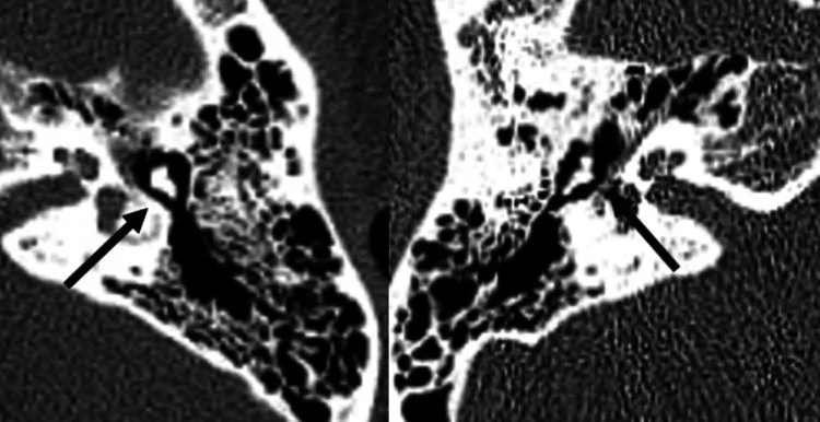
Fasiyal sinir kanalı dehisansı, kemik kanalın özellikle oval pencere üstündeki timpanik segmentinde örtünün seçilememesidir ve çoğu kez tesadüfen saptanır. İnce kesit kontrastsız temporal kemik BT'si sinirin kendisini değil, onu çevreleyen kemik kanalın sürekliliğini gösterir; cerrahi öncesinde bu anatomiyi tanımak önemlidir. Orta kulak kolesteatomu veya inflamasyonu dehisans alanına uzanabilir ve siniri yaralanmaya açık bırakabilir. İnce kemik duvarı gerçek açıklık sanılırsa bulgu abartılabilir; gerçek dehisans atlanırsa otolojik cerrahide sinir hasarı riski gözden kaçabilir.
Faz ve pencere
- Kontrastsız tanısal
- Kontrastsız ince kesit temporal kemik BT'sinde, özellikle oval pencere üstündeki timpanik segmentte kemik kanalın fokal kortikal devamlılık kaybı aranır. Kaynak kesitler ve çok düzlemli kemik rekonstrüksiyonları kanalı, oval pencereyi, kokleariform çıkıntıyı ve ikinci genuyu gösterir. İnce veya parsiyel hacim etkili korteks, ardışık kesitlerde ve ortogonal düzlemde devamlılık izlenerek gerçek açıklıktan ayırt edilir; İV kontrast gerekmez.
Önerilen pencereler: Kemik (G 1800 / M 400).
BT bulguları
- Timpanik kanal örtüsü — oval pencerenin hemen üstünde kemik kanalın fokal olarak izlenememesi dehisansın tipik yeridir.
- Fasiyal sinirin timpanik (yatay) ve mastoid (düşey) segmentlerinin seyri temporal kemik BT'sinde değerlendirilir.
- Mastoid segment — düşey kemik kanalın mastoid içinde aşağı inen kortike sınırları değerlendirilir; timpanik dehisansla aynı bölge değildir.
- Orta kulak komşuluğu — kanal defektinin oval pencere nişi veya orta kulak yumuşak dokusuyla doğrudan ilişkisi aranır.
- İnce kemik duvarı — parsiyel hacim etkisi ve çok ince korteks, ardışık kesitlerde devamlılıkla açıklık iddiasından ayrılır.
- Eşlik eden erozyon — kolesteatom veya kronik otit varsa kemikçik, scutum ve kanal komşuluğundaki erozyonun yayılımı tarif edilir.
Normalde
Normal temporal kemik BT'sinde fasiyal kanal, labirentin, timpanik ve mastoid segmentler boyunca izlenen kemik kanalı ile tanımlanır. Aynı seviyede iki tarafın özellikle oval pencere üstündeki kemik örtüsünü karşılaştır; tek kesitte silik görünen çok ince duvarı, ardışık görüntülerde de yokluğu gösterilmeden dehisans sayma.
Ayırıcı tanı
- İnce fasiyal kanal duvarı
- kemik örtü çok ince görünür ancak ardışık kesitlerin bir bölümünde devamlılık seçilebilir.
- Kolesteatom erozyonu
- orta kulak yumuşak dokusu scutum veya kemikçiklerde erozyonla birlikte kanal duvarını bozabilir.
- Kronik otit sekeli
- sklerotik mastoid ve orta kulak değişiklikleri eşlik eder; izole gelişimsel açıklık görünümü beklenmez.
- Parsiyel hacim etkisi
- kanal eğimine dik olmayan tek kesitte duvar kaybolur gibi olur, ortogonal düzlemde yeniden izlenir.
Tuzaklar
- Oval pencere üzerindeki çok ince korteks BT çözünürlüğünün altında kalabilir; tek görüntüdeki görünmezliği kesin açıklık diye raporlama.
- Timpanik segmentin kavisli seyri aksiyel düzlemde aralıklıymış gibi görünür; kanalın uzun eksenine uygun oblik koronal rekonstrüksiyonda devamlılığı kontrol et.
- Kanal çevresindeki orta kulak opasifikasyonu tek başına sinirin açığa çıktığını göstermez; önce kemik sınırdaki gerçek kaybı doğrula.
Kendini dene
Oval pencere düzeyindeki timpanik fasiyal kanalın kemik örtüsünde kesinti izleniyor ve dehisans siniri orta kulak içeriğine açık bırakıyor. Bu bulgu nedir?
Cevabı göster
Fasiyal kanal dehisansı. Fasiyal kanal dehisansı, kemik kanal yapısındaki kesintiyle tanımlanır; BT'de timpanik segmentin kemik örtüsünün eksik olduğu gösterilebilir.
Preoperatif temporal kemik BT'sinde fasiyal sinir kanalının kemik örtüsünü değerlendirmeyi güçleştirebilecek teknik sınırlılıklar nelerdir?
Cevabı göster
Dilim kalınlığı kısıtları ve parsiyel hacim artefaktları. Dilim kalınlığı kısıtları ve parsiyel hacim artefaktları küçük kemik defektlerini maskeleyebilir veya görüntüyü bozabilir. Kolesteatom olgularında BT, fasiyal kanal dehisansını intraoperatif değerlendirmeye kıyasla daha sık göstermiştir.
Kaynaklar
Bu sayfadaki 33 cümle ve 2 quiz sorusu aşağıdaki kaynaklardan alıntılarla otomatik olarak karşılaştırıldı; 3 cümle bu karşılaştırmada düzeltildi (2026-10-09).
- Recurrent unilateral facial nerve palsy in a child with dehiscent facial nerve canalpmc.ncbi.nlm.nih.gov
- Cerebellar abscess complicating chronic suppurative otitis media: An unusual route of intracranial spreadpmc.ncbi.nlm.nih.gov
- Middle ear malformations combined with cerebrospinal fluid otorrheapmc.ncbi.nlm.nih.gov
- The preoperative temporal bone CT: a mnemonic for reducing surgical riskpmc.ncbi.nlm.nih.gov
- Acute otitis media complicated by peripheral facial nerve palsy in an adult: A case reportpmc.ncbi.nlm.nih.gov
- Audiometric evaluation short and medium term in cochlear implants.pubmed.ncbi.nlm.nih.gov
- Recurrent transient facial nerve palsy on commercial air flights.pubmed.ncbi.nlm.nih.gov
- Labyrinthine Fistula in Chronic Otitis Media Surgery: Management and Outcomes.pubmed.ncbi.nlm.nih.gov
- Accuracy of High-Resolution Computed Tomography Compared to High-Definition Ear Endoscopy to Assess Cholesteatoma Extension.pubmed.ncbi.nlm.nih.gov
- Recurrent unilateral facial nerve palsy in a child with dehiscent facial nerve canal.pubmed.ncbi.nlm.nih.gov
- Bilateral carotid-cavernous fistulas following trauma: A rare case of diagnostic and management challenges.pubmed.ncbi.nlm.nih.gov
- Morphometric assessment of important landmarks on skull intended for Vidian nerve surgery.pubmed.ncbi.nlm.nih.gov
- Imaging of labyrinthine fistula after repair with bone pate.pubmed.ncbi.nlm.nih.gov
- Facial paralysis during air travel: case series and literature review.pubmed.ncbi.nlm.nih.gov
- Prevalence of facial canal dehiscence and other bone defects by computed tomography.pubmed.ncbi.nlm.nih.gov
- Prevalence of Cochlear-Facial Dehiscence in a Study of 1,020 Temporal Bone Specimens.pubmed.ncbi.nlm.nih.gov
- Audiovestibular Quantification in Rare Third Window Disorders in Children.pubmed.ncbi.nlm.nih.gov
- Radiologic classification of superior canal dehiscence: implications for surgical repair.pubmed.ncbi.nlm.nih.gov
- Computed tomography in chronic suppurative otitis media: value in surgical planning.pubmed.ncbi.nlm.nih.gov
- Middle ear malformations combined with cerebrospinal fluid otorrhea: A case report.pubmed.ncbi.nlm.nih.gov
- Congenital Anomalies and Variations of the Carotid and Vertebral Arteries: A Case-Based Imaging Review.pubmed.ncbi.nlm.nih.gov
- "Leave me alone": anatomical structures and variations seen on computed tomography of the temporal bone.pubmed.ncbi.nlm.nih.gov
- Anatomical Characteristics of the Facial Nerve in Patients With Cochlear Hypoplasia.pubmed.ncbi.nlm.nih.gov
- Assessment of the Oval Window-Facial Canal Corridor and Mastoid Pneumatization: A Preliminary HRCT-Based Radiological-Anatomical Framework.pubmed.ncbi.nlm.nih.gov
- The preoperative temporal bone CT: a mnemonic for reducing surgical risk.pubmed.ncbi.nlm.nih.gov
- Cerebellar abscess complicating chronic suppurative otitis media: An unusual route of intracranial spread.pubmed.ncbi.nlm.nih.gov
- Acute otitis media complicated by peripheral facial nerve palsy in an adult: A case report.pubmed.ncbi.nlm.nih.gov
- A Case of Acquired Pars Flaccida Cholesteatoma Extending to the Supralabyrinthine Petrous Apex With Preservation of the Otic Capsule.pubmed.ncbi.nlm.nih.gov
- Brazilian Society of Otology task force - Otosclerosis: evaluation and treatment.pubmed.ncbi.nlm.nih.gov
- International Society of Paediatric Surgical Oncology (IPSO) Surgical Practice Guidelines.pubmed.ncbi.nlm.nih.gov
- Total laparoscopic radical hysterectomy for cervical cancer in prolapsed uterus.pubmed.ncbi.nlm.nih.gov
- Key pre-operative CT parameters for cochlear implantation: a focused radiologic review with surgical relevance.pubmed.ncbi.nlm.nih.gov
- Rare Cause of Congenital Unilateral Facial Palsy: High-Resolution Computed Tomography (HRCT) Findings of Isolated Facial Canal Hypoplasia and Aplasia.pubmed.ncbi.nlm.nih.gov
- Facial Nerve Hemangioma in the Internal Auditory Canal: A Rare Localization and Therapeutic Challenge.pubmed.ncbi.nlm.nih.gov
- External auditory canal cholesteatoma presenting with facial nerve palsy: rare clinical entity.pubmed.ncbi.nlm.nih.gov
- Extensive Temporal Bone Necrosis in Petrous Bone Cholesteatoma Complicated by Actinomycosis: A Case Report.pubmed.ncbi.nlm.nih.gov
- Microsurgical Reconstruction of the Ear and Temporal Region: Structural and Functional Considerations Including Hearing Rehabilitation-A Narrative Review.pubmed.ncbi.nlm.nih.gov
- Multimodal imaging in neuro-ophthalmology.pubmed.ncbi.nlm.nih.gov
- Long-term Stable Unilateral Mandibular Deformity Associated With Ipsilateral Skull-base Soft-tissue Lesion and Degenerated Pterygoid Muscles in Neurofibromatosis Type 1.pubmed.ncbi.nlm.nih.gov
- The combined compartmental maxillary resection: technique codification and preliminary multicenter series analysis.pubmed.ncbi.nlm.nih.gov
- Pediatric Cholesteatoma Presenting as Persistent Otorrhea: A Case of Delayed Diagnosis Across Multiple ENT Specialists.pubmed.ncbi.nlm.nih.gov
- A Case of Petrous Apex Cholesteatoma Removed Using an Exoscope and an Endoscope Without Posterior Rerouting of the Facial Nerve.pubmed.ncbi.nlm.nih.gov
- Internal Acoustic Meatus: Morphological Peculiarities and Clinical Implications.pubmed.ncbi.nlm.nih.gov
- Radioanatomical assessment of the subtympanic sinus and its relationship to facial nerve position and temporal bone pneumatization in adults: CT study.pubmed.ncbi.nlm.nih.gov
- Novel TCOF1 Frameshift Variant and Phenotypic Heterogeneity in a Chinese Family With Treacher Collins Syndrome.pubmed.ncbi.nlm.nih.gov
- Surgical Techniques and Materials Used in the Treatment of Complicated Otomastoiditis: A Systematic Review.pubmed.ncbi.nlm.nih.gov
- Morphological and Epidemiological Analysis of the Sphenoid Sinus Based on Computed Tomography and Magnetic Resonance Imaging: A Narrative Review.pubmed.ncbi.nlm.nih.gov
- AI-Assisted Early Clinical Diagnosis of External Auditory Canal Squamous Cell Carcinoma: A Rare Case in a Young Male From Pakistan.pubmed.ncbi.nlm.nih.gov
- Successful radiosurgery for a growing vestibular schwannoma following partial resection of an ipsilateral transfundal inner ear schwannoma and cochlear implantation: illustrative case.pubmed.ncbi.nlm.nih.gov
- Antegrade submental artery osteomyocutaneous flap for the repair of maxillary alveolar process defects combined with personalized 3D-printed PEEK material for restoration of mandibular inferior border defects.pubmed.ncbi.nlm.nih.gov
- A feasibility study of deep learning-based segmentation of the inferior alveolar nerve on magnetic resonance neurography.pubmed.ncbi.nlm.nih.gov
- A 3D atlas of the trigeminal nerve and its relevance for comparative studies of the masticatory apparatus in rodents.pubmed.ncbi.nlm.nih.gov
- Comprehensive Review of External and Middle Ear Anatomy on Photon-Counting CT.pubmed.ncbi.nlm.nih.gov
- Clinical TNM Lung Cancer Staging: A Diagnostic Algorithm with a Pictorial Review.pubmed.ncbi.nlm.nih.gov
- Mastoid cortex osteoma associated with middle-ear and mastoid cholesteatoma: a case report.pubmed.ncbi.nlm.nih.gov
- Can imaging suggest the aetiology in skull base osteomyelitis? A systematic literature review.pubmed.ncbi.nlm.nih.gov
- Minimally invasive resection of a spheno-orbital tumor: Review of surgical techniques and a case report.pubmed.ncbi.nlm.nih.gov
- Cranial anatomy, palaeoneurology, palaeobiology and stratigraphic age of the large-bodied ornithopod, <i>Muttaburrasaurus langdoni</i> Bartholomai and Molnar, 1981, from the mid-Cretaceous of Australia.pubmed.ncbi.nlm.nih.gov
- Perigeniculate arachnoid cysts and CSF fistulae of the fallopian canal: Histopathologic correlates of a rare clinical entity.pubmed.ncbi.nlm.nih.gov
- Current Surgical Therapy of Locally Advanced cSCC: From Patient Selection to Microsurgical Tissue Transplant. Review.pubmed.ncbi.nlm.nih.gov
- Clinical utility of dental cone-beam computed tomography: current perspectives.pubmed.ncbi.nlm.nih.gov
- Traumatic perilymphatic fistula with stapes luxation into the vestibule and pneumolabyrinth: a case report.pubmed.ncbi.nlm.nih.gov
- Report of an Otic Capsule Disrupting Fracture of the Temporal Bone: Visualization of Pneumolabyrinth and Functional Assessment.pubmed.ncbi.nlm.nih.gov
- A review of brain regions and associated post-concussion symptoms.pubmed.ncbi.nlm.nih.gov
Eğitim amaçlıdır; hasta başında tanı ve tedavi kararının yerine geçmez, tıbbi tavsiye değildir. Metinler yapay zekâ desteğiyle yazıldı ve otomatik bir adımda kaynak alıntılarıyla karşılaştırıldı; bu bir tıbbi doğrulama değildir ve içerik uzman radyolog incelemesinden geçmedi. Klinik kararlarda güncel kılavuzları, kurum protokollerini ve radyoloji raporunu esas alın.