Özet
Konjenital oval pencere atrezisi veya stapes displazisi, çocuklukta ya da erişkinlikte iletim tipi işitme kaybının nadir gelişimsel nedenleri arasında yer alır. İnce kesit, kontrastsız temporal kemik BT; oval pencere nişini, stapesin küçük kruralarını ve komşu fasiyal sinir kanalını kemik ayrıntısıyla gösterir. Oval pencere kapanması stapes anomalisiyle, bazen de aşağı yerleşimli veya açıkta seyreden timpanik fasiyal sinir kanalıyla birliktedir. Bu anatominin atlanması doğuştan anomalinin edinsel hastalık sanılmasına ve cerrahi yaklaşımda sinir seyrinin hesaba katılmamasına neden olabilir.
Faz ve pencere
- Kontrastsız tanısal
- Yüksek çözünürlüklü temporal kemik BT, orta kulağın kemik yapılarını ve fasiyal sinir kanalındaki anomalileri değerlendirmede kullanılır.
Önerilen pencereler G genişlik, M merkez; değerler Hounsfield birimi (HU)
- KemikG 1800 / M +400
BT bulguları
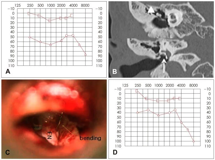
- Oval pencere atrezisi veya displazisinde nişte kemik lameller görülebilir ya da pencere aplazik veya ileri derecede displazik olabilir.
- İzole konjenital kemikçik deformiteleri içinde en sık stapes süperstrüktürü ve inkusun uzun çıkıntısı tutulur.
- Stapes anomalisi — Oval pencere atrezisine stapes malformasyonu veya yokluğu eşlik edebilir.
- Timpanik fasiyal sinir kanalının konum anomalisi — Fasiyal sinir oval pencerenin altında yerleşebilir veya oval pencereyi örtebilir; kanalın kemik örtüsünde açıklık da bulunabilir.
- Oval pencere atrezisine fasiyal sinir seyir anomalisi eşlik edebilir; sinirin oval pencerenin altında yerleşmesi veya pencereyi örtmesi BT'de araştırılmalıdır.
- Eşlik eden kemikçik anomalisi — inkusun lentiküler çıkıntısı ya da uzun kolu hipoplazik, yok veya aşağı yerleşimli olabilir.
- Karşı kulak ve dış kulak yolu — iki taraflı pencere/stapes gelişim kusuru veya dış kulak yolu darlığı-atrezisi gibi ek gelişimsel bulgular bulunabilir.
- Oval pencere atrezisine stapes malformasyonu veya yokluğu eşlik edebilir; stapes kruraları da değerlendirilmelidir.
- Konjenital kemikçik anomalileri Teunissen–Cremers yaklaşımında izole stapes ankilozu; diğer kemikçik anomalileriyle stapes ankilozu; hareketli stapes tabanıyla kemikçik zincir deformitesi; oval veya yuvarlak pencerenin ağır aplazi/displazisi gruplarında ele alınabilir.
Normalde
Oval pencere ve stapes gibi orta kulak kemik yapıları HRCT'de değerlendirilebilir. Timpanik fasiyal sinirin oval pencereye göre seyri değişken olabilir; HRCT'de bu ilişkiyi değerlendirin.
Normal BT ile kıyasla
Aynı bölgenin, kaynak veri setinde patoloji içermediği belirtilen bir BT incelemesini kesit kesit kaydırın; organ sınırlarını açarak anatomiyi hasta görüntüsüyle karşılaştırın. Küçük rastlantısal bulgular tümüyle dışlanmamıştır.
Kaynak: TotalSegmentator v2.01 veri seti (Wasserthal ve ark., Radiology: Artificial Intelligence 2023), CC BY 4.0, vaka s0849; organ sınırları veri setinin otomatik segmentasyonundan, birleştirilerek sadeleştirildi. Görüntüler 2,5 mm kesit aralığına yeniden örneklendi.
Ayırıcı tanı
- Fenestral otoskleroz
- fissula ante fenestra çevresinde fokal hipodens odak veya stapes tabanı çevresinde otik kapsül remodelingi görülür; gelişimsel niş kapanması ve eşlik eden stapes displazisi beklenen patern değildir.
- Timpanosklerozda orta kulak veya timpanik membran tutulumu görülebilir; oval pencere atrezisi ise doğuştan bir gelişim kusurudur.
- Konjenital stapes fiksasyonu
- oval pencere açıklığı ve stapes tabanı mevcut olabilir; yalnız kemik BT ile hareket kısıtlılığı doğrudan kanıtlanamaz.
- Orta kulak efüzyonu, çocuklarda iletim tipi işitme kaybının en sık nedenidir ve konjenital orta kulak anomalileriyle karışabilir.
- Kemikçik zincir travması
- Yüksek çözünürlüklü BT travmaya bağlı kemikçik bozulmalarını gösterebilir; konjenital orta kulak anomalileri de kemik yapıların yüksek çözünürlüklü BT ile değerlendirilmesinde incelenir.
- Otosklerozda HRCT'de oval pencerenin önünde hipodens odak görülebilir; konjenital oval pencere atrezisi BT'de gösterilebilir ve stapes veya fasiyal sinir anomalileriyle birlikte bulunabilir.
Tuzaklar
- Stapesin kemik yapılarının değerlendirilmesinde yüksek çözünürlüklü BT tercih edilir; kesin tanıyı en güvenilir biçimde eksploratuvar timpanotomi koyar.
- Orta kulak anomalileri seröz otitle karışabilir; orta kulak opasifikasyonu stapesi örtebileceğinden, stapesin seçilememesini yokluk olarak yorumlamayın.
- Oval pencerenin altında yerleşen fasiyal sinir, stapes cerrahisinde kullanılabilecek teknikleri etkileyebilir; ameliyat öncesinde sinirin seyri BT'de değerlendirilmelidir.
- Oval pencere atrezisi erişkinlerde otosklerozu klinik olarak taklit edebilir; yüksek çözünürlüklü BT'de atretik pencerenin aranması ayırıcı tanıya katkı sağlar.
Kendini dene
Teunissen–Cremers sınıflamasında oval veya yuvarlak pencerenin ağır aplazisi ya da displazisi hangi sınıftadır?
Cevabı göster
Sınıf 4: pencere aplazisi/displazisi. Sınıflama, oval veya yuvarlak pencerenin ağır aplazi ya da displazisini 4. gruba yerleştirir.
S1'de bildirilen birinci olgunun yüksek çözünürlüklü BT'sinde oval pencere ve stapes için hangi bulgu saptanmıştır?
Cevabı göster
Her ikisinin yokluğu. Kaynakta bildirilen olguda yüksek çözünürlüklü BT, oval pencere ve üzenginin yokluğunu göstermiştir.
İlgili konular
Kaynaklar
Bu sayfadaki 36 cümle ve 2 quiz sorusu aşağıdaki kaynaklardan alıntılarla otomatik olarak karşılaştırıldı; 23 cümle bu karşılaştırmada düzeltildi (2026-10-09).
- Congenital defects of the middle ear - uncommon cause of pediatric hearing loss ☆ ☆☆pmc.ncbi.nlm.nih.gov
- Persistent stapedial artery with stapes ankylosis.pubmed.ncbi.nlm.nih.gov
- Arthroscopic Talocalcaneal Coalition Resection in Children.pubmed.ncbi.nlm.nih.gov
- Endaural Laser-Assisted Single-Stage Inside-Out Cholesteatoma Surgery (LASIC) to Treat Advanced Congenital Cholesteatoma.pubmed.ncbi.nlm.nih.gov
- Outcomes Following Revision Stapes Surgery in Children: A Single-Surgeon, Single-Center 15-Year Experience.pubmed.ncbi.nlm.nih.gov
- Hypothalamic hamartomas and inner ear diverticula with X-linked stapes gusher syndrome - new associations?pubmed.ncbi.nlm.nih.gov
- Congenital familial fixation of the malleus.pubmed.ncbi.nlm.nih.gov
- Temporal Bone Histopathology in NOG-Symphalangism Spectrum Disorder.pubmed.ncbi.nlm.nih.gov
- Three-dimensional finite-element analysis of the cochlear hypoplasia.pubmed.ncbi.nlm.nih.gov
- Novel repair of stapedial footplate defect associated with recurrent meningitis.pubmed.ncbi.nlm.nih.gov
- Predictable prosthesis length on a high-resolution CT scan before a stapedotomy.pubmed.ncbi.nlm.nih.gov
- Stapedotomy in Congenital Fixation with Cochlear Hypoplasia: A New Concept Among the Treatment Options for Cochlear Hypoplasia.pubmed.ncbi.nlm.nih.gov
- [Effect of malleostapedotomy procedure on 17 cases of congenital middle ear malformation].pubmed.ncbi.nlm.nih.gov
- New Imaging Findings of Incomplete Partition Type III Inner Ear Malformation and Literature Review.pubmed.ncbi.nlm.nih.gov
- Congenital Aural Atresia: What the Radiologist Needs to Know?pubmed.ncbi.nlm.nih.gov
- Middle ear malformations combined with cerebrospinal fluid otorrhea: A case report.pubmed.ncbi.nlm.nih.gov
- Multimodal imaging in conductive hearing loss: Optimising CT, MRI and CTA for accurate diagnosis and management.pubmed.ncbi.nlm.nih.gov
- Radiological Perspectives in Congenital Sensorineural Hearing Loss: Insights from Cochlear Implant Candidates.pubmed.ncbi.nlm.nih.gov
- Cochlear Apex Triangulation Utilizing Ct Measures And Middle Ear Landmarks.pubmed.ncbi.nlm.nih.gov
- Developing a production workflow for 3D-printed temporal bone surgical simulators.pubmed.ncbi.nlm.nih.gov
- Brazilian Society of Otology task force - Otosclerosis: evaluation and treatment.pubmed.ncbi.nlm.nih.gov
- Anatomical Characteristics of the Facial Nerve in Patients With Cochlear Hypoplasia.pubmed.ncbi.nlm.nih.gov
- The preoperative temporal bone CT: a mnemonic for reducing surgical risk.pubmed.ncbi.nlm.nih.gov
- Pulsatile Tinnitus: A Comprehensive Clinical Approach to Diagnosis and Management.pubmed.ncbi.nlm.nih.gov
- Brazilian Society of Otology task force - cochlear implant ‒ recommendations based on strength of evidence.pubmed.ncbi.nlm.nih.gov
- Radiological diagnosis of the inner ear malformations in children with sensorineural hearing loss.pubmed.ncbi.nlm.nih.gov
- Beware of a Fragile Footplate: Lessons from Ossiculoplasty in Patients with Ossicular Anomalies Related to Second Pharyngeal Arch Defects.pubmed.ncbi.nlm.nih.gov
- Genetic Determinants of Non-Syndromic Enlarged Vestibular Aqueduct: A Review.pubmed.ncbi.nlm.nih.gov
- Aberrant Course of the Intratemporal Facial Nerve in Children with Congenital Hearing Loss.pubmed.ncbi.nlm.nih.gov
- Short-process incudo-stapedioplasty in congenital ear malformation.pubmed.ncbi.nlm.nih.gov
- Fusion of Middle Ear Optical Coherence Tomography and Computed Tomography.pubmed.ncbi.nlm.nih.gov
- The State of High-Resolution Imaging of the Human Inner Ear: A Look Into the Black Box.pubmed.ncbi.nlm.nih.gov
- Cranial anatomy, palaeoneurology, palaeobiology and stratigraphic age of the large-bodied ornithopod, <i>Muttaburrasaurus langdoni</i> Bartholomai and Molnar, 1981, from the mid-Cretaceous of Australia.pubmed.ncbi.nlm.nih.gov
- Case Report: Simultaneous repair of cerebrospinal fluid otorrhea and cochlear implantation in a patient with single-sided deafness and inner ear anomaly.pubmed.ncbi.nlm.nih.gov
- Relationship Between External Ear Severity and Temporal Bone Malformation in Microtia: A Computed Tomography Study.pubmed.ncbi.nlm.nih.gov
- Direct Acoustic Stimulation at the Lateral Canal: An Alternative Route to the Inner Ear?pubmed.ncbi.nlm.nih.gov
- Procedures for restoring vestibular disorders.pubmed.ncbi.nlm.nih.gov
- Prognostic Factors in Cochlear Implant in Otosclerosis: Radiologic and Clinical Variables: Cochlear Implant Predictors in Otosclerosis.pubmed.ncbi.nlm.nih.gov
- HRCT imaging of acquired cholesteatoma: a pictorial review.pubmed.ncbi.nlm.nih.gov
- Clinical High-Resolution Imaging of the Inner Ear by Using Magnetic Resonance Imaging (MRI) and Cone Beam Computed Tomography (CBCT).pubmed.ncbi.nlm.nih.gov
- New insights into the early morphological evolution of sea turtles by re-investigation of Nichollsemys baieri , a three-dimensionally preserved fossil stem chelonioid from the Campanian of Alberta, Canada.pubmed.ncbi.nlm.nih.gov
- Comprehensive Management of Cholesteatoma in Otitis Media: Diagnostic Challenges, Imaging Advances, and Surgical Outcome.pubmed.ncbi.nlm.nih.gov
- Management of Meningitis Due to Cystic Cochleovestibular Malformation: a Stitch in Time.pubmed.ncbi.nlm.nih.gov
- Role of CT Mastoids in the Diagnosis and Surgical Management of Chronic Inflammatory Ear Diseases.pubmed.ncbi.nlm.nih.gov
- Coexistence of Autoantibodies against the Golgi Complex and Ro52 Antigen in a Patient with Nonspecific Interstitial Pneumonia.pubmed.ncbi.nlm.nih.gov
- Long-term safety and effectiveness of the "OptEase" vena cava filter.pubmed.ncbi.nlm.nih.gov
- Bilateral Oval and Round Window Atresia on CT Temporal Bone: A Rare Anomaly Clinically Mimicking Otosclerosis in an Adult.pubmed.ncbi.nlm.nih.gov
- Rare Diseases of the Middle Ear and Lateral Skull Base.pubmed.ncbi.nlm.nih.gov
- Pre- and post-operative imaging of cochlear implants: a pictorial review.pubmed.ncbi.nlm.nih.gov
- Anatomical and Surgical Evaluation of the Common Marmoset as an Animal Model in Hearing Research.pubmed.ncbi.nlm.nih.gov
- Anatomy and cranial functional morphology of the small-bodied dinosaur Fruitadens haagarorum from the Upper Jurassic of the USA.pubmed.ncbi.nlm.nih.gov
- Commentary: an eye on PET quantification.pubmed.ncbi.nlm.nih.gov
- The Developing Human Sphenoid Bone: Linking Embryological Development to Adult Morphology.pubmed.ncbi.nlm.nih.gov
- Proceedings of the 15th International Congress on Circumpolar Health.pubmed.ncbi.nlm.nih.gov
- Anatomical and audiological considerations in branchiootorenal syndrome: A systematic review.pubmed.ncbi.nlm.nih.gov
- Bilateral Congenital Stapes Footplate Fixation With Absence of the Stapedial Tendon.pubmed.ncbi.nlm.nih.gov
- Stapes footplate's posterior border protrudes the vestibule in healthy ears: anatomical insights from ultra-high-resolution CT.pubmed.ncbi.nlm.nih.gov
- The Transmission of Sound to the Cochlea in Normal and Pathological Human Middle Ears.pubmed.ncbi.nlm.nih.gov
- Identification of Cerebrospinal Fluid Leakage by Cisternography in Recurrent Meningitis Associated With Inner Ear Malformation.pubmed.ncbi.nlm.nih.gov
- More Than Meets the Ear: Incidental Cerebrospinal Fluid Leak During Myringotomy in a Child With Common Cavity Deformity.pubmed.ncbi.nlm.nih.gov
- Facial Nerve Abnormalities in Congenital Middle Ear Malformations With Comments on Preoperative Detectability: A Report of Two Cases.pubmed.ncbi.nlm.nih.gov
- X-linked deafness/incomplete partition type 3: Radiological evaluation of temporal bone and intracranial findings.pubmed.ncbi.nlm.nih.gov
- Ear Surgery Training: A Comprehensive Review.pubmed.ncbi.nlm.nih.gov
- A White Spot Around the Fissula Ante Fenestrum: A New Diagnostic Indicator for Otosclerosis.pubmed.ncbi.nlm.nih.gov
- High Heterogeneity of Temporal Bone CT Aspects in Osteogenesis Imperfecta Is Not Linked to Hearing Loss.pubmed.ncbi.nlm.nih.gov
- Assessment of the Oval Window-Facial Canal Corridor and Mastoid Pneumatization: A Preliminary HRCT-Based Radiological-Anatomical Framework.pubmed.ncbi.nlm.nih.gov
- Comprehensive Review of Inner Ear Anatomy on Photon-Counting CT.pubmed.ncbi.nlm.nih.gov
- Head of the Syrian Hamster (<i>Mesocricetus auratus</i>): Planar Anatomy with Correlative Micro-Computed Tomography and Magnetic Resonance Imaging.pubmed.ncbi.nlm.nih.gov
- Mammalian middle ear mechanics: A review.pubmed.ncbi.nlm.nih.gov
- External and middle ear diseases: radiological diagnosis based on clinical signs and symptoms.pubmed.ncbi.nlm.nih.gov
- Traumatic perilymphatic fistula with stapes luxation into the vestibule and pneumolabyrinth: a case report.pubmed.ncbi.nlm.nih.gov
- Isolated Congenital Ossicular Chain Anomalies.pubmed.ncbi.nlm.nih.gov
- Diagnosing Middle Ear Malformation by Pure-Tone Audiometry Using a Three-Dimensional Finite Element Model: A Case-Control Study.pubmed.ncbi.nlm.nih.gov
- Realistic 3D computer model of the gerbil middle ear, featuring accurate morphology of bone and soft tissue structures.pubmed.ncbi.nlm.nih.gov
- Cranial osteology of the pampathere Holmesina floridanus (Xenarthra: Cingulata; Blancan NALMA), including a description of an isolated petrosal bone.pubmed.ncbi.nlm.nih.gov
- Comparative Anatomy of the Bony Labyrinth (Inner Ear) of Placental Mammals.pubmed.ncbi.nlm.nih.gov
- Endoscopic stapes surgery for otosclerosis: clinical, radiological, and audiological outcomes in a retrospective series of 40 patients.pubmed.ncbi.nlm.nih.gov
- The bone conduction threshold pattern may help to estimate the pathology underlying conductive hearing loss.pubmed.ncbi.nlm.nih.gov
- Sensorineural hearing loss and CT abnormalities in otosclerosis: Insights from otopathological specimens.pubmed.ncbi.nlm.nih.gov
- An Unusual Finding During Exploratory Tympanotomy in a Case of Conductive Hearing Loss Mimicking Clinical Otosclerosis: A Case Series.pubmed.ncbi.nlm.nih.gov
- Disrupted Vestibular Nuclei Neuron Development in a Chick Model for Congenital Vestibular Disorders.pubmed.ncbi.nlm.nih.gov
- Semiautomated Middle Ear Volume Measurement as a Predictor of Postsurgical Outcomes for Congenital Aural Atresia.pubmed.ncbi.nlm.nih.gov
- Masses of developmental and genetic origin affecting the paediatric craniofacial skeleton.pubmed.ncbi.nlm.nih.gov
- A brief history of otologic surgery: restoring hearing through the ages.pubmed.ncbi.nlm.nih.gov
- A distant global control region is essential for normal expression of anterior HOXA genes during mouse and human craniofacial development.pubmed.ncbi.nlm.nih.gov
- A Novel Clinical Feature in NOG Gene Mutation-Associated Syndrome.pubmed.ncbi.nlm.nih.gov
- Outcomes of Bonebridge Implantation in 10 Patients with Rare Genetic Syndromes and Difficult Anatomy.pubmed.ncbi.nlm.nih.gov
- Improved radiological imaging of congenital aural atresia using flat-panel volume CT.pubmed.ncbi.nlm.nih.gov
- Novel TCOF1 Frameshift Variant and Phenotypic Heterogeneity in a Chinese Family With Treacher Collins Syndrome.pubmed.ncbi.nlm.nih.gov
- Infant hearing loss: from diagnosis to therapy Official Report of XXI Conference of Italian Society of Pediatric Otorhinolaryngology.pubmed.ncbi.nlm.nih.gov
- The Efficacy of Acupuncture and Bee Venom Injections in Improving Hearing in a Patient with Idiopathic Conductive Hearing Loss: a case report.pubmed.ncbi.nlm.nih.gov
- The Audiological Aspect of Beckwith-Wiedemann Syndrome: A Systematic Review.pubmed.ncbi.nlm.nih.gov
- Pre-operative High-Resolution CT and MRI Evaluation in Pediatric Cochlear Implant Candidates: Correlation With Surgical Findings and Outcomes.pubmed.ncbi.nlm.nih.gov
- Neural crest contributions to the ear: Implications for congenital hearing disorders.pubmed.ncbi.nlm.nih.gov
- Congenital Anomalies and Variations of the Carotid and Vertebral Arteries: A Case-Based Imaging Review.pubmed.ncbi.nlm.nih.gov
- Persistent Stapedial Artery, Facial Nerve Overhang, and Calcified Stapedius Tendon Leading to Aborted Stapedotomy: An Unusual Middle Ear Anatomy.pubmed.ncbi.nlm.nih.gov
- Classification and diagnosis of ear malformations.pubmed.ncbi.nlm.nih.gov
- Comparative Morphometric Study of the Osseous Labyrinth in Humans and Animal Models Using Centerline Analysis.pubmed.ncbi.nlm.nih.gov
- "Leave me alone": anatomical structures and variations seen on computed tomography of the temporal bone.pubmed.ncbi.nlm.nih.gov
- Neuroimaging with photon-counting computed tomography: A review of clinical applications.pubmed.ncbi.nlm.nih.gov
- Reliability of high-resolution CT scan in diagnosis of ossicular tympanosclerosis.pubmed.ncbi.nlm.nih.gov
- Tuberous Sclerosis Complex: A Case Series from a Romanian Genetics Center and a Review of the Literature.pubmed.ncbi.nlm.nih.gov
- Intelligent diagnosis of ossicular chain malformations on CT: development and clinical efficacy of a cascaded AI framework.pubmed.ncbi.nlm.nih.gov
- Diagnosis and Management of Middle Ear Neuroendocrine Tumour (MeNET).pubmed.ncbi.nlm.nih.gov
- Intratympanic membrane cholesteatoma after traumatic tympanic membrane perforation: a case report.pubmed.ncbi.nlm.nih.gov
- Ventilation tubes (grommets) for otitis media with effusion (OME) in children.pubmed.ncbi.nlm.nih.gov
- Brazilian Society of Otology Task Force - bone-anchored hearing system - recommendations based on strength of evidence.pubmed.ncbi.nlm.nih.gov
- Endoscopic malleostapedotomy versus incudostapedotomy for stapes fixation with or without lateral chain fixation: A comparative outcomes study.pubmed.ncbi.nlm.nih.gov
- Comprehensive Review of External and Middle Ear Anatomy on Photon-Counting CT.pubmed.ncbi.nlm.nih.gov
- Radiologically Assessed Stapes Footplate Thickness and Audiologic Outcomes in Patients with Otosclerosis.pubmed.ncbi.nlm.nih.gov
- Photon-counting detector computed tomography for temporal bone: does higher resolution matter?pubmed.ncbi.nlm.nih.gov
Eğitim amaçlıdır; hasta başında tanı ve tedavi kararının yerine geçmez, tıbbi tavsiye değildir. Metinler yapay zekâ desteğiyle yazıldı ve otomatik bir adımda kaynak alıntılarıyla karşılaştırıldı; bu bir tıbbi doğrulama değildir ve içerik uzman radyolog incelemesinden geçmedi. Klinik kararlarda güncel kılavuzları, kurum protokollerini ve radyoloji raporunu esas alın.