Özet
Petroz apeks efüzyonu, havalanmış petroz apeks hücrelerinde sıvı birikmesidir ve çoğunlukla rastlantısal saptanır. İnce kesit kontrastsız temporal kemik BT, hücreler opaklaşmış olsa bile kemik septaların ve dış konturun korunup korunmadığını gösterir. Kontrastsız BT, petroz apeks efüzyonunu ekspansil lezyonlardan kemik yapısının korunmasıyla ayırır; doku karakterizasyonu için MRI tamamlayıcıdır. Efüzyonu eroziv kitle sanmak gereksiz endişeye, gerçek kemik yıkımını efüzyon kabul etmek ise önemli bir lezyonun gözden kaçmasına yol açabilir.
Faz ve pencere
- Kontrastsız tanısal
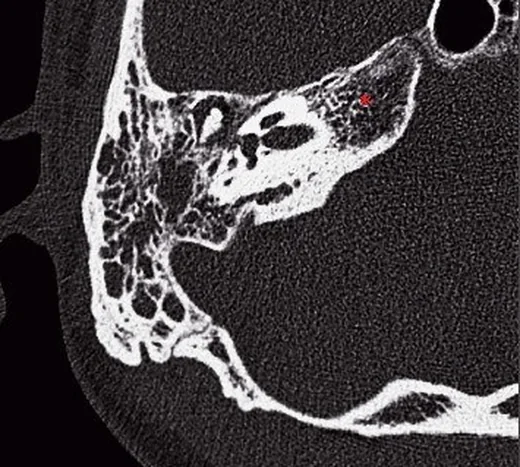
- Petroz apeksin havalı hücreleri yumuşak doku/sıvı yoğunluklu materyalle opaklaşır; opasite içinde ince kemik septalar ve hücre duvarları seçilir, apeks dış sınırında genişleme veya kortikal kesinti bulunmaz. Petroz apeks lezyonlarının değerlendirilmesinde BT ve MRI birbirini tamamlayan görüntüleme yöntemleridir.
- Kemik algoritması tanısal
- İnce kesit kemik rekonstrüksiyonları opasifiye hücrelerin iç mimarisini ve petroz apeksin dış konturunu ortaya koyar; trabeküler kafesin devamlılığı ve hacim artışının bulunmaması efüzyon lehinedir.
Önerilen pencereler: Kemik (G 1800 / M 400), Yumuşak doku (G 400 / M 40).
BT bulguları
- Petroz apeks hücrelerinde dolum — havalı hücreler homojen yumuşak doku/sıvı yoğunluklu görünüm alır.
- Septaların devamlılığı — opasite içinden ince kemik trabekülleri izlenebilir.
- Apeks hacminin korunması — karşı tarafla kıyasta petroz kemik genişlemiş görünmez.
- Dış korteks bütünlüğü — petroz apeksin periferik kemik sınırı kesintisizdir.
- Hücre sınırlarının seçilmesi — opak içerik çevresinde doğal havalı hücrelerin kortikal kenarları ayırt edilir.
Normalde
Normal havalanmış petroz apeks hücreleri düşük yoğunluklu hava içerir; hücreleri ayıran ince kemik septalar ve çevre korteks düzenli biçimde seçilir. Efüzyonlu tarafta hava yerini yumuşak doku yoğunluğuna bırakır, ancak aynı hücre mimarisi ve apeksin dış biçimi korunur; iki tarafı karşılaştırırken opasiteyi değil bu kemik çerçeveyi izleyin.
Normal BT ile kıyasla
Aynı bölgenin, kaynak veri setinde patoloji içermediği belirtilen bir BT incelemesini kesit kesit kaydırın; organ sınırlarını açarak anatomiyi hasta görüntüsüyle karşılaştırın. Küçük rastlantısal bulgular tümüyle dışlanmamıştır.
Kaynak: TotalSegmentator v2.01 veri seti (Wasserthal ve ark., Radiology: Artificial Intelligence 2023), CC BY 4.0, vaka s0103; organ sınırları veri setinin otomatik segmentasyonundan, birleştirilerek sadeleştirildi. Görüntüler 2,5 mm kesit aralığına yeniden örneklendi.
Ayırıcı tanı
- Kolesterol granülomu, petroz apeks hücrelerini genişletip kemik erozyonuna yol açabilen benign bir kitledir.
- Petroz kemik kolesteatomu
- havalı hücreleri genişleterek erozyon ve kemik yıkımı oluşturabilir.
- Petroz apisit, petroz apekste enfeksiyon ve iltihabi sıvı birikimine bağlı radyolojik opasifikasyon oluşturur.
- Petroz apeks mukoseli
- havalı hücreleri genişletip trabeküler veya kortikal erozyon oluşturabilir.
Tuzaklar
- Tek taraflı opasiteyi tek başına patolojik kitle saymayın; hücre septalarının opasite içinden sürmesi ve apeksin genişlememesi efüzyon lehinedir.
- Tek taraflı opasite her zaman patolojik değildir; kemik yapısının korunması ile ekspansiyon/erozyon arasındaki farkı karşılaştırarak değerlendirmelisiniz.
- BT yoğunluğu sıvının basit, proteinli veya enfekte olduğunu kesinleştirmez; klinik şüphe ya da atipik görünüm varsa MRI ve klinik bilgilerle tamamlayın.
Kendini dene
Petroz apeks hücreleri opak; iç kemik çizgileri sürüyor ve apeks genişlememiş. En uygun tanı hangisidir?
Cevabı göster
Petroz apeks efüzyonu. Efüzyon havalı hücreleri doldurur ancak kemik septaları ve apeks boyutunu korur. Kolesterol granülomu ve kolesteatom ekspansiyon/erozyon yapabilir. Apikal petrositiste iltihabi süreç ve nörolojik bulgular klinik ve radyolojik olarak izlenmelidir.
Sıvı sinyalli petroz apeks odağında ekspansiyon yok ve trabeküller korunmuş. En olası açıklama nedir?
Cevabı göster
Petroz apeks efüzyonu. Korunmuş hücre mimarisi ve genişleme bulunmaması hapsolmuş efüzyonu destekler. Ekspansil lezyonlar ve dejstrüktif süreçler arasında ayırıcı tanı yapılmalıdır.
İlgili konular
Kaynaklar
Bu sayfadaki 35 cümle ve 2 quiz sorusu aşağıdaki kaynaklardan alıntılarla tek tek karşılaştırıldı; 11 cümle bu karşılaştırmada düzeltildi (2026-10-08).
- Middle ear malformations combined with cerebrospinal fluid otorrheapmc.ncbi.nlm.nih.gov
- Gradenigo syndrome in a child: case report and literature reviewpmc.ncbi.nlm.nih.gov
- Repeated transitions between intracranial hypotension and intracranial hypertension in Gorham-Stout disease: illustrative casepmc.ncbi.nlm.nih.gov
- An Atypical Pediatric Headache: Outpatient Presentation of Petrous Apicitis With Cerebral Venous Sinus Thrombosispmc.ncbi.nlm.nih.gov
- A multicorridor strategy including the infralabyrinthine route for extensive skull base chondrosarcoma: illustrative casepmc.ncbi.nlm.nih.gov
- Bilateral symptomatic petrous apex effusion.pubmed.ncbi.nlm.nih.gov
- Endoscope Assisted Subcochlear Approach for Symptomatic Petrous Apex Effusion.pubmed.ncbi.nlm.nih.gov
- Giant Temporal Lobe Abscess in a Patient With Ectodermal Dysplasia.pubmed.ncbi.nlm.nih.gov
- A case of generalized lymphatic anomaly causing skull-base leakage and bacterial meningitis.pubmed.ncbi.nlm.nih.gov
- Spontaneous osteo-dural fistulae of petrous bone posterior wall.pubmed.ncbi.nlm.nih.gov
- Bilateral auditory ossicular expansions in a child with beta-thalassemia major: Case report and literature review.pubmed.ncbi.nlm.nih.gov
- Cholesterol granuloma of the middle ear invading the cochlea.pubmed.ncbi.nlm.nih.gov
- Exploratory operation in a patient with spontaneous temporal bone cerebrospinal fluid leaks: A case report.pubmed.ncbi.nlm.nih.gov
- A case of bilateral sensorineural hearing loss from Langerhans cell histiocytosis.pubmed.ncbi.nlm.nih.gov
- Eustachian Tube Dysfunction Due to Medial Pterygoid Nerve Impairment After Trigeminal Schwannoma Surgery.pubmed.ncbi.nlm.nih.gov
- Anatomy of the Middle Meningeal Artery: Implications for Middle Meningeal Artery Embolization in Chronic Subdural Hematoma.pubmed.ncbi.nlm.nih.gov
- Gradenigo Syndrome: A Case Report of a Rare Complication of Otitis Media.pubmed.ncbi.nlm.nih.gov
- Comprehensive Review of External and Middle Ear Anatomy on Photon-Counting CT.pubmed.ncbi.nlm.nih.gov
- Chronic Subdural Hematoma: Pathophysiology, Diagnosis, and the Emerging Role of Middle Meningeal Artery Embolization.pubmed.ncbi.nlm.nih.gov
- Endoscopic assisted minimally invasive resection of petrous bone epidermoid cyst via a presigmoid burr hole approach: illustrative case.pubmed.ncbi.nlm.nih.gov
- A Diagnostic Enigma: A Rare Case of Cholesterol Granuloma of Mastoid Masquerading as Acute Mastoiditis in a Young Female.pubmed.ncbi.nlm.nih.gov
- An Unusual Case of Petrous Apex Metastasis Revealed by Trigeminal and Abducens Nerve Palsies.pubmed.ncbi.nlm.nih.gov
- Refractory Pseudomonas Osteomyelitis of the Skull Base With Gradenigo's Syndrome: Early Dysphagia and Late Abducens Nerve Palsy.pubmed.ncbi.nlm.nih.gov
- Pre-treatment and post-treatment nasopharyngeal carcinoma imaging: imaging updates, pearls and pitfalls.pubmed.ncbi.nlm.nih.gov
- Critical infections in the head and neck: A pictorial review of acute presentations and complications.pubmed.ncbi.nlm.nih.gov
- Imaging of Skull Base Tumors.pubmed.ncbi.nlm.nih.gov
- Imaging for Pediatric Head and Neck Emergencies: Critical Insights.pubmed.ncbi.nlm.nih.gov
- Diagnostic Quandary of Granulomatosis With Polyangiitis Presenting First in the Petrous Apex.pubmed.ncbi.nlm.nih.gov
- Key pre-operative CT parameters for cochlear implantation: a focused radiologic review with surgical relevance.pubmed.ncbi.nlm.nih.gov
- Multimodal imaging in neuro-ophthalmology.pubmed.ncbi.nlm.nih.gov
- Neurolymphomatosis: A Comprehensive Review of Clinical and Imaging Features.pubmed.ncbi.nlm.nih.gov
- ESR Essentials: acute infections of the head and neck-practice recommendations by the European Society of Head and Neck Radiology.pubmed.ncbi.nlm.nih.gov
- Emerging and re-emerging infectious disease in otorhinolaryngology.pubmed.ncbi.nlm.nih.gov
- A Morphometric Analysis of the Middle Cranial Fossa in Mexican Adults for Dolenc and Kawase Approaches With Computed Tomography, 3D Reconstruction and Dry Skulls.pubmed.ncbi.nlm.nih.gov
- Morphometric anatomy of the middle cranial fossa via the anterior petrosal (Kawase) approach: a bilateral cadaveric study.pubmed.ncbi.nlm.nih.gov
- Clinician-Guided Deep Learning Segmentation of Skull Base Pneumatization on Computed Tomography Using 3D Slicer and MONAI Label.pubmed.ncbi.nlm.nih.gov
- Imaging of Temporal Bone Mass Lesions: A Pictorial Review.pubmed.ncbi.nlm.nih.gov
- Temporal Bone Fractures on High-Resolution CT: Bridging Radiologic Detail with Otologic Anatomy and Surgical Implications.pubmed.ncbi.nlm.nih.gov
- Radiological Classification and Management Algorithm of Petrous Apex Cholesterol Granuloma.pubmed.ncbi.nlm.nih.gov
- Surgical approaches to the petrous apex.pubmed.ncbi.nlm.nih.gov
- Middle ear malformations combined with cerebrospinal fluid otorrhea: A case report.pubmed.ncbi.nlm.nih.gov
- Structural Phenotype of High-Frequency Recurrent Facial Palsy.pubmed.ncbi.nlm.nih.gov
- Frontal Cholesterol Granuloma in a Pediatric Patient: A Case Report.pubmed.ncbi.nlm.nih.gov
- Third-ventricle cholesterol granuloma: An exceptional intraventricular localization.pubmed.ncbi.nlm.nih.gov
- Petroclival extra-adrenal myelolipoma: illustrative case.pubmed.ncbi.nlm.nih.gov
- Extensive Temporal Bone Necrosis in Petrous Bone Cholesteatoma Complicated by Actinomycosis: A Case Report.pubmed.ncbi.nlm.nih.gov
- Cerebellar abscess complicating chronic suppurative otitis media: An unusual route of intracranial spread.pubmed.ncbi.nlm.nih.gov
- Congenital Mastoid Cholesteatoma With Giant Intracranial Extension: A Case Report.pubmed.ncbi.nlm.nih.gov
- An Atypical Pediatric Headache: Outpatient Presentation of Petrous Apicitis With Cerebral Venous Sinus Thrombosis.pubmed.ncbi.nlm.nih.gov
- Intratemporal and Extratemporal Complications in a Rare Case of Acute Mastoiditis: A Case Report and Literature Review.pubmed.ncbi.nlm.nih.gov
- Beyond Otitis Media: When Mastoiditis Leads to Life-Threatening Complications in Children.pubmed.ncbi.nlm.nih.gov
- Clival Aneurysmal Bone Cyst With Cavernous Sinus Extension: A Diagnostic Challenge.pubmed.ncbi.nlm.nih.gov
- Isolated sphenoid sinus fungal mucoceles: A rare entity with a high propensity for causing neurological complications.pubmed.ncbi.nlm.nih.gov
- Blind spots in brain imaging: a pictorial essay.pubmed.ncbi.nlm.nih.gov
- An Uncommon Diagnosis of Necrotizing Mastoiditis Presenting as Bell's Palsy: A Case Report.pubmed.ncbi.nlm.nih.gov
- The Developing Human Sphenoid Bone: Linking Embryological Development to Adult Morphology.pubmed.ncbi.nlm.nih.gov
- The cavernous sinus: An anatomic study with clinical implication.pubmed.ncbi.nlm.nih.gov
- Patient cases with malignant otitis externa at the university clinic of Ludwig Maximilians University Munich from 2009 until 2020.pubmed.ncbi.nlm.nih.gov
- Extradural anterior transpetrosal approach: how I do it.pubmed.ncbi.nlm.nih.gov
- Imaging Evaluation for Jaw Deformities: Diagnostic Workup and Pre-Treatment Imaging Checklist for Orthognathic Surgery.pubmed.ncbi.nlm.nih.gov
- Detailed Radiomorphometric Analysis of the Surgical Corridor for the Suprageniculate Approach.pubmed.ncbi.nlm.nih.gov
- A Case of Acquired Pars Flaccida Cholesteatoma Extending to the Supralabyrinthine Petrous Apex With Preservation of the Otic Capsule.pubmed.ncbi.nlm.nih.gov
- Cholesterol granuloma of the sphenoid wing with long-term growth dynamics: illustrative case.pubmed.ncbi.nlm.nih.gov
- Nocardia Otomastoiditis in a Chemotherapy-Induced Neutropenic Patient With Diffuse Large B-cell Lymphoma: Successful Medical Management.pubmed.ncbi.nlm.nih.gov
- Surgical treatment of apical petrositis through the anterior transpetrosal approach: A case report.pubmed.ncbi.nlm.nih.gov
- Repeated transitions between intracranial hypotension and intracranial hypertension in Gorham-Stout disease: illustrative case.pubmed.ncbi.nlm.nih.gov
- Gradenigo syndrome in a child: case report and literature review.pubmed.ncbi.nlm.nih.gov
- Temporal bone mucocele with extension to mastoid in a 60-year-old male: a case report and literature review.pubmed.ncbi.nlm.nih.gov
- Rare Diseases of the Middle Ear and Lateral Skull Base.pubmed.ncbi.nlm.nih.gov
- "Leave me alone": anatomical structures and variations seen on computed tomography of the temporal bone.pubmed.ncbi.nlm.nih.gov
- A multicorridor strategy including the infralabyrinthine route for extensive skull base chondrosarcoma: illustrative case.pubmed.ncbi.nlm.nih.gov
- Skeletal manifestations in lymphatic diseases: nonenhanced magnetic resonance lymphography.pubmed.ncbi.nlm.nih.gov
- HRCT imaging of acquired cholesteatoma: a pictorial review.pubmed.ncbi.nlm.nih.gov
- Anatomical Step-by-Step Dissection of Complex Skull Base Approaches for Trainees: Surgical Anatomy of the Middle Fossa Approaches and Anterior Petrosectomy, Surgical Principles, and Illustrative Cases.pubmed.ncbi.nlm.nih.gov
- Clival and Paraclival Lesions: A Pictorial Review.pubmed.ncbi.nlm.nih.gov
- Skull base inverted papilloma: a comprehensive review.pubmed.ncbi.nlm.nih.gov
- Role of the Ear in Meningitis: A Narrative Review of Neuroimaging.pubmed.ncbi.nlm.nih.gov
- Beyond the otoscope: an imaging review of congenital cholesteatoma.pubmed.ncbi.nlm.nih.gov
- The Clinical Value of Cranial CT Venography for Predicting <i>Fusobacterium necrophorum</i> as the Causative Agent in Children with Complicated Acute Mastoiditis.pubmed.ncbi.nlm.nih.gov
Eğitim amaçlıdır; hasta başında tanı ve tedavi kararının yerine geçmez. İçerik yapay zekâ destekli bir süreçle güncel literatüre göre cümle cümle kaynaklarla karşılaştırıldı, ancak uzman radyolog incelemesinden geçmedi. Klinik kararlarda güncel kılavuzları, kurum protokollerini ve radyoloji raporunu esas alın.