Özet
Temporal ve petroz kemik metastazları, özellikle bilinen malignitesi olan hastada yeni otolojik yakınma, fasiyal güçsüzlük veya kafa tabanı nöropatisi geliştiğinde akılda tutulmalıdır. Kontrastsız ince kesit BT, litik, sklerotik ya da mikst kemik yanıtını ve kortikal destrüksiyonu gösterir; yumuşak doku ile kemik iliği yayılımını MRI daha iyi ortaya koyar. Petroz kemik, temporal kemik metastazlarında sık tutulan alanlardan biridir; tek bir lezyonun görünümü primer tümör tipine göre değişebilir. Foramen veya sinir kanalı boyunca yayılımın gözden kaçması kraniyal sinir bulgularının nedenini ve hastalığın gerçek sınırını eksik bırakabilir. Kraniyal nöropati şüphesinde kontrastlı MR ile kemik iliği, kafa tabanı yumuşak dokusu, dural uzanım ve ilgili kraniyal sinir/foramen boyunca yayılım değerlendirilir.
Faz ve pencere
- Kontrastsız tanısal
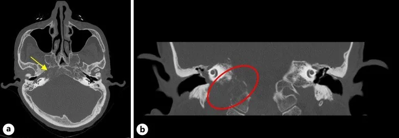
- Petroz kemik ve temporal kemik iliğinde litik, sklerotik veya mikst atenüasyon değişikliği; kortikal destrüksiyon, permeatif kemik kaybı ve eşlik eden yumuşak doku kitlesi aranır. Kontrast fazı kemik lezyonunun tipini belirlemek için gerekli değildir; tümör kitlesi ve kraniyal sinir/meningeal uzanım şüphesinde MRI tamamlayıcıdır.
- Kemik algoritması tanısal
- İnce kesit rekonstrüksiyonlar kemik iliği alanındaki skleroz veya litik yıkımı, kortikal bütünlüğü ve karotis kanalı, juguler foramen, internal akustik kanal ile mastoid sınırlarına uzanımı gösterir.
Önerilen pencereler: Kemik (G 1800 / M 400), Yumuşak doku (G 400 / M 40).
BT bulguları
- Litik odak — petroz kemik trabekülleri ve korteksi düşük atenüasyonlu yıkım alanıyla silinir.
- Sklerotik odak — normal petroz kemik iliğinin yerini yoğun kemik yanıtı alır; kenarları primer tümör öyküsüyle birlikte yorumlanır.
- Mikst kemik yanıtı — aynı lezyonda litik destrüksiyon ve reaktif skleroz birlikte bulunur.
- Permeatif kemik kaybı — kemik iliği boşluğundan ilerleyen, net sınırlı olmayan destrüksiyon paterni.
- Kortikal aşılma — dış kemik sınırında kesinti ve ekstraosseöz yumuşak doku komponenti.
- Kafa tabanı kanalı tutulumu — karotis kanalı, juguler foramen veya internal akustik kanal korteksindeki erozyon ya da genişleme.
- Çok odaklı kemik hastalığı — aynı incelemede klivus, sfenoid, diğer temporal kemik ve görüntü alanındaki kemiklerde benzer lezyonlar aranır.
- MR, kemik iliği ve ekstraosseöz yumuşak doku tutulumunun yaygınlığını göstermede BT’yi tamamlar.
Normalde
Normal petroz apeks kemik iliği düzgün trabeküler yapı gösterir; karotis kanalı, juguler foramen, internal akustik kanal ve kokleanın kortikal sınırları kesintisizdir. Karşı tarafla kıyaslarken fokal kemik yoğunluğu değişikliğinin yanı sıra trabekül kaybı, kortikal geçiş, kanal duvarı erozyonu ve yumuşak dokuya uzanımı değerlendirin.
Normal BT ile kıyasla
Aynı bölgenin, kaynak veri setinde patoloji içermediği belirtilen bir BT incelemesini kesit kesit kaydırın; organ sınırlarını açarak anatomiyi hasta görüntüsüyle karşılaştırın. Küçük rastlantısal bulgular tümüyle dışlanmamıştır.
Kaynak: TotalSegmentator v2.01 veri seti (Wasserthal ve ark., Radiology: Artificial Intelligence 2023), CC BY 4.0, vaka s0103; organ sınırları veri setinin otomatik segmentasyonundan, birleştirilerek sadeleştirildi. Görüntüler 2,5 mm kesit aralığına yeniden örneklendi.
Ayırıcı tanı
- Petroz apisit/osteomiyelit
- hücre opasifikasyonu, eşlik eden orta kulak-mastoid inflamasyonu ve klinik enfeksiyon bulguları kemik metastazı yerine enfeksiyonu düşündürür.
- Plazmasitom veya multipl miyelom
- ekspansil litik intraosseöz kitle yapabilir; iskeletin başka bölgelerindeki litik odaklar ve sistemik bağlam ayrım sağlar.
- Kolesterol granülomu
- petroz apekste düzgün sınırlı, kan ürünlerinin parçalanmasıyla oluşan genişleyici kistik odak; komşu kemikte erozyona yol açar ve MR'da T1 ile T2 görüntülerinde hiperintens sinyal verir.
- Jugulotimpanik paragangliom
- juguler foramen çevresinde düzensiz erozyon ve hipotimpanik kitle yapar; tipik vasküler karakteri MRI/MRA'da belirgindir.
- Kordoma veya kondrosarkom
- klivus/petroklival merkezli destrüktif yumuşak doku kitlesidir; matriks kalsifikasyonu ve merkez yerleşimi ayırıcı ipuçlarıdır.
- Kafa tabanı ayırıcı tanısında lenfoma ve plazmasitom da yer alır; plazmasitom klivusta litik lezyon oluşturabilir.
Tuzaklar
- Sklerotik görünümü metastaza özgü sayma; tedavi yanıtı, benign kemik odakları ve primer tümörün biyolojisi de yoğunluk değiştirir.
- Tek taraflı küçük litik odağı klinik öyküden bağımsız yorumlama; bilinen kanser, diğer kemik metastazları ve MRI kemik iliği sinyali tanısal olasılığı değiştirir.
- BT'de kemik korteksi sağlam diye erken kemik iliği yayılımını dışlama; kemik iliği sinyali ve yumuşak doku uzanımı için MRI gerekebilir.
Kendini dene
Bilinen akciğer kanseri olan hastada petroz apeks litik odağı korteksi aşarak yumuşak doku kitlesi oluşturuyor. En olası tanı nedir?
Cevabı göster
Petroz kemik metastazı. Bilinen primer malignite, litik kemik yıkımı ve ekstraosseöz kitle metastazı destekler. Apisit enfeksiyon/otomastoid bulgularla; kolesterol granülomu düzgün ekspansil kistik paternle; schwannom ise sinir kanalı merkezli düzgün genişlemeyle ayrılır.
Petroz kemikte sklerotik odak ve diğer kemiklerde benzer yoğunlukta lezyonlar görülmesi hangi tanı olasılığını destekler?
Cevabı göster
Temporal kemik metastazı. Farklı kemiklerde birden çok sklerotik odak ve malignite öyküsü metastazı düşündürür. Orta kulak efüzyonu sıvı birikimi ile opasifikasyon oluşturur; kolesterol granülomu petroz apekste kan ürünleri nedeniyle T1 ve T2'de hiperintensiteli genişleyici kistiktir; paragangliom ise juguler foramen bölgesinde yerleşen hipervasküler bir kitledir.
İlgili konular
- Petroz apeks kolesterol granülomu
- Petroz kemik kolesteatomu
- Jugulotimpanik paragangliom (glomus jugulare)
Kaynaklar
Bu sayfadaki 39 cümle ve 2 quiz sorusu aşağıdaki kaynaklardan alıntılarla otomatik olarak karşılaştırıldı; 4 cümle bu karşılaştırmada düzeltildi (2026-10-09).
- Case report: endolymphatic sac tumor masquerading as Meniere’s diseasepmc.ncbi.nlm.nih.gov
- Lung adenocarcinoma dural metastasis at the petroclival region mimicking meningioma: a frozen-section diagnostic pitfall. Illustrative casepmc.ncbi.nlm.nih.gov
- Diagnosis and Management of Middle Ear Neuroendocrine Tumour (MeNET)pmc.ncbi.nlm.nih.gov
- The preoperative temporal bone CT: a mnemonic for reducing surgical riskpmc.ncbi.nlm.nih.gov
- Metastatic Sinonasal Undifferentiated Carcinoma to the Temporal Bone: A Case Report and Literature Reviewpmc.ncbi.nlm.nih.gov
- Skull metastasis from intrahepatic cholangiocarcinoma: report of 3 cases and review of the literature.pubmed.ncbi.nlm.nih.gov
- Two cases of temporal bone metastases as presenting sign of lung cancer.pubmed.ncbi.nlm.nih.gov
- Lesions of the petrous ridge: metastatic pituitary carcinoma with discussion of differential diagnosis.pubmed.ncbi.nlm.nih.gov
- Post-aural ache: an unusual presentation of a metastatic temporal bone lesion from a primary adenocarcinoma of the lung.pubmed.ncbi.nlm.nih.gov
- [Metastasis to the temporal bone may cause acute peripheral vestibular syndrome and impaired hearing].pubmed.ncbi.nlm.nih.gov
- Metastatic Sinonasal Undifferentiated Carcinoma to the Temporal Bone: A Case Report and Literature Review.pubmed.ncbi.nlm.nih.gov
- Facial Nerve Hemangioma in the Internal Auditory Canal: A Rare Localization and Therapeutic Challenge.pubmed.ncbi.nlm.nih.gov
- Isolated abducens nerve involvement as a revealing sign of temporal bone fracture: A case report.pubmed.ncbi.nlm.nih.gov
- Skull Base Metastases: A Pictorial Review.pubmed.ncbi.nlm.nih.gov
- Skeletal manifestations in lymphatic diseases: nonenhanced magnetic resonance lymphography.pubmed.ncbi.nlm.nih.gov
- Review of imaging modalities and radiological findings of calvarial lesions.pubmed.ncbi.nlm.nih.gov
- Risk Factors for Pathologic Fracture in Dogs With Confirmed or Suspected Appendicular Osteosarcoma Treated With Low- or Standard-Dose-Rate Palliative External Beam Radiation Therapy.pubmed.ncbi.nlm.nih.gov
- Orbital malignant rhabdoid tumor with an aberrant immunophenotype: a case report.pubmed.ncbi.nlm.nih.gov
- Primary glioblastoma with spontaneous transdural invasion and contiguous calvarial/mastoid osteolysis: a case report and literature review.pubmed.ncbi.nlm.nih.gov
- Cavernous sinus syndrome from clival metastasis as the initial presentation of disseminated prostate adenocarcinoma: a case report and literature review.pubmed.ncbi.nlm.nih.gov
- Villaret Syndrome: A Systematic Review.pubmed.ncbi.nlm.nih.gov
- A rare case of chondroblastoma of the jugular foramen: Imaging and pathological features.pubmed.ncbi.nlm.nih.gov
- Lung adenocarcinoma dural metastasis at the petroclival region mimicking meningioma: a frozen-section diagnostic pitfall. Illustrative case.pubmed.ncbi.nlm.nih.gov
- A Rare Case of Oligosecretory Multiple Myeloma With Intracranial Plasmacytoma.pubmed.ncbi.nlm.nih.gov
- Congenital Mastoid Cholesteatoma With Giant Intracranial Extension: A Case Report.pubmed.ncbi.nlm.nih.gov
- The preoperative temporal bone CT: a mnemonic for reducing surgical risk.pubmed.ncbi.nlm.nih.gov
- Case report: Endolymphatic sac tumor with blurred vision.pubmed.ncbi.nlm.nih.gov
- Anatomical Reasons for an Impaired Internal Jugular Flow.pubmed.ncbi.nlm.nih.gov
- Multidisciplinary Exploration of Computed-Tomographic and Ancient-DNA Data of an Iron Age Skull From Latvia With Multiple Lytic Bone Lesions: Differential Diagnosis Between Metastatic Carcinoma, Multiple Myeloma and Skeletal Tuberculosis.pubmed.ncbi.nlm.nih.gov
- Imaging of Temporal Bone Mass Lesions: A Pictorial Review.pubmed.ncbi.nlm.nih.gov
- Head and neck manifestations of extramedullary plasmacytomas and their differential diagnoses: a pictorial review.pubmed.ncbi.nlm.nih.gov
- Chondrosarcoma: A comprehensive review of tumor biology, clinical behavior, and therapeutic advances.pubmed.ncbi.nlm.nih.gov
- Distant Cutaneous Metastasis of Chordoma: A Systematic Review and Individual-Participant-Data Meta-Analysis of 52 Cases, with an Illustrative Poorly Differentiated Index Case.pubmed.ncbi.nlm.nih.gov
- Giant neglected cutaneous squamous cell carcinoma of the scalp with full-thickness calvarial erosion and early fatal outcome: CT findings and treatment-planning implications.pubmed.ncbi.nlm.nih.gov
- Jugular Paraganglioma Presenting As Isolated Hypoglossal Nerve Palsy: An Anatomical Enigma.pubmed.ncbi.nlm.nih.gov
- Skull Base Metastasis As the Initial Manifestation of Occult Clear Cell Renal Cell Carcinoma: An Uncommon Cause of Acute Diplopia.pubmed.ncbi.nlm.nih.gov
- Microengineered bone models: advances and applications of bone-on-a-chip technology.pubmed.ncbi.nlm.nih.gov
- Photon-Counting CT in Musculoskeletal Radiology: Technical Principles, Clinical Applications, and Future Directions.pubmed.ncbi.nlm.nih.gov
- From Technological Innovation to Clinical Translation: Progress and Challenges in 3D Bioprinting for the Development of Breast Cancer Bone Metastasis Models.pubmed.ncbi.nlm.nih.gov
- Collet Sicard syndrome: A rare manifestation of jugular nerve schwannoma.pubmed.ncbi.nlm.nih.gov
- A Rare Case of Pediatric Cerebello-Pontine Meningioma With Extracranial Extension.pubmed.ncbi.nlm.nih.gov
- Imaging of cranial nerves: a pictorial overview.pubmed.ncbi.nlm.nih.gov
- Utidelone combined with radiotherapy followed by denosumab maintenance in a breast cancer patient with clivus metastasis: a long-term remission case report and literature review.pubmed.ncbi.nlm.nih.gov
- The unusual first sign of presentation of hepatocellular carcinoma: a rare case report.pubmed.ncbi.nlm.nih.gov
- Gradenigo syndrome in a child: case report and literature review.pubmed.ncbi.nlm.nih.gov
- Clinician-Guided Deep Learning Segmentation of Skull Base Pneumatization on Computed Tomography Using 3D Slicer and MONAI Label.pubmed.ncbi.nlm.nih.gov
- Chronic Subdural Hematoma: Pathophysiology, Diagnosis, and the Emerging Role of Middle Meningeal Artery Embolization.pubmed.ncbi.nlm.nih.gov
- Development of clinical-radiological prediction models to prioritize <i>ALK</i>, <i>ROS1</i>, and <i>RET</i> fusion testing in major driver alteration-negative non-small cell lung cancer.pubmed.ncbi.nlm.nih.gov
- Development of nasopharyngeal carcinoma target delineation: from two-dimensional radiotherapy to adaptive precision radiotherapy.pubmed.ncbi.nlm.nih.gov
- Steroid-Responsive Gradenigo's Syndrome Mimicking Subdural Hematoma.pubmed.ncbi.nlm.nih.gov
- Emerging and re-emerging infectious disease in otorhinolaryngology.pubmed.ncbi.nlm.nih.gov
- Skull-Base Plasmacytomas: A Systematic Review on Therapeutic Trends.pubmed.ncbi.nlm.nih.gov
- Operative diagnosis of a meningioma-like intraosseous skull lesion revealing multiple myeloma: Case report and literature review.pubmed.ncbi.nlm.nih.gov
- Cholesterol granuloma of the sphenoid wing with long-term growth dynamics: illustrative case.pubmed.ncbi.nlm.nih.gov
- Endoscopic assisted minimally invasive resection of petrous bone epidermoid cyst via a presigmoid burr hole approach: illustrative case.pubmed.ncbi.nlm.nih.gov
- Frontal Cholesterol Granuloma in a Pediatric Patient: A Case Report.pubmed.ncbi.nlm.nih.gov
- Case Series of Head and Neck Paragangliomas: Radiological Diagnosis and Clinical Characteristics.pubmed.ncbi.nlm.nih.gov
- A Recurrent Tympanicum Paraganglioma With a SDHD Pathogenic Variant: First Reported Case.pubmed.ncbi.nlm.nih.gov
- From Histology to Multi-Omics: Review of Chordoma Classification and Its Clinical Implications.pubmed.ncbi.nlm.nih.gov
- A Clival Mass Posing a Diagnostic Challenge: Pituitary Adenoma With Rathke Cleft Cyst Mimicking Chordoma.pubmed.ncbi.nlm.nih.gov
- Management of Tibial Adamantinoma: Balancing Oncologic Cure and Reconstructive Longevity.pubmed.ncbi.nlm.nih.gov
- Adrenal-derived factors drive progression of sclerotic prostate cancer in bone.pubmed.ncbi.nlm.nih.gov
- Calvarial Metastasis of Breast Carcinoma Manifesting as a New Daily Persistent Headache: A Case Report.pubmed.ncbi.nlm.nih.gov
- Orbital pseudotumor and skull base infiltration as the sole manifestation of granulomatosis with polyangiitis.pubmed.ncbi.nlm.nih.gov
- Case report: durable response to osimertinib, radiotherapy, and chemotherapy combination in advanced non-small cell lung cancer with atypical metastasis.pubmed.ncbi.nlm.nih.gov
- Recent AJCC Version 9 Staging of Nasopharyngeal Cancer.pubmed.ncbi.nlm.nih.gov
- T2 hypointense lesions in the parapharyngeal space: a diagnostic challenge.pubmed.ncbi.nlm.nih.gov
- Metastatic extra-axial medulloblastoma involving the trigeminal nerve: a rare prognostic entity with a comprehensive literature review.pubmed.ncbi.nlm.nih.gov
- Dual-energy CT for the detection of skull base invasion in nasopharyngeal carcinoma: comparison of simulated single-energy CT and MRI.pubmed.ncbi.nlm.nih.gov
- Skull Base Osteomyelitis: A Comprehensive Imaging Review.pubmed.ncbi.nlm.nih.gov
- Imaging findings in the temporomandibular joint complex following radiation therapy for head and neck cancer: a review.pubmed.ncbi.nlm.nih.gov
- Extra-axial chordoma in the distal fibula and the importance of ankle reconstruction.pubmed.ncbi.nlm.nih.gov
- Upper cervical spine chordoma of C2-C3.pubmed.ncbi.nlm.nih.gov
- Role of Surgery in the Multimodal Treatment of Pituitary Carcinoma: A Retrospective Single-Institution Case Series.pubmed.ncbi.nlm.nih.gov
- A Current Perspective of Two of the Most Aggressive Head and Neck Cancers: Pharyngeal and Laryngeal.pubmed.ncbi.nlm.nih.gov
- Challenges in diagnosing common tumors at uncommon sites.pubmed.ncbi.nlm.nih.gov
- Malignant external auditory canal and temporal bone tumours: Clinical outcomes in Cape Town, South Africa.pubmed.ncbi.nlm.nih.gov
- Facial nerve palsy from temporal bone metastasis in papillary carcinoma of thyroid: A case report.pubmed.ncbi.nlm.nih.gov
- Secondary aneurysmal bone cyst arising from polyostotic craniofacial fibrous dysplasia.pubmed.ncbi.nlm.nih.gov
- Prognostic Impact of Tumor Extension in Patients With Advanced Temporal Bone Squamous Cell Carcinoma.pubmed.ncbi.nlm.nih.gov
- Conquering the Rock-A Retrospective Single-Center Experience of the Transapical Petrosal Transtentorial (Kawase) Approach: Operative Technique and Impact on Cranial Nerve Function.pubmed.ncbi.nlm.nih.gov
- Survival Determinants in Temporal Bone Squamous Cell Carcinoma: A Systematic Review and Meta-Analysis.pubmed.ncbi.nlm.nih.gov
- Unveiling the uncommon: Cholesterol granuloma in the nasopharynx of a young patient.pubmed.ncbi.nlm.nih.gov
- Third-ventricle cholesterol granuloma: An exceptional intraventricular localization.pubmed.ncbi.nlm.nih.gov
- Petroclival extra-adrenal myelolipoma: illustrative case.pubmed.ncbi.nlm.nih.gov
- Decoding calvarial lesions: Contemporary neurosurgical management integrating radiological and histopathological features.pubmed.ncbi.nlm.nih.gov
- Perineural Spread and Base of Skull Involvement in Cutaneous Squamous Cell Carcinoma-A Critical Review from an Endemic Region.pubmed.ncbi.nlm.nih.gov
- Extracranial Extension of a Convexity Meningioma into the Temporal Fossa: A Rare Case Report and Literature Review.pubmed.ncbi.nlm.nih.gov
- Jugular Foramen Syndrome: Concurrent Neurological Deficits, Advanced Imaging Findings, Underlying Diagnoses, and Outcomes in 14 Dogs (2016-2024).pubmed.ncbi.nlm.nih.gov
- Clival Infiltrative Lesions: A Review of Differential Diagnosis Illustrated by a Rare Case of Coexisting Invasive Pituitary Adenoma and Clival Intravascular Lymphoma.pubmed.ncbi.nlm.nih.gov
- Spectrum of Neuroimaging Findings in CNS Lymphoproliferative Disorders.pubmed.ncbi.nlm.nih.gov
- Chronic Osteomyelitis or Squamous Cell Carcinoma of the Mandible: A Case Report With Diagnostic Challenges.pubmed.ncbi.nlm.nih.gov
- Imaging features of lacrimal gland disease.pubmed.ncbi.nlm.nih.gov
- Purely osteolytic bone lesions in the early osteoarticular phase of synovitis, acne, pustulosis, hyperostosis and osteitis syndrome.pubmed.ncbi.nlm.nih.gov
- Radiomics in spinal research: a narrative review.pubmed.ncbi.nlm.nih.gov
- Xanthogranulomatous Osteomyelitis as a Bone Tumor Mimic: A Systematic Review of Pre-biopsy Diagnostic Clues and Management Decision Points.pubmed.ncbi.nlm.nih.gov
Eğitim amaçlıdır; hasta başında tanı ve tedavi kararının yerine geçmez, tıbbi tavsiye değildir. Metinler yapay zekâ desteğiyle yazıldı ve otomatik bir adımda kaynak alıntılarıyla karşılaştırıldı; bu bir tıbbi doğrulama değildir ve içerik uzman radyolog incelemesinden geçmedi. Klinik kararlarda güncel kılavuzları, kurum protokollerini ve radyoloji raporunu esas alın.