Özet
Temporal kemik osteomiyeliti çoğunlukla nekrotizan dış kulak yolu enfeksiyonunun kafa tabanına ilerlemesiyle gelişir; merkezi/atipik kafa tabanı osteomiyeliti belirgin otolojik enfeksiyon olmadan da görülebilir; yaşlılık, diyabet ve bağışıklık baskılanması riski artırır. İnce kesit kontrastsız BT kortikal ve trabeküler yıkımı, sekestrasyonu ve mastoid/orta kulak opasifikasyonunu haritalar; kontrastlı yumuşak doku serisi çevre inflamasyonunu ve koleksiyonu gösterir. Erken hastalıkta BT kemik değişiklikleri henüz belirgin olmayabilir; MR kemik iliği ve yumuşak doku yayılımını daha duyarlı gösterir. Tanının gecikmesi, kraniyal sinir, damar, dural ve intrakraniyal yapılara yayılımın gözden kaçmasına yol açabilir.
Faz ve pencere
- Kontrastsız tanısal
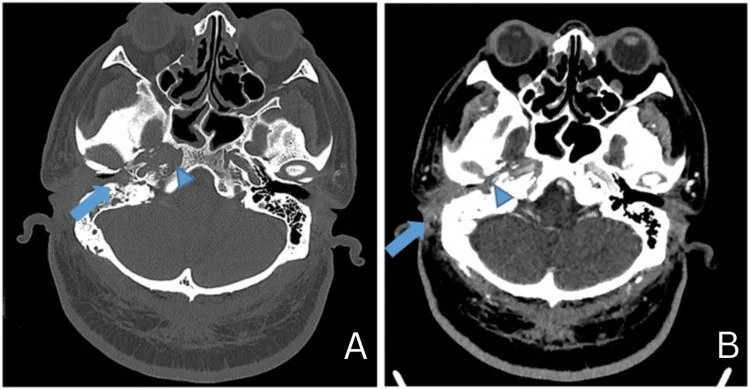
- İnce kesit kemik algoritmasında dış kulak yolu korteksinde düzensiz erozyon, mastoid ve petroz kemikte trabeküler destrüksiyon, sekestrum ve eşlik eden otomastoid opasifikasyon aranır.
- Kontrastlı tanısal
- İntravenöz kontrast sonrası yumuşak doku serisinde dış kulak yolu çevresi, retroauriküler alan, parotis, çiğneme boşluğu ve kafa tabanındaki inflamatuvar kalınlaşmanın yayılımı ile çevresel kontrastlanan koleksiyon araştırılır; bu seri, kemik penceresinde yapılan erozyon değerlendirmesinin yerine geçmez.
Önerilen pencereler: Kemik (G 1800 / M 400), Yumuşak doku (G 400 / M 40).
BT bulguları
- Dış kulak yolunda, özellikle kemik-kıkırdak bileşke çevresinde duvar kalınlaşması ve lümen yumuşak doku ile daralması görülebilir.
- Dış kulak yolu korteksinde fokal, düzensiz ve ardışık kesitlerde de izlenen erozyon, enfeksiyonun kemik tutulumu lehinedir.
- Mastoid korteks veya petroz kemikte kaba destrüksiyon ve trabeküler kayıp kafa tabanına yayılımı gösterir.
- Yoğun kemik parçacığı şeklindeki sekestrum, çevresindeki daha düşük dansiteli inflamatuvar dokudan ayrışabilir.
- Mastoid hava hücreleri ve orta kulak boşluğu sıvı/yumuşak doku dansitesiyle dolar; tek başına opasifikasyon osteomiyelit tanısı koydurmaz.
- Dış kulak yolu çevresinde retroauriküler, parotid veya çiğneme boşluğuna uzanan yumuşak doku kalınlaşması eşlik edebilir.
- Foramen jugulare, stilomastoid foramen ve petroklival kafa tabanı boyunca erozyon hastalığın anatomik yayılımını işaret eder.
- Tipik (otolojik) kafa tabanı osteomiyeliti dış kulak yolu/temporal kemik enfeksiyonundan ilerler; atipik (merkezi) biçim belirgin dış kulak yolu enfeksiyonu olmadan klivus ve sfenoid-basioksiput bölgesini tutabilir.
Normalde
Normal temporal kemikte dış kulak yolu korteksi düzgün ve kesintisizdir; mastoid hava hücreleri havalı, trabeküller korunmuş ve komşu retroauriküler yağ planı açıktır. Aynı düzeyde karşı tarafın korteksini, mastoid havalanmasını ve foramen jugulare/stilomastoid foramen çevresini kıyasla; inflamasyon asimetrisi ile gerçek kortikal kesintiyi ayrı ayrı tarif et.
Ayırıcı tanı
- Dış kulak yolu skuamöz hücreli karsinomu
- fokal kitle, düzensiz kemik yıkımı ve ilerleyici kanal daralması yapabilir; görüntüleme tek başına kesin ayırmaz.
- Akut otomastoidit
- mastoid hücre opasifikasyonu yapar; dış kulak yolu kaynaklı kortikal erozyon ve kafa tabanı yayılımı yoksa osteomiyelit daha az olasıdır.
- Kolesteatom
- sklerotik mastoid içinde orta kulak veya attik merkezli yumuşak doku ve kemikçik erozyonu görülür; dış kulak yolu korteksi başlangıç odağı olmayabilir.
- Nekrotizan dış kulak yolu enfeksiyonu
- dış kulak yolu çevresinde inflamasyonla başlar; osteomiyelit bu enfeksiyonun kemik tutulumu/yayılım evresini anlatır.
- Kafa tabanı metastazı
- litik destrüksiyon ve yumuşak doku kitlesi oluşturabilir; dış kulak yolu enfeksiyonu bulgusu olmadan da görülebilir.
Tuzaklar
- Mastoid opasifikasyonunu osteomiyelit olarak adlandırma; erozyon, kemik destrüksiyonu veya klinik bağlamla uyumlu kafa tabanı yayılımı ara.
- Erken enfeksiyonda korteks sağlam görünebilir; kontrastlı BT'de dış kulak yolu çevresi yağ planı ve yumuşak dokuyu da değerlendir, kemik iliği uzanımı için MR'ın tamamlayıcı rolünü belirt.
- Sekestrumu normal havalı hücre içindeki kemik septumundan ayırmak için parçanın çevresindeki destrüksiyon ve düzensiz sklerotik sınırları çok düzlemli incele.
- Dış kulak yolu tümörü ile enfeksiyon örtüşebilir; kitle benzeri asimetrik kalınlaşma veya tedaviye rağmen ilerleyen kemik yıkımında yalnız BT görünümüne dayanma.
- Dış kulak yolu veya kafa tabanı osteomiyeliti maligniteyi taklit edebilir; görüntüleme tek başına kesin ayrım sağlamayabileceğinden bazı olgularda biyopsi gerekebilir.
Kendini dene
BT'de dış kulak yolunda inflamatuvar yumuşak doku ve temporal kemikte erozyon görülüyor. Bu BT bulgularıyla en olası tanı hangisidir?
Cevabı göster
Temporal kemik osteomiyeliti. Dış kulak yolu enfeksiyonu temporal kemiğe yayılarak osteomiyelite yol açabilir; BT'de kanal çevresindeki inflamatuvar dokuya kemik erozyonunun eşlik etmesi bu tanıyla uyumludur. Görüntüleme bulguları kafa tabanı osteomiyelitini maligniteden ayırmada tek başına kesin olmayabilir.
Mastoid hücrelerde opasifikasyon var; korteks düzgün ve dış kulak yolu ya da kafa tabanında erozyon görülmüyor. En uygun yorum hangisi?
Cevabı göster
Otomastoid opasifikasyon. Mastoid hücre opasifikasyonu tek başına özgül değildir; kafa tabanı enfeksiyonlarının patognomonik görüntüleme bulgusu yoktur. Sekestrum ayrılmış yoğun kemik parçasıdır, karsinom ise kitle ve fokal yıkımla gündeme gelir.
Kaynaklar
Bu sayfadaki 39 cümle ve 2 quiz sorusu aşağıdaki kaynaklardan alıntılarla otomatik olarak karşılaştırıldı; 15 cümle bu karşılaştırmada düzeltildi (2026-10-09).
- A Comprehensive Review of Skull Base Osteomyelitis: Diagnostic and Therapeutic Challenges among Various Presentationspmc.ncbi.nlm.nih.gov
- Intrapetrous Internal Carotid Artery Pseudoaneurysm Secondary to Malignant Otitis Externa: A Case Reportpmc.ncbi.nlm.nih.gov
- Malignant External Otitis Complicated by Severe Allergic Reaction to Antibiotic Therapypmc.ncbi.nlm.nih.gov
- Temporal Bone Osteomyelitis Masquerading as Malignancy: A Diagnostic Challengepmc.ncbi.nlm.nih.gov
- Temporal Bone Osteomyelitis in a Child Closely Resembles Lateral Sinus Thrombosis: A Case Reportpmc.ncbi.nlm.nih.gov
- Skull base infections, their complications, and management.pubmed.ncbi.nlm.nih.gov
- Congenital tuberculosis presenting as otorrhoea in a preterm infant.pubmed.ncbi.nlm.nih.gov
- Bilateral skull base osteomyelitis in an immunocompetent patient.pubmed.ncbi.nlm.nih.gov
- Emergence of Refractory Otomastoiditis Due to Nontuberculous Mycobacteria: Institutional Experience and Review of the Literature.pubmed.ncbi.nlm.nih.gov
- Cefepime induced acute interstitial nephritis--a case report.pubmed.ncbi.nlm.nih.gov
- Pipeline embolization device and subsequent vessel sacrifice for treatment of a bleeding carotid pseudoaneurysm at the skull base: a case report.pubmed.ncbi.nlm.nih.gov
- Severe Daily Headache as an Uncommon Manifestation of Widespread Skull Base Osteomyelitis.pubmed.ncbi.nlm.nih.gov
- Clinical Challenges in the Diagnosis and Treatment of Temporal Bone Osteomyelitis.pubmed.ncbi.nlm.nih.gov
- Imaging Clinic: Destructive Temporal Bone Osteomyelitis in an Infant-CT and 3D Findings.pubmed.ncbi.nlm.nih.gov
- Temporal Bone Osteomyelitis in a Child Closely Resembles Lateral Sinus Thrombosis: A Case Report.pubmed.ncbi.nlm.nih.gov
- Intrapetrous Internal Carotid Artery Pseudoaneurysm Secondary to Malignant Otitis Externa: A Case Report.pubmed.ncbi.nlm.nih.gov
- Malignant External Otitis Complicated by Severe Allergic Reaction to Antibiotic Therapy.pubmed.ncbi.nlm.nih.gov
- Necrotising Otitis Externa: A Review of Imaging Modalities.pubmed.ncbi.nlm.nih.gov
- Diagnosis of Skull Base Osteomyelitis.pubmed.ncbi.nlm.nih.gov
- Necrotizing Otitis Externa Due to Scedosporium sp.: A Case Report.pubmed.ncbi.nlm.nih.gov
- Usefulness of <sup>99m</sup>Tc-HMPAO leukocyte scintigraphy in necrotizing external otitis/skull base osteomyelitis.pubmed.ncbi.nlm.nih.gov
- A Comprehensive Review of Skull Base Osteomyelitis: Diagnostic and Therapeutic Challenges among Various Presentations.pubmed.ncbi.nlm.nih.gov
- Imaging of Skull Base Tumors.pubmed.ncbi.nlm.nih.gov
- Imaging of Pathologies of the Temporal Bone and Middle Ear: Inflammatory Diseases, Their Mimics and Potential Complications-Pictorial Review.pubmed.ncbi.nlm.nih.gov
- Clival and Paraclival Lesions: A Pictorial Review.pubmed.ncbi.nlm.nih.gov
- Acute otitis media complicated by peripheral facial nerve palsy in an adult: A case report.pubmed.ncbi.nlm.nih.gov
- Bilateral Cavernous Sinus Thrombosis and Clival Osteomyelitis Complicating Pediatric Otitis Media.pubmed.ncbi.nlm.nih.gov
- Medication-related osteonecrosis of the external auditory canal and treatment with teriparatide.pubmed.ncbi.nlm.nih.gov
- Extensive Temporal Bone Necrosis in Petrous Bone Cholesteatoma Complicated by Actinomycosis: A Case Report.pubmed.ncbi.nlm.nih.gov
- Jugular Paraganglioma Presenting As Isolated Hypoglossal Nerve Palsy: An Anatomical Enigma.pubmed.ncbi.nlm.nih.gov
- Antibiotic-Impregnated Calcium Sulfate Beads for Local Therapy of Severe Skull Base Osteomyelitis.pubmed.ncbi.nlm.nih.gov
- Nerve-rattling infections! Clinical and radiological tracing of cranial nerve palsies to infectious skull base osteomyelitis.pubmed.ncbi.nlm.nih.gov
- The Challenge of Differentiating Mixed Histiocytosis: A Case Report.pubmed.ncbi.nlm.nih.gov
- The preoperative temporal bone CT: a mnemonic for reducing surgical risk.pubmed.ncbi.nlm.nih.gov
- Computed Tomographic Findings in Canine Otitis Externa.pubmed.ncbi.nlm.nih.gov
- Atypical Manifestation of Skull Base Osteomyelitis Secondary to Malignant Otitis Externa Following Radiotherapy: A Case Report.pubmed.ncbi.nlm.nih.gov
- Imaging for Pediatric Head and Neck Emergencies: Critical Insights.pubmed.ncbi.nlm.nih.gov
- Paediatric <i>Mycobacterium Avium</i> Complex Otomastoiditis: Case Series and Literature Review.pubmed.ncbi.nlm.nih.gov
- Chronic Osteomyelitis or Squamous Cell Carcinoma of the Mandible: A Case Report With Diagnostic Challenges.pubmed.ncbi.nlm.nih.gov
- A Case of Mandibular Osteomyelitis Occurring in a Patient With Parry-Romberg Syndrome.pubmed.ncbi.nlm.nih.gov
- When Ear Pain Signals More: Otomastoiditis-Related Bilateral Cranial Neuropathies in an Immunocompromised Patient.pubmed.ncbi.nlm.nih.gov
- Patient cases with malignant otitis externa at the university clinic of Ludwig Maximilians University Munich from 2009 until 2020.pubmed.ncbi.nlm.nih.gov
- Epidemiological and Clinical Changes in Pediatric Acute Mastoiditis Before and After the COVID-19 Pandemic: An Eight-Year Retrospective Study from a Tertiary-Level Center.pubmed.ncbi.nlm.nih.gov
- An Uncommon Diagnosis of Necrotizing Mastoiditis Presenting as Bell's Palsy: A Case Report.pubmed.ncbi.nlm.nih.gov
- Middle ear malformations combined with cerebrospinal fluid otorrhea: A case report.pubmed.ncbi.nlm.nih.gov
- Key pre-operative CT parameters for cochlear implantation: a focused radiologic review with surgical relevance.pubmed.ncbi.nlm.nih.gov
- Characteristics of the Facial Canal-Lateral Canal Dehiscence: The Under-recognized Third Window.pubmed.ncbi.nlm.nih.gov
- The Clinical Value of Cranial CT Venography for Predicting <i>Fusobacterium necrophorum</i> as the Causative Agent in Children with Complicated Acute Mastoiditis.pubmed.ncbi.nlm.nih.gov
- Aural Manifestations of Antineutrophil Cytoplasmic Antibody (ANCA)-Associated Vasculitis-Diagnosis, Symptoms, Treatment.pubmed.ncbi.nlm.nih.gov
- AI-Assisted Early Clinical Diagnosis of External Auditory Canal Squamous Cell Carcinoma: A Rare Case in a Young Male From Pakistan.pubmed.ncbi.nlm.nih.gov
- Trichoepithelioma of the External Auditory Canal Mimicking Cholesteatoma.pubmed.ncbi.nlm.nih.gov
- Benign Osteonecrosis of the External Auditory Canal: Diagnostic Challenges and Outcomes in a Case Series.pubmed.ncbi.nlm.nih.gov
- A Dilemma in the Diagnosis of Group A Streptococcus (GAS) Meningitis Versus Drug-Induced Aseptic Meningitis: A Case Report.pubmed.ncbi.nlm.nih.gov
- Diagnosis and Management of Middle Ear Neuroendocrine Tumour (MeNET).pubmed.ncbi.nlm.nih.gov
- External auditory canal cholesteatoma presenting with facial nerve palsy: rare clinical entity.pubmed.ncbi.nlm.nih.gov
- Congenital Mastoid Cholesteatoma With Giant Intracranial Extension: A Case Report.pubmed.ncbi.nlm.nih.gov
- The Silent Threat: Unveiling Pathogenicity and Antimicrobial Resistance in Nonfermenting Gram-Negative Bacilli.pubmed.ncbi.nlm.nih.gov
- Microbial spectrum, management challenges, and outcome in patients with otogenic skull base osteomyelitis.pubmed.ncbi.nlm.nih.gov
- Aspergillosis: An Update on Epidemiology, Risk Factors, Diagnosis, Susceptibility, and Treatment.pubmed.ncbi.nlm.nih.gov
- Skull Base Metastases: A Pictorial Review.pubmed.ncbi.nlm.nih.gov
- Operative diagnosis of a meningioma-like intraosseous skull lesion revealing multiple myeloma: Case report and literature review.pubmed.ncbi.nlm.nih.gov
- Aggressive metastatic parotid squamous cell carcinoma with pulmonary, cerebral, and hepatic metastases: A radiologic case report.pubmed.ncbi.nlm.nih.gov
- Orbital pseudotumor and skull base infiltration as the sole manifestation of granulomatosis with polyangiitis.pubmed.ncbi.nlm.nih.gov
- From Conductive Hearing Loss to Whipple's Surgery: A Case Report and Literature Review on Neuroendocrine Tumors of the Middle Ear.pubmed.ncbi.nlm.nih.gov
- Role of the Ear in Meningitis: A Narrative Review of Neuroimaging.pubmed.ncbi.nlm.nih.gov
- Critical infections in the head and neck: A pictorial review of acute presentations and complications.pubmed.ncbi.nlm.nih.gov
- Emerging and re-emerging infectious disease in otorhinolaryngology.pubmed.ncbi.nlm.nih.gov
- Rhinosinusitis: Evidence and experience - 2024.pubmed.ncbi.nlm.nih.gov
- Autoimmune Diseases and the Vestibular and Oculomotor System: Clinical Presentation, Diagnosis, and Treatment.pubmed.ncbi.nlm.nih.gov
- Comparison of 11 staging classifications in carcinoma of the external auditory canal.pubmed.ncbi.nlm.nih.gov
- Utility and Challenges of Imaging in Peripheral Vestibular Disorder Diagnosis: A Narrative Review.pubmed.ncbi.nlm.nih.gov
- Parametric Physically Grounded Rendering of Otoscopic Morphology for Synthetic Medical Image Generation.pubmed.ncbi.nlm.nih.gov
- Tympanic Membrane Involvement of Multiple Myeloma: Report of a Case.pubmed.ncbi.nlm.nih.gov
- Multimodal imaging in conductive hearing loss: Optimising CT, MRI and CTA for accurate diagnosis and management.pubmed.ncbi.nlm.nih.gov
- Can imaging suggest the aetiology in skull base osteomyelitis? A systematic literature review.pubmed.ncbi.nlm.nih.gov
- Primary tuberculous osteomyelitis of the maxilla mimicking a refractory post-endodontic infection: A diagnostic challenge.pubmed.ncbi.nlm.nih.gov
- Purely osteolytic bone lesions in the early osteoarticular phase of synovitis, acne, pustulosis, hyperostosis and osteitis syndrome.pubmed.ncbi.nlm.nih.gov
- Context-dependent roles of the NLRP3 inflammasome in osteomyelitis and strategies for precision modulation.pubmed.ncbi.nlm.nih.gov
- Imaging of Temporal Bone Mass Lesions: A Pictorial Review.pubmed.ncbi.nlm.nih.gov
- "Leave me alone": anatomical structures and variations seen on computed tomography of the temporal bone.pubmed.ncbi.nlm.nih.gov
- Odontogenic, atypical skull-base osteomyelitis: diagnostic pitfalls and therapeutic insights-a case report and mini-review.pubmed.ncbi.nlm.nih.gov
- Primary Intracranial Cholesteatoma Presenting as a Cerebellar Mass Without Otologic Symptoms.pubmed.ncbi.nlm.nih.gov
- Infected Nonunion of the Tibia Due to Paenibacillus turicensis in a Healthy Young Adult After an All-Terrain Vehicle Accident.pubmed.ncbi.nlm.nih.gov
- Tuberculosis of the spine.pubmed.ncbi.nlm.nih.gov
- Review of Myeloma Therapies and Their Potential for Oral and Maxillofacial Side Effects.pubmed.ncbi.nlm.nih.gov
- Multidisciplinary Exploration of Computed-Tomographic and Ancient-DNA Data of an Iron Age Skull From Latvia With Multiple Lytic Bone Lesions: Differential Diagnosis Between Metastatic Carcinoma, Multiple Myeloma and Skeletal Tuberculosis.pubmed.ncbi.nlm.nih.gov
- Necrotizing Otitis Externa: Insights From a Nationwide Database on In-Hospital Outcomes and Characteristics.pubmed.ncbi.nlm.nih.gov
- Eight months of misdiagnosis: Intra-articular osteoid osteoma in a 48-year-old woman.pubmed.ncbi.nlm.nih.gov
- A rare case report: MRSA osteomyelitis after CRPP for pediatric distal both-bone forearm fracture.pubmed.ncbi.nlm.nih.gov
- Surgical Techniques and Materials Used in the Treatment of Complicated Otomastoiditis: A Systematic Review.pubmed.ncbi.nlm.nih.gov
- The cavernous sinus: An anatomic study with clinical implication.pubmed.ncbi.nlm.nih.gov
- Fungal necrotizing otitis externa; challenges in diagnosis and therapy.pubmed.ncbi.nlm.nih.gov
- The diagnostic conundrum in necrotizing otitis externa.pubmed.ncbi.nlm.nih.gov
- Case report: endolymphatic sac tumor masquerading as Meniere's disease.pubmed.ncbi.nlm.nih.gov
- Skull Base Osteomyelitis and Parapharyngeal Abscess as Complications of Otitis Media in a Young Immunocompetent Man.pubmed.ncbi.nlm.nih.gov
- Clinical Features and Surgical Outcomes in Patients With Central Skull Base Osteomyelitis.pubmed.ncbi.nlm.nih.gov
- Pre-treatment and post-treatment nasopharyngeal carcinoma imaging: imaging updates, pearls and pitfalls.pubmed.ncbi.nlm.nih.gov
- Review of imaging modalities and radiological findings of calvarial lesions.pubmed.ncbi.nlm.nih.gov
Eğitim amaçlıdır; hasta başında tanı ve tedavi kararının yerine geçmez, tıbbi tavsiye değildir. Metinler yapay zekâ desteğiyle yazıldı ve otomatik bir adımda kaynak alıntılarıyla karşılaştırıldı; bu bir tıbbi doğrulama değildir ve içerik uzman radyolog incelemesinden geçmedi. Klinik kararlarda güncel kılavuzları, kurum protokollerini ve radyoloji raporunu esas alın.