Özet
Metastaz, plazma hücreli miyelom ve lenfoma mandibula ya da maksillada tek odaklı veya çok odaklı kemik iliği lezyonları yapabilir; çene metastazı bilinen kanserin yayılımını veya nadiren henüz saptanmamış primeri gösterebilir. Kontrastsız kemik BT'si litik veya sklerotik yıkım, yumuşak doku komponenti ve kortikal bütünlüğün bozulması riskini gösterir; miyelom hastalarında iyotlu kontrast böbrek hasarı riski nedeniyle genellikle kaçınılmalıdır. Lezyonların görünümü histolojik olarak özgül değildir; primer kanser öyküsü, tam iskelet görüntüleri ve doku tanısı ayrımı tamamlar. Alt dudak-çene uyuşması, kanal ya da mental foramen tutulumu ile birlikteyse nörolojik yayılım bulgusu olarak ciddiye alınmalı; tek çene odağına bakmak sistemik çoklu hastalığı gözden kaçırabilir.
Okuma sırası
- Mandibula medullası. Kemik penceresinde mandibula korpusunu iki tarafta karşılaştır; homojen trabeküler yapıdan ayrılan litik ya da sklerotik medüller odağı ve tek mi çoklu mu olduğunu belirle.
- Mandibular kanal. Mandibular kanalı boyunca lezyonla ilişkisini değerlendir ve kanal tutulumu olup olmadığını ara.
- Mental foramen. Kanalın ön ucundaki mental foramen çevresinde kemik konturunu ve yumuşak doku yoğunluğunu kontrol et; alt dudak ya da çene uyuşması varsa bu bölgeyi özellikle ilişkilendir.
- Ekstraosseöz dokular ve çevre kemikler. Yumuşak doku penceresinde korteksin dışındaki komponenti çiğneme kasları ve ağız tabanına göre tarif et; son olarak patolojik kırık ve görüntü alanındaki diğer kemik odaklarını ara.
Faz ve pencere
- Kontrastsız tanısal
- Kemik algoritmasında medüller litik odaklar, delik deşik görünüm, yaygın osteopeni, sklerotik metastatik odak, kortikal kesinti ve patolojik kırık aranır; mandibular kanal ve mental foramenin kemik sınırları iki taraflı kıyaslanır. İyotlu kontrast kemik iliği litik yıkımını göstermek için gerekli değildir.
- Kontrastlı tanısal
- Yumuşak doku penceresinde korteks dışına çıkan kitle, mandibular kanal boyunca yumuşak doku değişikliği ve boyun lenf düğümleri incelenir. Lenfoma veya metastazda yumuşak doku komponenti eşlik edebilir; bu görünüm tek başına tümör tipini belirlemez.
Önerilen pencereler: Kemik (G 1800 / M 400), Yumuşak doku (G 400 / M 40).
BT bulguları
- Tek veya çok sayıda medüller litik odak; sınırlar keskin delik delik, belirsiz permeatif veya geniş destrüktif olabilir.
- Miyelomda genellikle kenarında sklerotik halka bulunmayan çok sayıda keskin litik odak veya yaygın medüller osteopeni izlenir; osteoskleroz nadirdir.
- Lenfoma ve metastazda korteks dışı yumuşak doku komponenti görülebilir; görüntüleme görünümü histolojik olarak özgül değildir.
- Miyelomda genellikle litik odaklar görülürken, osteoskleroz nadirdir; metastazlarda ise litik, sklerotik veya karışık kemik yıkım paternleri izlenebilir.
- Mandibular kanalın genişlemesi, kortikal sınırlarının kaybı veya mental foramen çevresindeki destrüksiyon alt dudak-çene parestezisiyle korele olabilir.
- Diş yuvalarında lamina dura kaybı, dişleri destekleyen kemikte silinme veya diş mobilitesi görülebilir.
- Korteksin dışına geçen kitle, yumuşak doku invazyonu veya patolojik kırık ileri lokal kemik tutulumunu gösterir.
- Birden fazla mandibular, maksiller veya kafatası odağı sistemik plazma hücre hastalığı ya da yaygın metastaz lehinedir.
Normalde
Normal anatomide dişleri destekleyen kemikte lamina dura bütündür ve mandibular kanal kortikal sınırları kesintisizdir; bu yapıların malignite nedeniyle yıkılması veya düzensizleşmesi patolojik kabul edilir. Lezyonun yerini, sınırını, iç yapısını ve alt mandibular sinir gibi önemli anatomik yapılara etkisini değerlendirmelisiniz.
Normal BT ile kıyasla
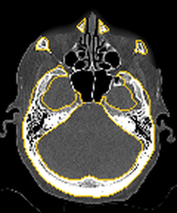
Aynı bölgenin, kaynak veri setinde patoloji içermediği belirtilen bir BT incelemesini kesit kesit kaydırın; organ sınırlarını açarak anatomiyi hasta görüntüsüyle karşılaştırın. Küçük rastlantısal bulgular tümüyle dışlanmamıştır.
Kaynak: TotalSegmentator v2.01 veri seti (Wasserthal ve ark., Radiology: Artificial Intelligence 2023), CC BY 4.0, vaka s0849; organ sınırları veri setinin otomatik segmentasyonundan, birleştirilerek sadeleştirildi. Görüntüler 2,5 mm kesit aralığına yeniden örneklendi.
Ayırıcı tanı
- Çene osteomiyeliti veya odontojenik kökenli enfeksiyonlarda litik yıkım, periostal reaksiyon ve yumuşak doku tutulumu gibi bulgular görülebilir.
- Kronik osteomiyelit, düzensiz kortikal ve medüller değişiklikler ile periostal yeni kemik üretimi göstererek primer kemik tümörlerini veya metastazı taklit edebilir.
- Osteosarkom
- agresif karışık litik-osteoid matriks, spiküler periostal kemik ve periodontal aralık genişlemesi görülebilir.
- Stafne kemik boşluğu
- kanal altında mandibulanın lingual yüzünde konkavite yapar; medüller destrüksiyon veya gerçek yumuşak doku kitlesi değildir.
- Odontojenik kist
- diş kökü veya gömülü kronla ilişkili, çoğu kez düzgün sınırlı ekspansil boşluk oluşturur; çoklu medüller odak paterni yapmaz.
Tuzaklar
- 'Delik deşik' (punched-out) litik alanlar tek başına miyelom olarak kabul edilmemelidir; metastaz ve lenfoma benzer görünebilir, bu nedenle eşlik eden tüm iskelet bulguları ve malignite öyküsü birlikte değerlendirilmelidir.
- Sklerotik görünüm metastazı dışlamaz; primer tümörün osteoblastik veya osteolitik yanıt oluşturma eğilimi paterni değiştirir.
- Mandibular kanal tutulumu tek başına tanı koydurmaz; malign metastaz veya lenfoma gibi durumlarda perinöral yayılım belirtisi olan alt dudak-çene uyuşması (Numb Chin Syndrome), kortikal erozyon ve klinik öykü birlikte değerlendirilmelidir.
- Yalnız çene kesitlerine odaklanmayın; kafatasında veya diğer görüntü alanındaki çoklu kemik odaklarını ve patolojik kırığı ayrıca arayın.
Kendini dene
Kanser öyküsü olan hastada mandibular kanal çevresinde belirsiz sınırlı litik odak ve alt dudak uyuşması var. En olası açıklama nedir?
Cevabı göster
Mandibula metastazı. Bilinen malignite, kanal çevresi yıkım ve mental sinir dağılımında uyuşma mandibular metastazı kuvvetle düşündürür. Dentigeröz kist gömülü diş kronuna tutunur; Stafne boşluğu lingual korteks konkavitesidir; apse diş apeksinde enfeksiyon bulgularıyla merkezlenir.
Çene ve kalvaryumda çok sayıda keskin litik odak, reaktif sklerotik halka olmadan izleniyor. Hangi hastalık özellikle düşünülür?
Cevabı göster
Multipl miyelom. Çene ve kafatasındaki çoklu keskin, sklerotik kenarsız litik odaklar multipl miyelom için bilinen bir örüntüdür. Osteosarkom genellikle tek agresif primer odak ve osteoid üretimi yapar; Stafne boşluğu tek anatomik konkavitedir; keratokist odontojenik bölgede sınırlı kistik lezyondur.
İlgili konular
Kaynaklar
Bu sayfadaki 40 cümle ve 2 quiz sorusu aşağıdaki kaynaklardan alıntılarla tek tek karşılaştırıldı; 10 cümle bu karşılaştırmada düzeltildi (2026-10-08).
- Multiple Myeloma: A Review of Imaging Features and Radiological Techniquespmc.ncbi.nlm.nih.gov
- A Pattern-based Imaging Approach to Pediatric Jaw Lesionspmc.ncbi.nlm.nih.gov
- Cone-Beam Computed Tomography Imaging of Primary Non-Hodgkin Lymphoma of the Jaws: A 6-Case Seriespmc.ncbi.nlm.nih.gov
- Neurosensory Disturbances Related to the Inferior Alveolar Nerve Amongst Patients With Mandibular Medication-Related Osteonecrosis of the Jaw (MRONJ): A Clinical and Radiological Overviewpmc.ncbi.nlm.nih.gov
- Quantitative CT texture analysis of the mandibular condyle in medication-related osteonecrosis of the jawpmc.ncbi.nlm.nih.gov
- Mandibular lymphoma.pubmed.ncbi.nlm.nih.gov
- Leptomeningeal Metastasis: A Review of the Pathophysiology, Diagnostic Methodology, and Therapeutic Landscape.pubmed.ncbi.nlm.nih.gov
- Multiple myeloma.pubmed.ncbi.nlm.nih.gov
- Maxillofacial lymphomas.pubmed.ncbi.nlm.nih.gov
- Classification, imaging, biopsy and staging of osteosarcoma.pubmed.ncbi.nlm.nih.gov
- ACR appropriateness criteria on metastatic bone disease.pubmed.ncbi.nlm.nih.gov
- Imaging of skull vault tumors in adults.pubmed.ncbi.nlm.nih.gov
- Diffuse Large B-Cell Lymphoma of the Mandible Diagnosed by Metagenomic Sequencing: A Case Report.pubmed.ncbi.nlm.nih.gov
- [Malignant paraganglioma mimicking multiple myeloma].pubmed.ncbi.nlm.nih.gov
- The role of PET/CT in the evaluation of skeletal metastases.pubmed.ncbi.nlm.nih.gov
- Differential diagnostic value of 18 F-FDG PET/CT in osteolytic lesions.pubmed.ncbi.nlm.nih.gov
- Therapeutic Efficacy of 213 Bi-labeled sdAbs in a Preclinical Model of Ovarian Cancer.pubmed.ncbi.nlm.nih.gov
- Primary testicular plasmocytoma: A five year follow-up.pubmed.ncbi.nlm.nih.gov
- PET/CT in benign and malignant musculoskeletal tumors and tumor-like conditions.pubmed.ncbi.nlm.nih.gov
- Osteomyelitis of the Jaw Bones and Its Mimics: Resolving the Diagnostic Enigma.pubmed.ncbi.nlm.nih.gov
- How does medication-related osteonecrosis of the jaw (MRONJ) influence the health-related quality of life after surgery?pubmed.ncbi.nlm.nih.gov
- Mesenchymal Chondrosarcoma of the Mandible, a Big Dilemma: Report of a Rare Case in Mesenchymal Chondrosarcoma of the Mandible-Report of a Case With Discussion of Diagnostic and Therapeutic Dilemmas.pubmed.ncbi.nlm.nih.gov
- Imaging approach for jaw and maxillofacial bone tumors with updates from the 2022 World Health Organization classification.pubmed.ncbi.nlm.nih.gov
- Multimodal Imaging of Osteosarcoma: From First Diagnosis to Radiomics.pubmed.ncbi.nlm.nih.gov
- Primary Aggressive Oral Lymphomas (PAOL): A Narrative Review of Diagnosis, Molecular Features, Therapeutic Approaches, and the Integrated Role of Dentists and Hematologists.pubmed.ncbi.nlm.nih.gov
- The Immune Checkpoint Inhibitors Journey: From Early Promise to Lasting Impact.pubmed.ncbi.nlm.nih.gov
- The CXCR4-targeted theranostics era: a comprehensive review of a decade of progress (2015-2025).pubmed.ncbi.nlm.nih.gov
- Metastatic gallbladder cancer presenting as numb chin syndrome: A case report and literature review.pubmed.ncbi.nlm.nih.gov
- Numb Chin Syndrome in Sickle Cell Disease: A Systematic Review and Recommendations for Investigation and Management.pubmed.ncbi.nlm.nih.gov
- Intraosseous synovial sarcoma of the mandible: A case report and review of the literature.pubmed.ncbi.nlm.nih.gov
- "Genetic tumor syndromes of the head and neck: Update in the genomic era".pubmed.ncbi.nlm.nih.gov
- Mandibular Inflammatory Myofibroblastic Tumors: A Literature Review with a New Case Presentation.pubmed.ncbi.nlm.nih.gov
- Management of Tibial Adamantinoma: Balancing Oncologic Cure and Reconstructive Longevity.pubmed.ncbi.nlm.nih.gov
- Giant Cell Tumor of the Sacrum With Secondary Changes of an Aneurysmal Bone Cyst: A Case Report.pubmed.ncbi.nlm.nih.gov
- Follicular Thyroid Carcinoma Metastasis to the Retromolar Trigone: A Case Report.pubmed.ncbi.nlm.nih.gov
- Surgical Management of Advanced Mandibular Osteonecrosis Utilizing a Contemporary Mandibular Reconstruction Plate in Patients Unsuitable for Free Flap Reconstruction-Preliminary Study and Case Series.pubmed.ncbi.nlm.nih.gov
- Neuroblastoma Metastasis to the Mandible in Children: A Case Report and Focused Narrative Review of Reported Cases.pubmed.ncbi.nlm.nih.gov
- Giant Cell Tumor of the Sacrum: A Narrative Review of Management Challenges and Modalities.pubmed.ncbi.nlm.nih.gov
- Osteosarcoma: A Comprehensive Morphological and Molecular Review with Prognostic Implications.pubmed.ncbi.nlm.nih.gov
- Review of imaging modalities and radiological findings of calvarial lesions.pubmed.ncbi.nlm.nih.gov
- Beyond the mouth: Uncovering non-secretory multiple myeloma through oral symptoms.pubmed.ncbi.nlm.nih.gov
- Imaging technologies in the differential diagnosis and follow-up of brown tumor in primary hyperparathyroidism: Case report and review of the literature.pubmed.ncbi.nlm.nih.gov
- Radiologic and Pathologic Insights into Medication-Related Osteonecrosis of the Jaw in Myeloma Patients: A Report of 3 Cases.pubmed.ncbi.nlm.nih.gov
- Cone-Beam Computed Tomography Imaging of Primary Non-Hodgkin Lymphoma of the Jaws: A 6-Case Series.pubmed.ncbi.nlm.nih.gov
- Radiographic Patterns and Clinical Correlates of Medication-Related Osteonecrosis of the Jaw (MRONJ): A Retrospective Analysis.pubmed.ncbi.nlm.nih.gov
- Uncovering jaw-specific radiographic differences in medication related osteonecrosis of the jaws (MRONJ): a case-control study.pubmed.ncbi.nlm.nih.gov
- Giant Cell Tumor of Bone: Biology, Pathophysiology, and Histopathology in the Era of H3F3A.pubmed.ncbi.nlm.nih.gov
- Giant cell tumor of bone and secondary osteoarthritis.pubmed.ncbi.nlm.nih.gov
- Chondrosarcoma: A Clinical Review.pubmed.ncbi.nlm.nih.gov
- Chondrosarcoma: A comprehensive review of tumor biology, clinical behavior, and therapeutic advances.pubmed.ncbi.nlm.nih.gov
- Prognostic Value of Cranial Nerve Invasion in T4-Stage Nasopharyngeal Carcinoma: A Retrospective Cohort Study With a Median Follow-Up of 136 Months.pubmed.ncbi.nlm.nih.gov
- Radiographic Assessment of Normal Periapical Bone Density: A Descriptive Analysis Based on Digital Imaging.pubmed.ncbi.nlm.nih.gov
- Anatomical and Systemic Risk Factors for Recurrence in Medication-Related Osteonecrosis of the Jaw (MRONJ): A Retrospective Study of 812 Patients.pubmed.ncbi.nlm.nih.gov
- Temporomandibular Joint Defects: Etiology and Reconstruction Strategy: A Narrative Review.pubmed.ncbi.nlm.nih.gov
- Association between systemic osteoporosis and mandibular condyle characteristics: A comparative imaging study.pubmed.ncbi.nlm.nih.gov
- Primary Diffuse Large B-Cell Lymphoma Mimicking a Dental Abscess: A Case Report.pubmed.ncbi.nlm.nih.gov
- Masked invaders: A case series on uncommon manifestations of extrapulmonary tuberculosis.pubmed.ncbi.nlm.nih.gov
- Chronic Osteomyelitis or Squamous Cell Carcinoma of the Mandible: A Case Report With Diagnostic Challenges.pubmed.ncbi.nlm.nih.gov
- Xanthogranulomatous Osteomyelitis as a Bone Tumor Mimic: A Systematic Review of Pre-biopsy Diagnostic Clues and Management Decision Points.pubmed.ncbi.nlm.nih.gov
- Orbital Exenteration in Maxillary Osteosarcoma: A Case Report.pubmed.ncbi.nlm.nih.gov
- Exploring Paediatric Jaw Osteosarcoma: A Detailed Case Report of a 12-Year-Old Girl and Review of the Literature.pubmed.ncbi.nlm.nih.gov
- A Rare Case of Anterior Stafne Bone Cavity in the Mandibular Region.pubmed.ncbi.nlm.nih.gov
- Stafne Bone Cavity: A systematic review.pubmed.ncbi.nlm.nih.gov
- Atypical variant of Stafne bone defect mimicking odontogenic cyst of the jaw.pubmed.ncbi.nlm.nih.gov
- Primary Intraosseous Mucoepidermoid Carcinoma of the Mandible: A Report of a Rare Case.pubmed.ncbi.nlm.nih.gov
- Malignant Odontogenic Tumors: A Clinicopathological Study of 53 Cases in Brazil.pubmed.ncbi.nlm.nih.gov
- Ameloblastic Carcinoma: A Case Report with Emphasis on Relevant Diagnostic Aspects.pubmed.ncbi.nlm.nih.gov
- Oral squamous cell carcinoma vs. medication-related osteonecrosis of the jaw in patients assuming bone-modifying agents: a diagnostic challenge in a multi-hospital case series.pubmed.ncbi.nlm.nih.gov
- A Case of Primary Intraosseous Adenoid Cystic Carcinoma of the Mandible.pubmed.ncbi.nlm.nih.gov
- Precision management of gastrointestinal tumor-associated osteoporosis driven by cutting-edge technologies: Current status, challenges, and future prospects.pubmed.ncbi.nlm.nih.gov
- Perioperative considerations for patients with multiple myeloma.pubmed.ncbi.nlm.nih.gov
- Machine learning based differential diagnosis of SAPHO syndrome and secondary bone tumors using whole body bone scintigraphy.pubmed.ncbi.nlm.nih.gov
- Skull Base Metastases: A Pictorial Review.pubmed.ncbi.nlm.nih.gov
- Neurosensory Disturbances Related to the Inferior Alveolar Nerve Amongst Patients With Mandibular Medication-Related Osteonecrosis of the Jaw (MRONJ): A Clinical and Radiological Overview.pubmed.ncbi.nlm.nih.gov
- Late recurrence of Burkitt's lymphoma in the jaw: numb chin syndrome as the only symptom.pubmed.ncbi.nlm.nih.gov
- MASCC/ISOO Clinical Practice Statement: imaging and clinical laboratory tests in the diagnosis and management of medication-related osteonecrosis of the jaw.pubmed.ncbi.nlm.nih.gov
- Metastasis of follicular carcinoma of the thyroid gland to the mandible: A report of a rare case.pubmed.ncbi.nlm.nih.gov
- Radiolucent Jaw Lesions: Imaging Approach.pubmed.ncbi.nlm.nih.gov
- Radiolucent lesions of the mandible: a pattern-based approach to diagnosis.pubmed.ncbi.nlm.nih.gov
- Cone-beam computed tomography-based analysis of jawbone destruction patterns in multiple myeloma: Associations with clinical data in an observational study.pubmed.ncbi.nlm.nih.gov
- A Giant-Cell Lesion with Cellular Cannibalism in the Mandible: Case Report and Review of Brown Tumors in Hyperparathyroidism.pubmed.ncbi.nlm.nih.gov
- Hedgehog signaling in tissue homeostasis, cancers, and targeted therapies.pubmed.ncbi.nlm.nih.gov
- Evidence supporting endodontic therapy as a safe strategy in patients previously treated with head and neck radiotherapy: a scoping review.pubmed.ncbi.nlm.nih.gov
- Comparative assessment of METastasis Reporting and Data System for Prostate Cancer versus Prostate Cancer Clinical Trials Working Group 3 Criteria in assessing treatment response in patients with metastatic castration-resistant prostate cancer.pubmed.ncbi.nlm.nih.gov
- Comparison of Diagnostic Performance of Whole-Body 360° CZT Bone SPECT/CT and Planar Bone Scintigraphy in Detecting Bone Metastasis in Small Cell Lung Cancer.pubmed.ncbi.nlm.nih.gov
- Osteoblastic skeletal metastases mimicking high-bone-turnover metabolic bone disease in gastric adenocarcinoma.pubmed.ncbi.nlm.nih.gov
- The usefulness of different imaging modalities in mandibular osteonecrosis and osteomyelitis diagnosis.pubmed.ncbi.nlm.nih.gov
- Preliminary Application of Bilateral Submandibular Horizontal Incision in the Treatment of Upper Cervical Tumors.pubmed.ncbi.nlm.nih.gov
- Diagnostic challenge of the brown tumors in developing country: A case series.pubmed.ncbi.nlm.nih.gov
- Ozone Infiltration for Osteonecrosis of the Jaw Therapy: A Case Series.pubmed.ncbi.nlm.nih.gov
- Rare Diseases of the Orbit.pubmed.ncbi.nlm.nih.gov
- Imaging of Skull Base Tumors.pubmed.ncbi.nlm.nih.gov
- A Pattern-based Imaging Approach to Pediatric Jaw Lesions.pubmed.ncbi.nlm.nih.gov
- Perineural spread in head and neck malignancies: imaging findings - an updated literature review.pubmed.ncbi.nlm.nih.gov
- Numb Chin Syndrome Leading to a Diagnosis of Salivary Ductal Adenocarcinoma: A Case Report and Review of the Literature.pubmed.ncbi.nlm.nih.gov
- Spinal lesions: a comprehensive radiologic overview.pubmed.ncbi.nlm.nih.gov
- Solitary plasmacytoma of the calvarium: A successful management of a long-standing large lesion with a long-term follow-up.pubmed.ncbi.nlm.nih.gov
- Imaging findings in a case of leptomeningeal myelomatosis, a rare but critical central nervous system complication of multiple myeloma.pubmed.ncbi.nlm.nih.gov
- Tooth Transplantation With Sticky Bone for Vertical Bone Defects: A 4-Year Follow-Up Case Report.pubmed.ncbi.nlm.nih.gov
- Incidence and predictors of root resorption after delayed replantation of permanent teeth.pubmed.ncbi.nlm.nih.gov
- Tooth Auto-Transplantation in Patients with Cleft Lip and/or Palate: A Systematic Review and Preliminary Clinical Protocol Proposal.pubmed.ncbi.nlm.nih.gov
- Follow-up of idiopathic osteosclerosis on cone-beam computed tomography: a retrospective longitudinal study.pubmed.ncbi.nlm.nih.gov
- Efficiency and Safety of Dental Implantation in the Area of Hyperdense Jaw Lesions: A Narrative Review.pubmed.ncbi.nlm.nih.gov
- The cavernous sinus: An anatomic study with clinical implication.pubmed.ncbi.nlm.nih.gov
- Post-COVID-19 Jaw Osteonecrosis: A Narrative Review.pubmed.ncbi.nlm.nih.gov
- Musculoskeletal Manifestations of T-Cell Lymphomas: Clinical Presentation, Imaging Features, Pathobiology, and Orthopedic Considerations-A Narrative Review.pubmed.ncbi.nlm.nih.gov
- Primary bone lymphoma with extensive imaging-defined necrosis on magnetic resonance imaging and <sup>18</sup>F-fluorodeoxyglucose positron emission tomography: A diagnostic pitfall.pubmed.ncbi.nlm.nih.gov
- Multidisciplinary Exploration of Computed-Tomographic and Ancient-DNA Data of an Iron Age Skull From Latvia With Multiple Lytic Bone Lesions: Differential Diagnosis Between Metastatic Carcinoma, Multiple Myeloma and Skeletal Tuberculosis.pubmed.ncbi.nlm.nih.gov
- Mandibular Follicular Lymphoma: A Rare Extranodal Presentation With Initial Facial Bone Involvement.pubmed.ncbi.nlm.nih.gov
- Multiple Ossifying Fibroma: A Case Presentation with Comprehensive Review of Literature.pubmed.ncbi.nlm.nih.gov
- Sporadic Burkitt Lymphoma First Presenting as Painful Gingival Swellings and Tooth Hypermobility: A Life-Saving Referral.pubmed.ncbi.nlm.nih.gov
- 2025 American Thyroid Association Management Guidelines for Adult Patients with Differentiated Thyroid Cancer.pubmed.ncbi.nlm.nih.gov
- Diagnosis and treatment of Paget's disease of bone: position paper from the Italian Society of Osteoporosis, Mineral Metabolism and Skeletal Diseases (SIOMMMS).pubmed.ncbi.nlm.nih.gov
- Mechanism of bisphosphonate-related osteonecrosis of the jaw (BRONJ) revealed by targeted removal of legacy bisphosphonate from jawbone using competing inert hydroxymethylene diphosphonate.pubmed.ncbi.nlm.nih.gov
- Chinese expert consensus on the diagnosis and clinical management of medication-related osteonecrosis of the jaw.pubmed.ncbi.nlm.nih.gov
Eğitim amaçlıdır; hasta başında tanı ve tedavi kararının yerine geçmez. İçerik yapay zekâ destekli bir süreçle güncel literatüre göre cümle cümle kaynaklarla karşılaştırıldı, ancak uzman radyolog incelemesinden geçmedi. Klinik kararlarda güncel kılavuzları, kurum protokollerini ve radyoloji raporunu esas alın.