Özet
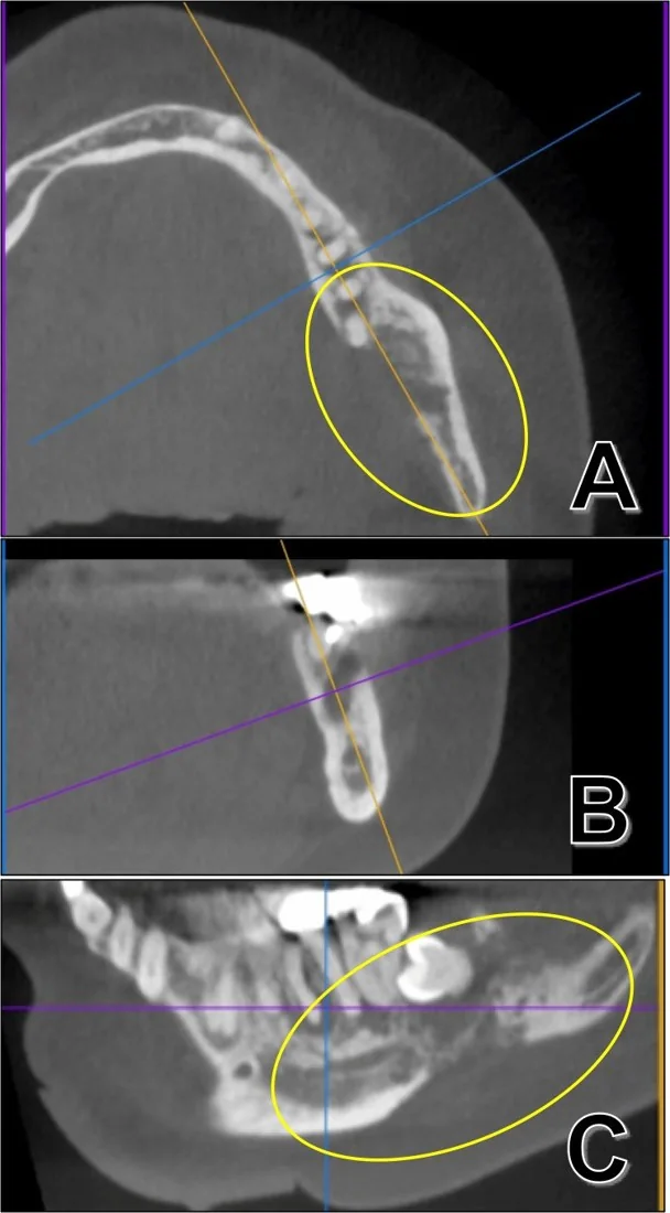
Primer intraosseöz karsinom, çene kemiğinin içinde gelişen ve çoğu kez odontojenik epitel kökenli olan nadir bir malignitedir; odontojenik karsinomlar ise histolojik olarak farklı alt tipleri kapsar. BT, medüller yıkımın sınırını, kortikal geçişi ve kemik dışı uzanımı haritalar; görüntü tek başına alt tipi veya primer intraosseöz kökeni kanıtlamaz. Diş kökü çevresinde görülen düzensiz bir lezyonu sıradan apikal hastalık saymak tanıyı geciktirebilir. Mandibular kanal boyunca yayılım, patolojik kırık veya çevre yumuşak doku tutulumu atlanırsa hastalığın yerel kapsamı eksik kalır.
Faz ve pencere
- Kontrastsız tanısal
- İnce kesit kontrastsız BT'nin kemik algoritmasında medüller trabekül kaybı, geçiş zonunun belirsizliği, kortikal yıkım ve olası patolojik kırık seçilir; odontojenik karsinom için doğrulanmış özgül bir HU eşiği yoktur.
Önerilen pencereler: Kemik (G 1800 / M 400), Yumuşak doku (G 400 / M 40).
BT bulguları
- Primer intraosseöz skuamöz hücreli karsinom, görüntülemede iyi sınırlı benign benzeri kitlelerden sınırları belirsiz ve kemik yıkımlı litik lezyonlara kadar değişen görünüm gösterebilir. Sınır görünümü tek başına tanıyı dışlamaz veya doğrulamaz.
- Lingual, bukkal ya da inferior korteks boyunca kesinti; destrüksiyon tek bir kortikal yüzle sınırlı kalmayabilir.
- Kemik defektini aşan, komşu çiğneme kası veya ağız tabanına uzanan solid yumuşak doku bileşeni.
- Mandibular kanal korteksinde silinme veya asimetrik genişleme; mental foramen çevresine uzanım eşlik edebilir.
- Lezyon komşuluğundaki dişlerde lamina dura kaybı ya da periodontal aralığın genişlemesi ile birlikte 'yüzen diş' görünümü izlenebilir.
- Önceden kistik ya da odontojenik lezyon olarak izlenen alanda yeni kortikal yıkım veya yumuşak doku uzanımı.
- İleri destrüksiyonda mandibula sürekliliğinin bozulması ve patolojik kırık hattı.
- Primer intraosseöz karsinom NOS tanısı, çene kemiğinde merkezlenen skuamöz karsinomun histopatolojik doğrulanmasını ve mukozal köken, metastaz, diğer malign odontojenik tümörler ile intraosseöz tükürük bezi tümörlerinin dışlanmasını gerektirir; görüntüleme tek başına tanı koydurmaz.
Ölçütler ve sınıflamalar
- Primer intraosseöz karsinom, NOS
- Başka bir odontojenik tümör, intraosseöz tükürük bezi tümörü, metastaz veya komşu mukozal karsinomla açıklanamayan primer kemik içi karsinom kategorisidir.
- Ameloblastik karsinom
- Ameloblastom benzeri histoloji taşıyan primer odontojenik karsinomdur; BT görünümü tek başına tanı koydurmaz.
- Berrak hücreli odontojenik karsinom
- WHO sınıflamasındaki malign odontojenik tümörlerden biridir; berrak hücreli metastaz ve tükürük bezi tümörleri dışlanmalıdır.
- Hayalet hücreli odontojenik karsinom
- Malign odontojenik tümör sınıfındadır; kistik veya solid odontojenik öncül lezyonla ilişkisi histopatolojik olarak belirlenir.
- Sklerozan odontojenik karsinom
- Belirgin stromal skleroz ve yerel infiltrasyonla tanımlanan çok nadir odontojenik karsinomdur.
- Odontojenik karsinosarkom (ayrı malign odontojenik tümör)
- Malign epitel ve mezenkimal bileşenleri olan çok nadir bir odontojenik tümördür; WHO 2022 sınıflamasında ayrı bir başlıkta yer alsa da bazı çalışmalar onu terminal bir fenotip olarak ele almaktadır.
Normalde
Normal mandibulada trabeküler yapı süreklidir, kortikal plaklar tamdır ve mandibular kanal belirgin, düzenli sınırlarla izlenir. Maksillada alveoler süreçlerin ve sinüs duvarlarının bütünlüğü korunmuş olmalıdır. BT'de mandibula kortikal düzensizliğini ve çevre yumuşak doku uzanımını değerlendirin.
Normal BT ile kıyasla
Aynı bölgenin, kaynak veri setinde patoloji içermediği belirtilen bir BT incelemesini kesit kesit kaydırın; organ sınırlarını açarak anatomiyi hasta görüntüsüyle karşılaştırın. Küçük rastlantısal bulgular tümüyle dışlanmamıştır.
Kaynak: TotalSegmentator v2.01 veri seti (Wasserthal ve ark., Radiology: Artificial Intelligence 2023), CC BY 4.0, vaka s0849; organ sınırları veri setinin otomatik segmentasyonundan, birleştirilerek sadeleştirildi. Görüntüler 2,5 mm kesit aralığına yeniden örneklendi.
Ayırıcı tanı
- Odontojenik osteomiyelit
- diş apeksinden devam eden inflamasyon, periostal yeni kemik ve çevre yağ dokusunda inflamatuvar değişiklikler tanıyı destekler.
- Mandibula metastazı
- bilinen primer tümör, çoklu kemik odakları veya kanal ve mental foramen boyunca infiltrasyon öne çıkar.
- Lenfoma
- geniş kemik iliği tutulumu ve yumuşak doku kitlesi, korteks görece korunurken de görülebilir.
- Ameloblastom ekspansil ve multiloküler görünebilir; karsinomla ayrımı görüntüleme bulgularına dayanarak kesinleştirilemez ve histopatolojik değerlendirme gerekir.
- Oral mukoza skuamöz hücreli karsinomu
- yumuşak doku kitlesi mukozada merkezlenir ve kemiğe dıştan ilerler; primer intraosseöz köken dışlama tanısıdır.
Tuzaklar
- Kök apeksine komşuluk maligniteyi dışlamaz; lamina dura ve periodontal aralığın düzensiz kaybını, lezyonun diş apeksini aşan medüller yayılımıyla birlikte arayın.
- Kanal genişlemesini tek başına perinöral tümör yayılımı saymayın; kanal korteksinin silinmesi ve mental foramen çevresindeki doku değişikliğiyle doğrulayın.
- Tanı konulabilmesi için oral mukozadan kaynaklı olması ve başka bir primer tümörden metastaz niteliğinde olması kesinlikle dışlanmalıdır; yalnızca kemik merkezli yıkım kökeni kanıtlamaz.
- Dental konik ışınlı BT'nin dar görüş alanı kemik yıkımını gösterse de ekstraosseöz uzanımı ve boyun lenf düğümlerini eksik kapsayabilir.
Kendini dene
Mandibula ramusunda düzensiz medüller yıkım korteksi geçerek solid yumuşak doku uzanımı oluşturuyor. En uygun tanı grubu hangisidir?
Cevabı göster
Odontojenik karsinom. İnfiltratif medüller yıkımın korteksi aşıp solid dokuya devam etmesi malign odontojenik tümörü düşündürür; BT tek başına alt tipi veya primer intraosseöz kökeni kanıtlamaz. Ameloblastom tipik olarak ekspansil ve septalıdır; osteomiyelit diş odağı ve inflamatuvar değişikliklerle desteklenir; Stafne boşluğu lingual korteks konkavitesidir.
Ağız mukozasında merkezlenen ülseratif kitle mandibulaya dış yüzeyden ilerliyor. Bu görünüm hangi tanıya daha uygundur?
Cevabı göster
Oral skuamöz hücreli karsinom. Kitle mukozada merkezlenip korteksi dıştan geçiyorsa oral skuamöz hücreli karsinomun kemiğe invazyonu önceliklidir. Primer intraosseöz karsinomda kemik içi başlangıç gösterilmeli ve mukozal primer dışlanmalıdır; diğer iki seçenek bu mukozal başlangıç örüntüsünü açıklamaz.
İlgili konular
Kaynaklar
Bu sayfadaki 51 cümle ve 2 quiz sorusu aşağıdaki kaynaklardan alıntılarla otomatik olarak karşılaştırıldı; 8 cümle bu karşılaştırmada düzeltildi (2026-10-09).
- The World Health Organization Classification of Odontogenic Lesions: A Summary of the Changes of the 2022 (5 th ) Editionpmc.ncbi.nlm.nih.gov
- Primary intraosseous squamous cell carcinoma of the mandible: a case with atypical imaging featurespmc.ncbi.nlm.nih.gov
- Cone-Beam Computed Tomography Imaging of Primary Non-Hodgkin Lymphoma of the Jaws: A 6-Case Seriespmc.ncbi.nlm.nih.gov
- Clinicopathological and genetic analysis of primary intraosseous carcinoma not otherwise specified (PIOC NOS)pmc.ncbi.nlm.nih.gov
- Ameloblastic Carcinoma: A Case Report with Emphasis on Relevant Diagnostic Aspectspmc.ncbi.nlm.nih.gov
- Primary Intraosseous Mucoepidermoid Carcinoma of the Maxilla.pubmed.ncbi.nlm.nih.gov
- Delayed diagnosis of a primary intraosseous squamous cell carcinoma: A case report.pubmed.ncbi.nlm.nih.gov
- Primary intraosseous carcinoma arising from an odontogenic cyst: A case report.pubmed.ncbi.nlm.nih.gov
- Squamous cell carcinoma arising from a keratocystic odontogenic tumor: a case report.pubmed.ncbi.nlm.nih.gov
- Primary Central Intraosseous Mucoepidermoid Carcinoma of the Mandible.pubmed.ncbi.nlm.nih.gov
- Primary intraosseous squamous cell carcinoma of the jaw: two new cases and review of the literature.pubmed.ncbi.nlm.nih.gov
- Primary intraosseous carcinoma of the maxilla arising from an odontogenic keratocyst: a case report and review of the literature.pubmed.ncbi.nlm.nih.gov
- Malignant Odontogenic Tumors: A Clinicopathological Study of 53 Cases in Brazil.pubmed.ncbi.nlm.nih.gov
- Solid type primary intraosseous squamous cell carcinoma in a long hair cat.pubmed.ncbi.nlm.nih.gov
- When Cancer Mimics Pain: Maxillary Primary Intraosseous Carcinoma Misdiagnosed as Trigeminal Neuralgia.pubmed.ncbi.nlm.nih.gov
- Primary squamous cell carcinoma of the parotid gland: A rare case with diagnostic challenges.pubmed.ncbi.nlm.nih.gov
- Pattern of Mandibular Bone Invasion as a Prognostic Factor.pubmed.ncbi.nlm.nih.gov
- Benign and malignant lesions of the mandible and maxilla: imaging findings and systematic approach.pubmed.ncbi.nlm.nih.gov
- An Odontogenic Cutaneous Sinus Tract Mimicking a Mandibular Neoplasm: Diagnostic Challenges and Radiologic Correlation.pubmed.ncbi.nlm.nih.gov
- Isolated Mental Nerve Neuropathy as the First Clinical Sign of Nodal Marginal Zone Lymphoma: A Diagnostic Challenge.pubmed.ncbi.nlm.nih.gov
- Asia-Pacific consensus statement on medication-related osteonecrosis of the jaw in patients with osteoporosis.pubmed.ncbi.nlm.nih.gov
- Imaging-Guided Surgical Decision-Making and Bone Healing in Mandibular Cystic and Tumor-like Lesions: A Case-Based Radiologic Observation.pubmed.ncbi.nlm.nih.gov
- Neurolymphomatosis: A Comprehensive Review of Clinical and Imaging Features.pubmed.ncbi.nlm.nih.gov
- Expert consensus on the management of third molar health.pubmed.ncbi.nlm.nih.gov
- State-of-the-Art Imaging Techniques in Metastatic Spinal Cord Compression.pubmed.ncbi.nlm.nih.gov
- Oral and maxillofacial pain as the first sign of metastasis of an occult primary tumour: A fifteen-year retrospective study.pubmed.ncbi.nlm.nih.gov
- Cone-Beam Computed Tomography Imaging of Primary Non-Hodgkin Lymphoma of the Jaws: A 6-Case Series.pubmed.ncbi.nlm.nih.gov
- Ameloblastic Carcinoma: A Case Report with Emphasis on Relevant Diagnostic Aspects.pubmed.ncbi.nlm.nih.gov
- Rare mandibular metastasis of neuroblastoma in a child: a case report.pubmed.ncbi.nlm.nih.gov
- Maxillary calcifying epithelial odontogenic tumor with sinus and buccal vestibule extension: a case report and immunohistochemical study.pubmed.ncbi.nlm.nih.gov
- Novel Insights into the Management of Painful Trigeminal Neuropathy: Targeting Peripheral and Central Pathomechanisms.pubmed.ncbi.nlm.nih.gov
- "Genetic tumor syndromes of the head and neck: Update in the genomic era".pubmed.ncbi.nlm.nih.gov
- The cavernous sinus: An anatomic study with clinical implication.pubmed.ncbi.nlm.nih.gov
- Different Approaches to the Treatment of Radicular and Related Cysts Associated with Nasal Mucosa in the Maxilla: A Case Series.pubmed.ncbi.nlm.nih.gov
- Early Detection of Jaw Malignancies: Insights From Three Clinical Case Reports for Dental Professionals.pubmed.ncbi.nlm.nih.gov
- Mandibular Inflammatory Myofibroblastic Tumors: A Literature Review with a New Case Presentation.pubmed.ncbi.nlm.nih.gov
- Endoscopic management of uncommon benign maxillary sinus lesions: a case series.pubmed.ncbi.nlm.nih.gov
- A Rare Presentation of Mandibular Bone Metastasis from Primarily Diagnosed Hepatocellular Carcinoma: A Case Report.pubmed.ncbi.nlm.nih.gov
- Xanthogranulomatous Osteomyelitis as a Bone Tumor Mimic: A Systematic Review of Pre-biopsy Diagnostic Clues and Management Decision Points.pubmed.ncbi.nlm.nih.gov
- From radiology to microscopy: Unveiling the mandibular - Molar angle ramus pathologies: A retrospective study.pubmed.ncbi.nlm.nih.gov
- The Curious Case of a 'Maverick' Cementoblastoma!pubmed.ncbi.nlm.nih.gov
- Pathophysiology of Medication-Related Osteonecrosis of the Jaw-A Minireview.pubmed.ncbi.nlm.nih.gov
- Lingual Mandibular Bone Depression.pubmed.ncbi.nlm.nih.gov
- Three-Dimensional Morphometric Analysis of the Maxillary Sinus-Nasal Complex in Adolescents With Unilateral Cleft Lip and Palate Using Cone‑Beam Computed Tomography.pubmed.ncbi.nlm.nih.gov
- Panoramic Landmarks: Comparing LLM-Assisted, Manual Tracing, and Self-Directed Learning in Dental Education.pubmed.ncbi.nlm.nih.gov
- Oronasal fistula following spontaneous exfoliation of a maxillary third molar in a patient receiving concurrent CPAP therapy: a case report.pubmed.ncbi.nlm.nih.gov
- Root canal and nasopalatine canal morphology in a Guangxi population: a cone-beam CT study on regional variations.pubmed.ncbi.nlm.nih.gov
- Integrated management of mandibular fractures and traumatic dental injuries: a literature review and clinical algorithm proposal.pubmed.ncbi.nlm.nih.gov
- How Clinical and Radiological Findings in Chronic Mandibular Osteomyelitis Do Not Always Correlate: Diagnostic Dilemmas in Dental-Related Bone Inflammations.pubmed.ncbi.nlm.nih.gov
- Intraosseous lesions of the jaw: a clinicohistological study.pubmed.ncbi.nlm.nih.gov
- Intraosseous Mucoepidermoid Carcinoma of the Maxilla Mimicking an Odontogenic Lesion.pubmed.ncbi.nlm.nih.gov
- Diagnostic Concordance Between Clinical and Histopathologic Diagnoses of Oral Mucosal Lesions: A Retrospective Study.pubmed.ncbi.nlm.nih.gov
- Giant Unicystic Ameloblastoma in a Teenager of Azande Tribe: A Clinical Case Report.pubmed.ncbi.nlm.nih.gov
- Ameloblastic Carcinoma: A 40-Year Scoping Review of the Literature.pubmed.ncbi.nlm.nih.gov
- Sclerosing Odontogenic Carcinoma: A Unique Odontogenic Carcinoma with Metastatic Potential.pubmed.ncbi.nlm.nih.gov
- A contemporary revision of the undergraduate oral and maxillofacial pathology syllabus and best practice recommendations.pubmed.ncbi.nlm.nih.gov
- CLEAR CELL VARIANT OF AMELOBLASTIC CARCINOMA OF THE MAXILLA: A CASE REPORT AND REVIEW OF DIFFERENTIAL DIAGNOSTIC AND THERAPEUTIC APPROACH.pubmed.ncbi.nlm.nih.gov
- Proceedings of the 2025 North American Society of Head and Neck Pathology Companion Meeting, Boston, MA, March 23, 2025: Diagnostic Borderlands in ENT Pathology-Jagulars, Heffalumps, and Cheshire Cats in Odontogenic Cysts and Tumors-How not to Feel Like Eeyore When Facing Diagnostic Odontogenic Borderlands.pubmed.ncbi.nlm.nih.gov
- Challenging pitfalls in frozen section pathology: a case of mandible ghost cell odontogenic carcinoma and the literature review.pubmed.ncbi.nlm.nih.gov
- Correlation of amyloid and ameloblast-associated proteins to odontogenic cysts and tumors: A cross-sectional study.pubmed.ncbi.nlm.nih.gov
- Sclerosing mucoepidermoid carcinoma of salivary glands.pubmed.ncbi.nlm.nih.gov
- Update of newly-recognized salivary gland neoplasms: molecular and immunohistochemical findings and clinical importance.pubmed.ncbi.nlm.nih.gov
- Odontogenic Carcinosarcoma: Clinicopathologic and Molecular Features of Three Cases, a Literature Review and Nomenclature Proposal.pubmed.ncbi.nlm.nih.gov
- Rethinking Ameloblastic Fibroma and Fibro-odontoma: A Serie of 6 Cases and Reclassification Proposal.pubmed.ncbi.nlm.nih.gov
- Ameloblastic Fibrosarcoma Transformation From Recurrent Ameloblastic Fibroma: A Case Report From Morocco.pubmed.ncbi.nlm.nih.gov
- Odontogenic Sarcomas: Clinicopathologic Analysis and Immunohistochemical Expression of BRAF and SOX9 in a Multicenter Series of 10 Cases.pubmed.ncbi.nlm.nih.gov
- Effectiveness of Biphasic Calcium Sulfate Mineral Cement in the Treatment of Advanced Peri-Implantitis: A Case Report.pubmed.ncbi.nlm.nih.gov
- Odontogenic Abscesses in Pet Rabbits: A Comprehensive Review of Pathogenesis, Diagnosis, and Treatment Advances.pubmed.ncbi.nlm.nih.gov
- Rare Diseases of the Orbit.pubmed.ncbi.nlm.nih.gov
- Review of imaging modalities and radiological findings of calvarial lesions.pubmed.ncbi.nlm.nih.gov
- Comprehensive review of post-treatment imaging in head and neck cancers: from expected to unexpected and beyond.pubmed.ncbi.nlm.nih.gov
- Osteoarthritis: Epidemiology, Diagnosis, and Treatment.pubmed.ncbi.nlm.nih.gov
- Primary Intraosseous Squamous Cell Carcinoma of the Mandible: A Rare and Challenging Case.pubmed.ncbi.nlm.nih.gov
- A Rare Case Report of Extraosseous Ameloblastic Carcinoma and Review Article on Diagnosis and Treatment Dilemma.pubmed.ncbi.nlm.nih.gov
- Importance of 3-dimensional imaging in the early diagnosis of chondroblastic osteosarcoma.pubmed.ncbi.nlm.nih.gov
- Radiolucent Jaw Lesions: Imaging Approach.pubmed.ncbi.nlm.nih.gov
- Sinonasal Inflammation or Neoplasm: Raise the Red Flagsǃ-A Pictorial Review.pubmed.ncbi.nlm.nih.gov
- Perineural spread in head and neck malignancies: imaging findings - an updated literature review.pubmed.ncbi.nlm.nih.gov
- Common conditions associated with mandibular canal widening: A literature review.pubmed.ncbi.nlm.nih.gov
- Recent AJCC Version 9 Staging of Nasopharyngeal Cancer.pubmed.ncbi.nlm.nih.gov
- Commentary: an eye on PET quantification.pubmed.ncbi.nlm.nih.gov
- Investigating the Role of Ultrasound in the Diagnosis of Oral Lesions: A Scoping Review.pubmed.ncbi.nlm.nih.gov
- Prognostic factors in advanced pharyngeal and oral cavity cancer; significance of multimodality imaging in terms of 7th edition of TNM.pubmed.ncbi.nlm.nih.gov
- Overview of Ultrasound in Dentistry for Advancing Research Methodology and Patient Care Quality with Emphasis on Periodontal/Peri-implant Applications.pubmed.ncbi.nlm.nih.gov
- Oral Carcinoma Cuniculatum: Diagnostic Challenges and Emerging Perspectives-Case Report.pubmed.ncbi.nlm.nih.gov
- Clinico‑pathological profile of oral squamous cell carcinoma: a five‑year analysis of 150 cases at Alexandria University.pubmed.ncbi.nlm.nih.gov
- Advanced Maxillary Squamous Cell Carcinoma With Extension to the Infratemporal Fossa Masquerading as Extensive Osteomyelitis: A Case Report.pubmed.ncbi.nlm.nih.gov
- Masses of developmental and genetic origin affecting the paediatric craniofacial skeleton.pubmed.ncbi.nlm.nih.gov
- International Harmonization of Nomenclature and Diagnostic Criteria (INHAND): Non-proliferative and Proliferative Lesions of the Non-human Primate ( M. fascicularis ).pubmed.ncbi.nlm.nih.gov
- Clinicopathological and genetic analysis of primary intraosseous carcinoma not otherwise specified (PIOC NOS).pubmed.ncbi.nlm.nih.gov
- Chronic Osteomyelitis or Squamous Cell Carcinoma of the Mandible: A Case Report With Diagnostic Challenges.pubmed.ncbi.nlm.nih.gov
- Imaging findings in the temporomandibular joint complex following radiation therapy for head and neck cancer: a review.pubmed.ncbi.nlm.nih.gov
- Integration of Photon-Counting CT into the Surgical Workflow of Complex Maxillofacial Reconstruction: A Pilot Feasibility Study.pubmed.ncbi.nlm.nih.gov
- Osteomyelitis of the Jaw Bones and Its Mimics: Resolving the Diagnostic Enigma.pubmed.ncbi.nlm.nih.gov
- Guidelines for the Surgical Management of Oral Cancer: Korean Society of Thyroid-Head and Neck Surgery.pubmed.ncbi.nlm.nih.gov
- International Society of Paediatric Surgical Oncology (IPSO) Surgical Practice Guidelines.pubmed.ncbi.nlm.nih.gov
- Radiological review of skull lesions.pubmed.ncbi.nlm.nih.gov
- Surgical Management of a Large Dentigerous Cyst Associated With an Impacted Supernumerary Tooth: A Rare Case Report.pubmed.ncbi.nlm.nih.gov
- Case Series of Mandibular Glandular Odontogenic Cysts: Radiographic and Histopathological Evaluation.pubmed.ncbi.nlm.nih.gov
- Evaluation of Jaw Pathologies by Computed Tomography.pubmed.ncbi.nlm.nih.gov
- Evaluation of early trabecular changes around implants using fractal analysis and panoramic indices.pubmed.ncbi.nlm.nih.gov
- Imaging Evaluation for Jaw Deformities: Diagnostic Workup and Pre-Treatment Imaging Checklist for Orthognathic Surgery.pubmed.ncbi.nlm.nih.gov
- Patient-Specific Subperiosteal Implants for Oral and Maxillofacial Rehabilitation: A Scoping Review Across Indications, from Established Full-Arch Use to Emerging Single-Tooth and Oncologic Applications.pubmed.ncbi.nlm.nih.gov
- CAD/CAM-guided vascularized fibula graft for anterior maxillary reconstruction: a case report.pubmed.ncbi.nlm.nih.gov
- New reconstruction of DAN5 cranium (Gona, Ethiopia) supports complex emergence of Homo erectus.pubmed.ncbi.nlm.nih.gov
- Sarcomatoid squamous cell carcinoma of the mandible: Radiologic-pathologic correlation and diagnostic pitfalls of an aggressive osteolytic lesion.pubmed.ncbi.nlm.nih.gov
- Case Report: Treatment of an unresectable mucoepidermoid carcinoma with osimertinib.pubmed.ncbi.nlm.nih.gov
- Skin Basal Cell Carcinosarcoma in a Trichoblastic Guise-Molecular Characterization of a Novel Entity Mimicking a Pyoderma Gangrenosum.pubmed.ncbi.nlm.nih.gov
- Integrating genotype-phenotype correlations into the diagnostic workflow of odontogenic tumors and cysts: insights from a 69 lesion cohort.pubmed.ncbi.nlm.nih.gov
- Retrospective clinicopathological analysis of 193 cases of bone and soft tissue tumors in the foot and ankle.pubmed.ncbi.nlm.nih.gov
- Two-year follow-up outcomes of neoadjuvant therapy in oral squamous cell carcinoma: an exploratory systematic review and meta-analysis.pubmed.ncbi.nlm.nih.gov
- Antegrade submental artery osteomyocutaneous flap for the repair of maxillary alveolar process defects combined with personalized 3D-printed PEEK material for restoration of mandibular inferior border defects.pubmed.ncbi.nlm.nih.gov
- Decompression, an Unusual Treatment Option for Multicystic Ameloblastoma: Concepts and Controversies.pubmed.ncbi.nlm.nih.gov
- Mandibular Reconstruction after Resection of Ameloblastoma by Custom-Made CAD/CAM Mandibular Titanium Prosthesis: Two Case Reports, Finite Element Analysis and Discussion of the Technique.pubmed.ncbi.nlm.nih.gov
- Magnetic resonance imaging versus cone beam computed tomography in diagnosis of periapical pathosis - A systematic review.pubmed.ncbi.nlm.nih.gov
Eğitim amaçlıdır; hasta başında tanı ve tedavi kararının yerine geçmez, tıbbi tavsiye değildir. Metinler yapay zekâ desteğiyle yazıldı ve otomatik bir adımda kaynak alıntılarıyla karşılaştırıldı; bu bir tıbbi doğrulama değildir ve içerik uzman radyolog incelemesinden geçmedi. Klinik kararlarda güncel kılavuzları, kurum protokollerini ve radyoloji raporunu esas alın.