
Özet
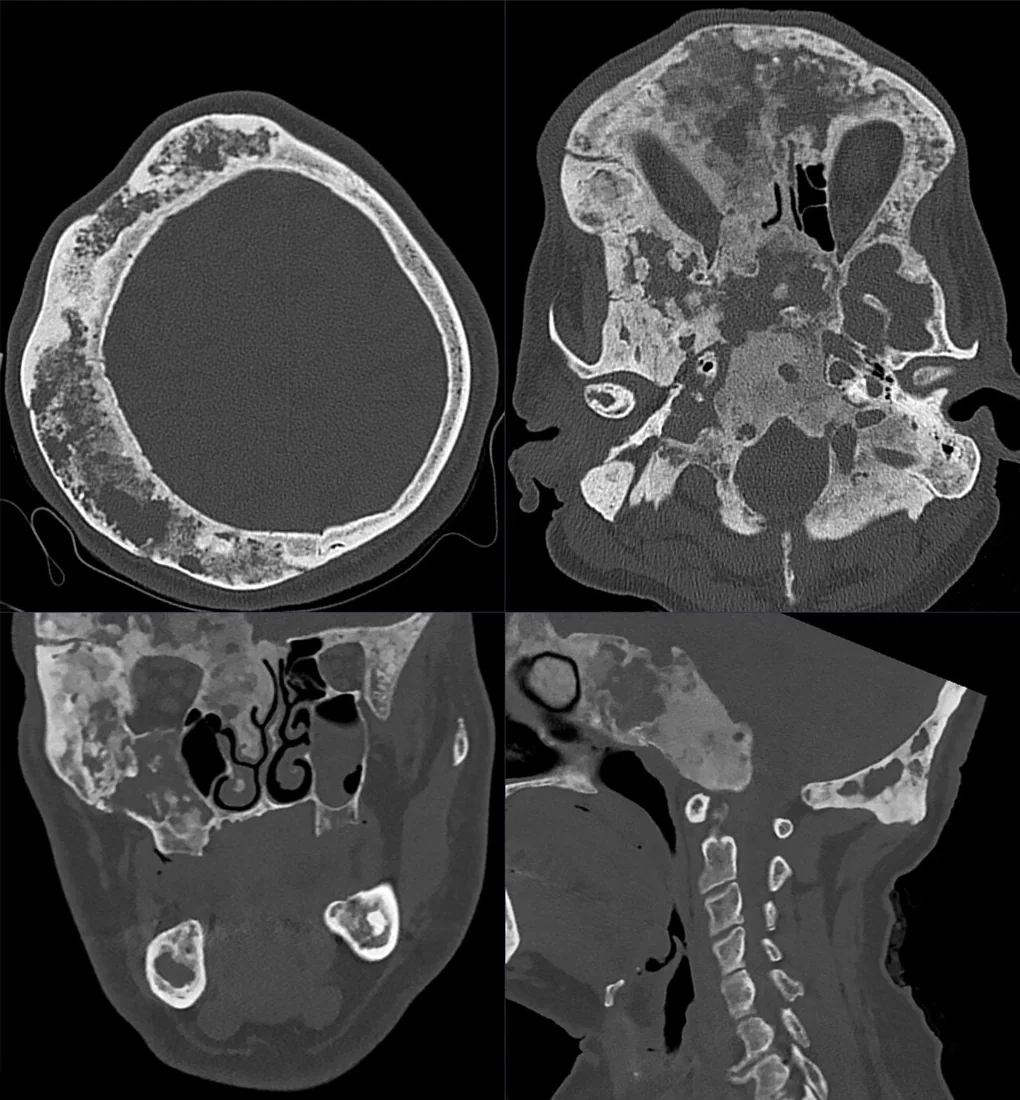
Çene fibröz displazisi, normal kemik dokusunun fibröz stroma ve düzensiz örgü kemikle yer değiştirdiği benign gelişimsel bir kemik hastalığıdır; tek çene kemiğini ya da daha geniş kraniofasiyal alanı tutabilir. BT, buzlu cam veya karışık matriksi ve kemik genişlemesinin kafa tabanı, sinüsler, mandibular kanal ve diş arkıyla ilişkisini haritalamak için önemlidir. Lezyon çoğunlukla çevre kemiğe geçişli sınıra sahiptir; bu özellik keskin sınırlı ossifiye fibromdan ayrımda yararlıdır ancak atipik görünümde histoloji gerekebilir. Geniş kraniofasiyal tutulum ve kanal ya da sinüs daralması fark edilmezse anatomik etkilenim eksik raporlanabilir.
Faz ve pencere
- Kontrastsız tanısal
- İntramedüller alanda buzlu cam benzeri homojen veya yamalı yoğunluk artışı; daha az mineralize olgularda karışık litik-opak matriks görülebilir. BT, çene fibröz displazisinin yaygınlığını ve lezyon matriksini değerlendirmede kullanılır.
- Kemik algoritması tanısal
- Bölgesel kemik genişlemesi, ince fakat devamlı korteks, komşu kemiğe geçişli sınır, sinüs boşluğunun daralması ve mandibular kanalın yer değiştirmesi değerlendirilir.
Önerilen pencereler G genişlik, M merkez; değerler Hounsfield birimi (HU)
- KemikG 1800 / M +400
BT bulguları
- Buzlu cam matriks — normal trabeküler yapının yerini alan, ince ve yaygın mineralize granüler atenüasyon.
- Karışık fibro-osseöz görünüm — daha az olgun alanda litik ve opak bileşenler aynı kemik segmentinde bir arada bulunabilir.
- Geçişli lezyon sınırı — anormal matriks normal kemiğe keskin bir kapsül oluşturmadan karışır.
- Fusiform kemik genişlemesi — maksilla veya mandibula dış konturu genişler; korteks incelse de çoğunlukla devamlı kalır.
- Maksiller sinüs daralması — maksilladaki genişleme sinüs boşluğunu küçültebilir veya duvarlarını yeniden şekillendirebilir.
- Diş arkı ve kök etkisi — alveoler kontur bozulabilir, dişler yer değiştirebilir; belirgin kök rezorpsiyonu tipik değildir.
- Mandibular kanal deplasmanı — kanal genişlemiş kemik içinde yukarı, aşağı veya yana ötelenebilir.
Ölçütler ve sınıflamalar
- Klinik yaygınlık
- Fibröz displazi monostotik veya poliostotik olarak sınıflandırılır; maksiller kraniofasiyal tutulum sütürleri aşarak sfenoid, zigoma, kafa tabanı ve frontonazal kemiklere uzanabilir.
- BT matriks görünümü
- Buzlu cam, homojen yoğun sklerotik ve kistik paternler tarif edilmiştir; görünüm yaş ve mineralizasyon olgunluğuyla değişebilir.
Normalde
Normal maksilla ve mandibulada trabeküler kemik düzenli ve çevre korteksi pürüzsüzdür; maksiller sinüs havalı, mandibular kanal beklenen seyirdedir. Karşı taraf ve komşu kemik segmentleriyle kıyaslayarak buzlu cam matriksin sınırının nerede eridiğini, kemik konturunun nasıl genişlediğini ve diş arkı ya da kanalın yer değiştirip değiştirmediğini takip edin.
Ayırıcı tanı
- Santral ossifiye fibrom
- çevre kemikten keskin ayrılır ve fokal merkez etrafında daha belirgin konsantrik ekspansiyon yapar.
- Semento-osseöz displazi
- çoğunlukla mandibular diş apeksleri çevresinde odaksal ya da çok odaklı olgunlaşan opasiteler yapar; kemiğin uzun segmentini homojen genişletmez.
- Osteoma, iyi sınırlı radyopak bir çene lezyonu olarak görülebilir; fibröz displazi ise çoğunlukla belirgin sınırları olmayan, çevre kemiğe geçişli bir görünüm sergiler.
- Osteosarkom, çene fibröz displazisinin ayırıcı tanısında yer alır; kortikal destrüksiyon gibi agresif görüntüleme bulguları ek patolojik değerlendirmeyi gerektirebilir.
- Çene fibröz displazisinin ayırıcı tanısında Paget hastalığı ve düşük dereceli intramedüller osteosarkom da yer alır; hızlı büyüme ve radyografik görünümde değişiklik malignite düşündürebilir.
Tuzaklar
- Buzlu cam matriks fibröz displazide görülebilse de diğer fibro-osseöz lezyonlarla radyolojik benzerlik nedeniyle lezyon sınırları ve tutulum kapsamı birlikte değerlendirilmelidir.
- Kraniofasiyal yayılımı yalnız mandibula veya maksilla düzeyinde kesme; ince kesitlerde kafa tabanı ve komşu sinüs kemiklerini de takip et.
- Çocuk ve erişkin görüntülerini aynı matriks beklentisiyle yorumlama; mineralizasyon paterni zaman içinde homojen buzlu camdan daha karışık ya da yoğun görünüme değişebilir.
Kendini dene
Maksillada buzlu cam matriksli genişleme, ince devamlı korteks ve komşu kemiğe belirsiz geçiş görülüyor. En olası tanı nedir?
Cevabı göster
Çene fibröz displazisi. Geçişli sınır, buzlu cam matriks ve incelmiş korteks fibröz displaziyi destekler. Santral ossifiye fibrom ise lezyon ile normal kemik arasında belirgin ayrışma gösterir [S2].
Mandibulada çevresinden net ayrılan, belirgin merkezli ekspansil ve karışık mineralize lezyon görülüyor. Lezyonun normal kemikten keskin biçimde ayrılması hangi fibro-osseöz tanıyla daha uyumludur?
Cevabı göster
Santral ossifiye fibrom. Normal kemikten keskin ayrılma santral ossifiye fibromla uyumludur; fibröz displazide lezyon sınırları çoğunlukla çevre kemikle geçişlidir. Fibröz displazi ise çevre kemiğe geçişli sınıra sahiptir [S2].
Kaynaklar
Bu sayfadaki 41 cümle ve 2 quiz sorusu aşağıdaki kaynaklardan alıntılarla otomatik olarak karşılaştırıldı; 8 cümle bu karşılaştırmada düzeltildi (2026-10-09).
- Impact of Multimodal Rehabilitation Protocol in a 20-Year-Old Patient With Cherubism Undergone Facial Surgery: A Rare Case Reportpmc.ncbi.nlm.nih.gov
- Fibrous Dysplasia of the Jaw: Advances in Imaging and Treatmentpmc.ncbi.nlm.nih.gov
- Massive Mandibular Fibrous Dysplasia Mimicking Calcifying Epithelial Odontogenic Tumor Radiologically: A Case Reportpmc.ncbi.nlm.nih.gov
- Fibrous Dysplasia Presenting as an Exophytic Gingival Mass: A Rare Clinical Presentationpmc.ncbi.nlm.nih.gov
- Endoscopic management of uncommon benign maxillary sinus lesions: a case seriespmc.ncbi.nlm.nih.gov
- Ameloblastoma: An Updated Narrative Review of an Enigmatic Tumor.pubmed.ncbi.nlm.nih.gov
- Maxillofacial fibro-osseous lesions.pubmed.ncbi.nlm.nih.gov
- Fibrous Dysplasia Characterization Using Lacunarity Analysis.pubmed.ncbi.nlm.nih.gov
- Imaging of Mixed and Radiopaque Jaw Lesions.pubmed.ncbi.nlm.nih.gov
- Innovative Surgical Treatment of Severe Cherubism.pubmed.ncbi.nlm.nih.gov
- Cherubism: panoramic and CT features in adults.pubmed.ncbi.nlm.nih.gov
- Bone-related disorders of the jaw: A clinico-radiological diagnostic algorithm.pubmed.ncbi.nlm.nih.gov
- Haemophilic pseudotumour in two parts of the maxilla: case report.pubmed.ncbi.nlm.nih.gov
- Diagnosis and Treatment of a Benign Pediatric Mandible Tumor.pubmed.ncbi.nlm.nih.gov
- Nonfamilial cherubism: A case report and review of literature.pubmed.ncbi.nlm.nih.gov
- Computed Tomography in Craniofacial Fibrous Dysplasia: A Case Series with Review of Literature and Classification Update.pubmed.ncbi.nlm.nih.gov
- A Novel ANO5 Mutation Causing Gnathodiaphyseal Dysplasia With High Bone Turnover Osteosclerosis.pubmed.ncbi.nlm.nih.gov
- Multiple periapical dysplasias analyzed by cone-beam-computer tomography and 99 Tcm-Scintigraphy.pubmed.ncbi.nlm.nih.gov
- Imaging in the diagnosis of cemento-ossifying fibroma: a case series.pubmed.ncbi.nlm.nih.gov
- Mandibular Cemento-Osseous Dysplasia: A Report of Three Cases in Indian Women With Emphasis on Diagnostic Challenges.pubmed.ncbi.nlm.nih.gov
- Fibrous Dysplasia Presenting as an Exophytic Gingival Mass: A Rare Clinical Presentation.pubmed.ncbi.nlm.nih.gov
- Massive Mandibular Fibrous Dysplasia Mimicking Calcifying Epithelial Odontogenic Tumor Radiologically: A Case Report.pubmed.ncbi.nlm.nih.gov
- Aggressive Juvenile Trabecular Ossifying Fibroma in a 4-Year-Old: Case Report Bringing Diagnostic Challenges and Surgical Management.pubmed.ncbi.nlm.nih.gov
- Endoscopic management of uncommon benign maxillary sinus lesions: a case series.pubmed.ncbi.nlm.nih.gov
- How Clinical and Radiological Findings in Chronic Mandibular Osteomyelitis Do Not Always Correlate: Diagnostic Dilemmas in Dental-Related Bone Inflammations.pubmed.ncbi.nlm.nih.gov
- The solitary skull mass: An atypical presentation of anaplastic large cell lymphoma.pubmed.ncbi.nlm.nih.gov
- When plasmacytoma defies expectations: An unusual maxillary presentation with mixed imaging features in a young patient.pubmed.ncbi.nlm.nih.gov
- Benign and malignant lesions of the mandible and maxilla: imaging findings and systematic approach.pubmed.ncbi.nlm.nih.gov
- Failure rate and treatment plan options of dental implants in patients with fibro-osseous lesions: a systematic review.pubmed.ncbi.nlm.nih.gov
- Bilateral Mandibular Simple Bone Cysts: A Report of an Unusual Case.pubmed.ncbi.nlm.nih.gov
- Imaging-Guided Surgical Decision-Making and Bone Healing in Mandibular Cystic and Tumor-like Lesions: A Case-Based Radiologic Observation.pubmed.ncbi.nlm.nih.gov
- Cyst-Resembling Fibrous Dysplasia of the Mandible: A Case Report.pubmed.ncbi.nlm.nih.gov
- Management of Tibial Adamantinoma: Balancing Oncologic Cure and Reconstructive Longevity.pubmed.ncbi.nlm.nih.gov
- Fibrous dysplasia: Clinical features, deformities, management, and outcomes.pubmed.ncbi.nlm.nih.gov
- Fibrous Dysplasia of Sphenoid: A Case Report.pubmed.ncbi.nlm.nih.gov
- Decoding calvarial lesions: Contemporary neurosurgical management integrating radiological and histopathological features.pubmed.ncbi.nlm.nih.gov
- Clinical, radiographic and histopathological analysis of bone-forming tumors of the oral and maxillofacial region: A 53-year retrospective study.pubmed.ncbi.nlm.nih.gov
- A modified surgical strategy for proximal femur benign tumors: a retrospective analysis of 100 patients.pubmed.ncbi.nlm.nih.gov
- Case Report: A primary breast collision tumor composed of myeloid sarcoma and invasive ductal carcinoma.pubmed.ncbi.nlm.nih.gov
- Conventional radiography for the assessment of focal bone lesions of the appendicular skeleton: fundamental concepts in the modern imaging era.pubmed.ncbi.nlm.nih.gov
- Intraosseous tumours of the knee.pubmed.ncbi.nlm.nih.gov
- Intraosseous schwannoma of the olecranon: A case report.pubmed.ncbi.nlm.nih.gov
- Surgically Treated Benign Bone Tumors and Tumor-like Conditions in the Pediatric Population-A 10-Year Institutional Experience.pubmed.ncbi.nlm.nih.gov
- Pediatric triad of craniofacial fibrous dysplasia, Chiari malformation type I and syringomyelia: a case report.pubmed.ncbi.nlm.nih.gov
- Prolyl 3-hydroxylase 1 (P3H1) deficient osteogenesis imperfecta with vascular malformations: a rare disorder with atypical features.pubmed.ncbi.nlm.nih.gov
- Unusual Manifestations of Sinonasal Osteomas: A Narrative Review.pubmed.ncbi.nlm.nih.gov
- Mandibular Inflammatory Myofibroblastic Tumors: A Literature Review with a New Case Presentation.pubmed.ncbi.nlm.nih.gov
- An Unusual Case of Periapical Cemento-Osseous Dysplasia.pubmed.ncbi.nlm.nih.gov
- The Vital Illusion: Cone-Beam Computed Tomography (CBCT)-Tracked Evolution of Periapical Cemento-Osseous Dysplasia.pubmed.ncbi.nlm.nih.gov
- Pediatric Desmoplastic Fibroma of the Jaws: A Comprehensive Review of Clinical Presentation, Management, and Outcomes.pubmed.ncbi.nlm.nih.gov
- Conservative Treatment of Pediatric Fractures-Narrative Review of Acceptable Deformity and Remodeling Potential.pubmed.ncbi.nlm.nih.gov
- Freeman-Burian syndrome.pubmed.ncbi.nlm.nih.gov
- McCune-Albright syndrome.pubmed.ncbi.nlm.nih.gov
- Fibrous dysplasia imitating malignancy.pubmed.ncbi.nlm.nih.gov
- Intracranial aneurysmal bone cysts in association with fibrous dysplasia: Case report and scoping review of case reports.pubmed.ncbi.nlm.nih.gov
- A rare case of odontogenic myxofibroma of the maxilla in a young patient: a challenging diagnostic and differential diagnosis.pubmed.ncbi.nlm.nih.gov
- Rare Presentation of Cemento-Ossifying Fibroma in the Anterior Maxilla: A Case Report.pubmed.ncbi.nlm.nih.gov
- A mouse-tracking study on visualization of panoramic radiographs among Oral & Maxillofacial Radiology faculty.pubmed.ncbi.nlm.nih.gov
- Orbital pseudotumor and skull base infiltration as the sole manifestation of granulomatosis with polyangiitis.pubmed.ncbi.nlm.nih.gov
- Perineurioma-like EMA-positive calvarial neoplasms: clinicopathological study of eight cases.pubmed.ncbi.nlm.nih.gov
- Malignant Transformation of Musculoskeletal Lesions with Imaging-Pathology Correlation-Part 1: Bone Lesions.pubmed.ncbi.nlm.nih.gov
- Efficiency and Safety of Dental Implantation in the Area of Hyperdense Jaw Lesions: A Narrative Review.pubmed.ncbi.nlm.nih.gov
- Alveolar Rhabdomyosarcoma of Nasopharynx and Paranasal Sinuses in Children Diagnosis and Treatment-Review of the Literature and Case Report.pubmed.ncbi.nlm.nih.gov
- Calvarial lesions: overview of imaging features and neurosurgical management.pubmed.ncbi.nlm.nih.gov
- Xanthogranulomatous Osteomyelitis as a Bone Tumor Mimic: A Systematic Review of Pre-biopsy Diagnostic Clues and Management Decision Points.pubmed.ncbi.nlm.nih.gov
- Giant Fibrous Dysplasia of the Sixth Rib Managed in a Rural, Resource-Limited Setting: A Case Report.pubmed.ncbi.nlm.nih.gov
- Familial florid cemento-osseous dysplasia in two young sisters: case report and literature review.pubmed.ncbi.nlm.nih.gov
- Small ethmoid mucocele with intracranial extension associated with fibrous dysplasia: A rare case report.pubmed.ncbi.nlm.nih.gov
- Identification of a Hidden Lesion in Meckel's Cave: A Diagnostic Odyssey of Unilateral Facial Pain in a Nonagenarian.pubmed.ncbi.nlm.nih.gov
- Orthopaedic diagnostic pitfalls in fibroblast growth factor 23-mediated hypophosphatemic rickets/osteomalacia in fibrous dysplasia/McCune-Albright syndrome: Two burosumab-treated cases.pubmed.ncbi.nlm.nih.gov
- A phase 2 trial of burosumab for treatment of fibroblast growth factor-23-mediated hypophosphatemia in children and adults with fibrous dysplasia.pubmed.ncbi.nlm.nih.gov
- Cavernous venous malformation of the frontal sinus: CT and MRI imaging findings.pubmed.ncbi.nlm.nih.gov
- Ossifying Fibroma of the Frontal Sinus: A Case Report and Literature Review.pubmed.ncbi.nlm.nih.gov
- Temporomandibular Joint Defects: Etiology and Reconstruction Strategy: A Narrative Review.pubmed.ncbi.nlm.nih.gov
- Bone lesions of the tibia: Multimodal iconographic review and diagnostic algorithms, Part 2: Metaphyseal and epiphyseal lesions.pubmed.ncbi.nlm.nih.gov
- Ossifying fibroma of the maxilla: A case report with literature review.pubmed.ncbi.nlm.nih.gov
- Orbital infantile myofibroma: a case report and clinicopathologic review of 24 cases from the literature.pubmed.ncbi.nlm.nih.gov
- A Pattern-based Imaging Approach to Pediatric Jaw Lesions.pubmed.ncbi.nlm.nih.gov
- Craniovertebral Junction Instability after Oncological Resection: A Narrative Review.pubmed.ncbi.nlm.nih.gov
- Osteosarcoma: A Comprehensive Morphological and Molecular Review with Prognostic Implications.pubmed.ncbi.nlm.nih.gov
- Review of imaging modalities and radiological findings of calvarial lesions.pubmed.ncbi.nlm.nih.gov
- Sclerotic Lesions of the Jaw: A Pictorial Review.pubmed.ncbi.nlm.nih.gov
- A needle arthroscopy approach to the temporomandibular joints in dogs.pubmed.ncbi.nlm.nih.gov
- Multimodal Imaging of Osteosarcoma: From First Diagnosis to Radiomics.pubmed.ncbi.nlm.nih.gov
- Current concepts of craniofacial fibrous dysplasia: pathophysiology and treatment.pubmed.ncbi.nlm.nih.gov
- Non-Ossifying Fibromas: A 2025 Review.pubmed.ncbi.nlm.nih.gov
- Osteomyelitis of the Jaw Bones and Its Mimics: Resolving the Diagnostic Enigma.pubmed.ncbi.nlm.nih.gov
- The Occurrence and Outcomes of Cemento-Osseous Dysplasias (COD) in the Jaw Bones of the Population of Lower Silesia, Poland.pubmed.ncbi.nlm.nih.gov
- Are there atypical sites of IgG4 related disease in head and neck region? Personal experience and literature review.pubmed.ncbi.nlm.nih.gov
- Cystic bone Lesions: Diagnostic pitfalls and therapeutic considerations.pubmed.ncbi.nlm.nih.gov
- Ossifying Fibroma of Non-odontogenic Origin: A Fibro-osseous Lesion in the Craniofacial Skeleton to be (Re-)considered.pubmed.ncbi.nlm.nih.gov
- Radiological Society of North America (RSNA) 3D printing Special Interest Group (SIG): guidelines for medical 3D printing and appropriateness for clinical scenarios.pubmed.ncbi.nlm.nih.gov
- The 2022 On-site Padua Days on Muscle and Mobility Medicine hosts the University of Florida Institute of Myology and the Wellstone Center, March 30 - April 3, 2022 at the University of Padua and Thermae of Euganean Hills, Padua, Italy: The collection of abstracts.pubmed.ncbi.nlm.nih.gov
- 10th Anniversary of the European Association for Predictive, Preventive and Personalised (3P) Medicine - EPMA World Congress Supplement 2020.pubmed.ncbi.nlm.nih.gov
- Diagnosis and treatment of Paget's disease of bone: position paper from the Italian Society of Osteoporosis, Mineral Metabolism and Skeletal Diseases (SIOMMMS).pubmed.ncbi.nlm.nih.gov
- Facial Asymmetry-Demystifying the Entity.pubmed.ncbi.nlm.nih.gov
- Individualized antiresorptive therapy in fibrous dysplasia and McCune-Albright syndrome: A retrospective cohort study.pubmed.ncbi.nlm.nih.gov
- Exploring Paediatric Jaw Osteosarcoma: A Detailed Case Report of a 12-Year-Old Girl and Review of the Literature.pubmed.ncbi.nlm.nih.gov
Eğitim amaçlıdır; hasta başında tanı ve tedavi kararının yerine geçmez, tıbbi tavsiye değildir. Metinler yapay zekâ desteğiyle yazıldı ve otomatik bir adımda kaynak alıntılarıyla karşılaştırıldı; bu bir tıbbi doğrulama değildir ve içerik uzman radyolog incelemesinden geçmedi. Klinik kararlarda güncel kılavuzları, kurum protokollerini ve radyoloji raporunu esas alın.