Özet
Santral ossifiye fibrom, çene kemiğinde fibröz doku içinde değişen miktarda kemik ya da sement benzeri mineral birikimi oluşturan, iyi sınırlı benign bir tümördür. Semento-ossifiye fibrom benign odontojenik fibro-osseöz neoplazmdır. Kontrastsız ince kesit BT veya KBKT, lezyonun çene içindeki yerini, iç mineralizasyonunu ve korteks ile diş köklerine etkisini belirlemede temel görüntüleme yöntemidir. Görünüm fibröz displazi ve diğer fibro-osseöz lezyonlarla örtüşebilir; kesin tanı klinik-radyolojik ve histopatolojik korelasyon gerektirir. Lezyonun komşu kemik ve yumuşak doku uzanımı, BT veya KBKT ile kortikal bütünlük, genişleme ve kök yer değiştirmelerinin değerlendirilmesiyle doğrulanmalıdır.
Faz ve pencere
- Kontrastsız tanısal
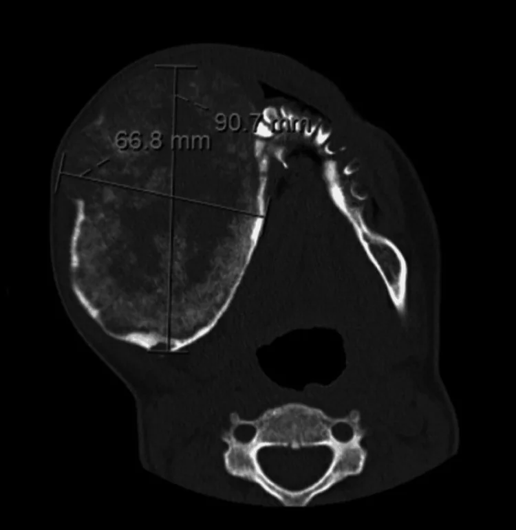
- Çene medüllasında keskin sınırlı, ekspansil, radyolüsentten karışık atenüasyonlu veya yoğun mineralize görünüme değişebilen kitle; lezyon içindeki kemik/sement benzeri odaklar maturasyonla belirginleşir.
- Kemik algoritması tanısal
- İnce kesitlerde çevre kemikten ayrılan net sınır, konsantrik kemik genişlemesi, kortikal incelme veya perforasyon ve kök deplasmanı/rezorpsiyonu değerlendirilir; iç mineralize trabeküller lezyon merkezinde ya da periferinde dağılabilir.
Önerilen pencereler: Kemik (G 1800 / M 400), Yumuşak doku (G 400 / M 40).
BT bulguları
- Keskin lezyon sınırı — ekspansil medüller kitle çevre trabeküler kemikten belirgin biçimde ayrılır; ince sklerotik kenar bulunabilir.
- Konsantrik ekspansiyon — mandibula konturu lezyon merkezinden dışa doğru genişler ve normal kemik şeklini değiştirebilir.
- Değişken mineralize matriks — erken lezyon daha litik; olgun lezyon karışık veya belirgin opak olabilir.
- İç kalsifikasyon/trabekül — dağınık yoğun odaklar, ince kemiksi çizgiler veya lezyon kenarına uzanan trabeküller görülebilir.
- Kortikal incelme — bukkal ve lingual plaklar ekspansiyon boyunca kâğıt inceliğine gelebilir.
- Kortikal incelme — lezyonun büyümesiyle birlikte bukkal veya lingual korteks kâğıt inceliğine gelerek dışa doğru bombeleşebilir.
- Diş kökü etkisi — komşu köklerde ayrışma, deplasman ve bazen rezorpsiyon gelişebilir.
- Raporlamada lezyonun mandibular kanal ve diş kökleriyle ilişkisini, kortikal incelme veya perforasyonu ayrı ayrı belirtin.
Ölçütler ve sınıflamalar
- WHO 2022 sınıflaması
- Semento-ossifiye fibrom benign odontojenik fibro-osseöz neoplazmdır.
Normalde
Santral ossifiye fibromda lezyon genellikle çevre kemikten iyi sınırlı ve kortike görünür; mandibulada ekspansil kitle oluşturabilir. Kıyaslamada lezyonun çevre kemikten ayrık sınırına, mandibula konturunu merkezden dışa değiştirmesine ve köklerin yer değiştirmesine odaklanın.
Ayırıcı tanı
- Çene fibröz displazisi
- lezyon sınırı komşu kemiğe belirsiz biçimde karışırken, ossifiye fibromda sınır daha iyi tanımlanmış ve genellikle kortike görünür.
- Semento-osseöz displazi
- diş apeksleri çevresinde olgunlaşan, çoğu kez az ekspansil odaklar yapar; belirgin konsantrik çene deformasyonu beklenmez.
- Ameloblastom posterior mandibulada özellikle sık görülür; ossifiye fibrom ise iyi sınırlı, ekspansil ve iç radyopasitesi değişken bir kitle olarak görülebilir, bu nedenle sınır ve matriks özellikleri ayırıcı tanıda yardımcı olur.
- Kalsifiye epiteliyal odontojenik tümör
- gömülü dişle ilişkili radyolüsent kitle içinde kalsifikasyonlar bulunabilir; koronal diş ilişkisi tanısal ipucudur.
- Fibröz displazide sınır çoğunlukla normal kemiğe geçişlidir; ossifiye fibromda iyi sınırlı, ekspansil ve çevre kemikten ayrılan kitle görünümü daha tipiktir.
Tuzaklar
- İyi sınırlı her fibro-osseöz lezyonu ossifiye fibrom olarak adlandırma; semento-osseöz displazi diş apekslerine, fibröz displazi ise geçişli sınıra ve buzlu cam matrikse göre ayrılır.
- BT görüntüsünde iç yoğunluk değişkenlik gösterebilir; olgun lezyon tanısında yalnızca mineralizasyona değil, sınırın belirginliğine, lezyonun merkezi konumuna ve çene konturundaki konsantrik deformasyona odaklanılmalıdır.
- Kortikal bütünlük tek bir eksende değerlendirilmemelidir; bukkal ve lingual plakların incelmesi veya perforasyonu, lezyonun gerçek ekspansiyonunu göstermek için koronal ve aksiyal düzlemlerde ayrıca doğrulanmalıdır.
Kendini dene
Mandibula arka gövdede keskin sınırlı, mineralize odaklar içeren ekspansil kitle korteksi inceltiyor ve kökleri ayırıyor. En olası tanı nedir?
Cevabı göster
Santral ossifiye fibrom. Net sınır, konsantrik ekspansiyon ve değişken mineralize matriks ossifiye fibromu destekler. Fibröz displazi çevre kemiğe geçişlidir; ameloblastom çoğunlukla litik çok odacıklıdır; kondanse osteit ekspansil kitle yapmaz.
Çene lezyonunda buzlu cam matriks var ve sınır normal kemiğe belirsiz biçimde karışıyor. Hangi tanı daha uygundur?
Cevabı göster
Monostotik fibröz displazi. Monostotik fibröz displazide buzlu cam matriks, normal kemiğe geçişli sınırla birlikte görülebilir. Santral dev hücreli granülom ve anevrizmal kemik kisti daha çok litik, ekspansil odaklar oluşturur; kronik osteomiyelitte skleroz, sekestr veya periostal yeni kemik eşlik edebilir.
Kaynaklar
Bu sayfadaki 40 cümle ve 2 quiz sorusu aşağıdaki kaynaklardan alıntılarla otomatik olarak karşılaştırıldı; 12 cümle bu karşılaştırmada düzeltildi (2026-10-09).
- Three types of ossifying fibroma: A report of 4 cases with an analysis of CBCT featurespmc.ncbi.nlm.nih.gov
- A systematic review of the clinical and radiographic features of hybrid central giant cell granuloma lesions of the jaws.pubmed.ncbi.nlm.nih.gov
- Central ossifying fibroma of mandible.pubmed.ncbi.nlm.nih.gov
- Juvenile central ossifying fibroma of the mandible: a case report.pubmed.ncbi.nlm.nih.gov
- Expansive Central Ossifying Fibroma of the Right Mandible.pubmed.ncbi.nlm.nih.gov
- Bimaxillary presentation of central ossifying fibroma: a unique aggressive entity.pubmed.ncbi.nlm.nih.gov
- Extensive central ossifying fibroma of the maxilla: a case report.pubmed.ncbi.nlm.nih.gov
- Asymptomatic, nonexpansile radiopacity of the jaw associated with external root resorption: a diagnostic dilemma.pubmed.ncbi.nlm.nih.gov
- Peritrabecular clefting in fibrous dysplasia of the jaws: an important histopathologic feature for differentiating fibrous dysplasia from central ossifying fibroma.pubmed.ncbi.nlm.nih.gov
- Maxillofacial fibro-osseous lesions.pubmed.ncbi.nlm.nih.gov
- Central cementifying fibroma of maxilla.pubmed.ncbi.nlm.nih.gov
- Psammomatoid and trabecular juvenile ossifying fibroma: two distinct radiologic entities.pubmed.ncbi.nlm.nih.gov
- Central calcifying epithelial odontogenic tumour in the posterior maxilla: a case report.pubmed.ncbi.nlm.nih.gov
- Amyloid Variant of Central Odontogenic Fibroma in the Mandible: A Case Report and Literature Review.pubmed.ncbi.nlm.nih.gov
- Incidental Detection of Ossifying Fibroma of the Frontal Sinus on 99mTc-MDP Bone Scan.pubmed.ncbi.nlm.nih.gov
- Maxillofacial Cemento-Ossifying Fibroma: A Rare Case Report.pubmed.ncbi.nlm.nih.gov
- Fibrous Dysplasia Presenting as an Exophytic Gingival Mass: A Rare Clinical Presentation.pubmed.ncbi.nlm.nih.gov
- A Case Report on the Diagnostic Challenge of Mandibular Osteolipoma.pubmed.ncbi.nlm.nih.gov
- Ossifying Fibroma of Non-odontogenic Origin: A Fibro-osseous Lesion in the Craniofacial Skeleton to be (Re-)considered.pubmed.ncbi.nlm.nih.gov
- [Florid cemento-osseous dysplasia: A case report].pubmed.ncbi.nlm.nih.gov
- Frequency of Odontogenic Tumors: A Single Center Study of 1089 Cases in Japan and Literature Review.pubmed.ncbi.nlm.nih.gov
- Cemento-ossifying fibroma: A South American multicenter study.pubmed.ncbi.nlm.nih.gov
- A Case of Peripheral Odontogenic Myxofibroma Arising in the Palatal Gingiva of the Maxillary Second Premolar Region: A Case Report.pubmed.ncbi.nlm.nih.gov
- Benign and malignant lesions of the mandible and maxilla: imaging findings and systematic approach.pubmed.ncbi.nlm.nih.gov
- Rapidly progressive angiomatous epulis: How to differentiate a benign gingival lesion from malignancy?pubmed.ncbi.nlm.nih.gov
- Peripheral Giant Cell Granuloma Mimicking Central Giant Cell Granuloma in a Pediatric Patient: A Diagnostic Challenge.pubmed.ncbi.nlm.nih.gov
- Desmoplastic Fibroma of the Maxilla: A Report of a Rare Case.pubmed.ncbi.nlm.nih.gov
- Pediatric Desmoplastic Fibroma of the Jaws: A Comprehensive Review of Clinical Presentation, Management, and Outcomes.pubmed.ncbi.nlm.nih.gov
- Case reports with literature review of an aneurysmal bone cyst in the maxilla and mandible of two juvenile dogs.pubmed.ncbi.nlm.nih.gov
- Imaging approach for jaw and maxillofacial bone tumors with updates from the 2022 World Health Organization classification.pubmed.ncbi.nlm.nih.gov
- Uncommon Nasal Mass Presentation: A Radiological Case Series.pubmed.ncbi.nlm.nih.gov
- Imaging Features of Mandibular Ramus Osteoma on Computed Tomography: A Case Report With Literature Analysis.pubmed.ncbi.nlm.nih.gov
- Diagnostic and Treatment Strategies for Adult Non-Ossifying Fibromas: A Case Report and Literature Review.pubmed.ncbi.nlm.nih.gov
- Three types of ossifying fibroma: A report of 4 cases with an analysis of CBCT features.pubmed.ncbi.nlm.nih.gov
- Giant cemento-ossifying fibroma of the mandible.pubmed.ncbi.nlm.nih.gov
- Conventional radiography for the assessment of focal bone lesions of the appendicular skeleton: fundamental concepts in the modern imaging era.pubmed.ncbi.nlm.nih.gov
- Peripheral cemento-ossifying fibroma - Clinicopathological diagnosis, management, and literature review.pubmed.ncbi.nlm.nih.gov
- Recount of Clinicopathological Features of Squamous Odontogenic Tumor: A Case Report with Review.pubmed.ncbi.nlm.nih.gov
- Calcifying Epithelial Odontogenic Tumor: A Report of a Rare Case With Clinical and Radiographic Findings.pubmed.ncbi.nlm.nih.gov
- Radiological Landmarks as an Aid in the Interpretation of Rodent Skull Extra-Oral Projections.pubmed.ncbi.nlm.nih.gov
- Integrating bioprinting, cell therapies and drug delivery towards in vivo regeneration of cartilage, bone and osteochondral tissue.pubmed.ncbi.nlm.nih.gov
- A Narrative Review on the Effectiveness of Bone Regeneration Procedures with OsteoBiol ® Collagenated Porcine Grafts: The Translational Research Experience over 20 Years.pubmed.ncbi.nlm.nih.gov
- Assessment of Deep Convolutional Neural Network Models for the Classification of Benign Fibro-Osseous Lesions of the Jaws.pubmed.ncbi.nlm.nih.gov
- Modality-specific quantitative radiologic characterization of diffuse sclerosing osteomyelitis of the mandible: a pilot study.pubmed.ncbi.nlm.nih.gov
- Proceedings of the 2025 North American Society of Head and Neck Pathology Companion Meeting, Boston, MA, March 23, 2025: Diagnostic Borderlands in ENT Pathology-Jagulars, Heffalumps, and Cheshire Cats in Odontogenic Cysts and Tumors-How not to Feel Like Eeyore When Facing Diagnostic Odontogenic Borderlands.pubmed.ncbi.nlm.nih.gov
- Histopathologic spectrum and clinical correlation of lesions of jaw - a series of 60 cases.pubmed.ncbi.nlm.nih.gov
- Efficiency and Safety of Dental Implantation in the Area of Hyperdense Jaw Lesions: A Narrative Review.pubmed.ncbi.nlm.nih.gov
- Performance of AI Chatbots in Preliminary Diagnosis of Maxillofacial Pathologies.pubmed.ncbi.nlm.nih.gov
- Rare Presentation of Cemento-Ossifying Fibroma in the Anterior Maxilla: A Case Report.pubmed.ncbi.nlm.nih.gov
- Glandular odontogenic cyst: a ten-case series and literature review with emphasis on radiological diagnostic challenges.pubmed.ncbi.nlm.nih.gov
- Adenomatoid odontogenic tumor: clinicopathological analysis of 34 cases from Karachi, Pakistan.pubmed.ncbi.nlm.nih.gov
- Masses of developmental and genetic origin affecting the paediatric craniofacial skeleton.pubmed.ncbi.nlm.nih.gov
- Non-Ossifying Fibromas: A 2025 Review.pubmed.ncbi.nlm.nih.gov
- Why was the concordance rate of imaging and clinical diagnosis in cemento-osseous dysplasia low? A retrospective study of 55 cases.pubmed.ncbi.nlm.nih.gov
- Clinical and radiologic outcomes of dental implants in cemento-osseous dysplasia: a systematic review and retrospective case series.pubmed.ncbi.nlm.nih.gov
- Conversion of Aneurysmal Bone Cyst into Fibrous Dysplasia: A Rare Pediatric Case Report.pubmed.ncbi.nlm.nih.gov
- International Harmonization of Nomenclature and Diagnostic Criteria (INHAND): Non-proliferative and Proliferative Lesions of the Non-human Primate ( M. fascicularis ).pubmed.ncbi.nlm.nih.gov
- Unique Imaging Findings of a Calvarial Ossifying Fibroma Successfully Managed With Surgery and Definitive Intent Radiotherapy.pubmed.ncbi.nlm.nih.gov
- Ossifying Fibroma of the Frontal Sinus: A Case Report and Literature Review.pubmed.ncbi.nlm.nih.gov
- Radiomics-Based Diagnosis in Dentomaxillofacial Radiology: A Systematic Review.pubmed.ncbi.nlm.nih.gov
- Intraosseous Venous Malformations of the Head and Neck.pubmed.ncbi.nlm.nih.gov
- Case Series of Mandibular Glandular Odontogenic Cysts: Radiographic and Histopathological Evaluation.pubmed.ncbi.nlm.nih.gov
- Dental Topics for Plastic Surgeons, Part Three: Infection and Trauma of the Dentition.pubmed.ncbi.nlm.nih.gov
- Benign fibro-osseous lesions of the craniofacial complex. A review.pubmed.ncbi.nlm.nih.gov
- Pathological Fracture in the Metatarsal Bone Due to an Intraosseous Schwannoma: A Case Report.pubmed.ncbi.nlm.nih.gov
- Radiographic Assessment of Normal Periapical Bone Density: A Descriptive Analysis Based on Digital Imaging.pubmed.ncbi.nlm.nih.gov
- Uncovering jaw-specific radiographic differences in medication related osteonecrosis of the jaws (MRONJ): a case-control study.pubmed.ncbi.nlm.nih.gov
- Anatomical Evaluation of Localized Mandibular Trabecular Texture Differences in Children with Hypodontia Using Radiographic Fractal Analysis.pubmed.ncbi.nlm.nih.gov
- Radiolucent Lesions of the Jaws: An Attempted Demonstration of the Use of Co-Word Analysis to List Main Similar Pathologies.pubmed.ncbi.nlm.nih.gov
- Juvenile psammomatoid ossifying fibroma with aneurysmal bone cyst in the mandible: A report of a rare case.pubmed.ncbi.nlm.nih.gov
- Primary Stability of Dental Implants in Human Jawbones: Experiments & FE Analyses.pubmed.ncbi.nlm.nih.gov
- Histopathological and radiographic characteristics of central giant cell granulomas: a retrospective study of 45 cases.pubmed.ncbi.nlm.nih.gov
- The Curious Case of a 'Maverick' Cementoblastoma!pubmed.ncbi.nlm.nih.gov
- Cemento-Osseous Dysplasia of the Jaw: Demographic and Clinical Analysis of 191 New Cases.pubmed.ncbi.nlm.nih.gov
- Maxillofacial Bone Involvement in Fibro-Osseous Lesions: Emphasizing the Significance of Differential Diagnosis.pubmed.ncbi.nlm.nih.gov
- Sturge-Weber syndrome with cemento-ossifying fibroma in the maxilla and giant odontoma in the mandible: A case report.pubmed.ncbi.nlm.nih.gov
- Synchronous cemento-ossifying fibromas: a systematic review.pubmed.ncbi.nlm.nih.gov
- Malignant Odontogenic Tumors: A Clinicopathological Study of 53 Cases in Brazil.pubmed.ncbi.nlm.nih.gov
- Evaluation of Jaw Pathologies by Computed Tomography.pubmed.ncbi.nlm.nih.gov
- Aggressive Central Giant Cell Granuloma: A Case Report.pubmed.ncbi.nlm.nih.gov
- Idiopathic Osteosclerosis of the Jaw in a Brazilian Population: a Retrospective Study.pubmed.ncbi.nlm.nih.gov
- Bone-Preserving Management of Maxillary Cemento-Ossifying Fibroma With Orbital Floor Thinning: A Case Report.pubmed.ncbi.nlm.nih.gov
- Dilated Odontoma of the Mandible: A Rare Case Report with Discussion about the Differential Diagnosis.pubmed.ncbi.nlm.nih.gov
- When plasmacytoma defies expectations: An unusual maxillary presentation with mixed imaging features in a young patient.pubmed.ncbi.nlm.nih.gov
- Aggressive Juvenile Trabecular Ossifying Fibroma in a 4-Year-Old: Case Report Bringing Diagnostic Challenges and Surgical Management.pubmed.ncbi.nlm.nih.gov
- Cyst-Resembling Fibrous Dysplasia of the Mandible: A Case Report.pubmed.ncbi.nlm.nih.gov
- "Maxillary juvenile ossifying fibroma: A case study of severe tooth displacement and orbital involvement in a pediatric patient".pubmed.ncbi.nlm.nih.gov
- Radiolucent rim as a possible diagnostic aid for differentiating jaw lesions.pubmed.ncbi.nlm.nih.gov
- Monostotic fibrous dysplasia of jaw bones: a case series.pubmed.ncbi.nlm.nih.gov
- Trabecular Juvenile Ossifying Fibroma Involving the Maxilla: A Rare Case.pubmed.ncbi.nlm.nih.gov
- Musculoskeletal tumors and tumor-like lesions with "dark" signal intensity on T2-weighted MR images: A pictorial review.pubmed.ncbi.nlm.nih.gov
- A Case of an Ossifying Fibroma of the Mandible Suspected as a Static Bone Cavity.pubmed.ncbi.nlm.nih.gov
- Myxoma Unveiled: Insights from Central and Peripheral Variants through Two Rare Cases.pubmed.ncbi.nlm.nih.gov
- Imaging of the temporomandibular joint: An update.pubmed.ncbi.nlm.nih.gov
- Deep circumflex iliac artery flap maxillary reconstruction for juvenile trabecular ossifying fibroma: a case report and systematic review.pubmed.ncbi.nlm.nih.gov
- Giant cementoblastoma involving multiple teeth: A rare case report and review of differential diagnoses.pubmed.ncbi.nlm.nih.gov
- Sclerosing Odontogenic Carcinoma: A Unique Odontogenic Carcinoma with Metastatic Potential.pubmed.ncbi.nlm.nih.gov
- Aggressive Aneurysmal Bone Cyst of the Mandible: A Rare Case of Rapid Expansion and Surgical Management.pubmed.ncbi.nlm.nih.gov
- Surgically Treated Benign Bone Tumors and Tumor-like Conditions in the Pediatric Population-A 10-Year Institutional Experience.pubmed.ncbi.nlm.nih.gov
- Evaluation of primary physicians' awareness levels of non-ossifying fibromas and fibrous cortical defects by using referral documents.pubmed.ncbi.nlm.nih.gov
- Primary Ewing's Sarcoma of the Sinonasal Region: A Rare Clinical Encounter.pubmed.ncbi.nlm.nih.gov
Eğitim amaçlıdır; hasta başında tanı ve tedavi kararının yerine geçmez, tıbbi tavsiye değildir. Metinler yapay zekâ desteğiyle yazıldı ve otomatik bir adımda kaynak alıntılarıyla karşılaştırıldı; bu bir tıbbi doğrulama değildir ve içerik uzman radyolog incelemesinden geçmedi. Klinik kararlarda güncel kılavuzları, kurum protokollerini ve radyoloji raporunu esas alın.