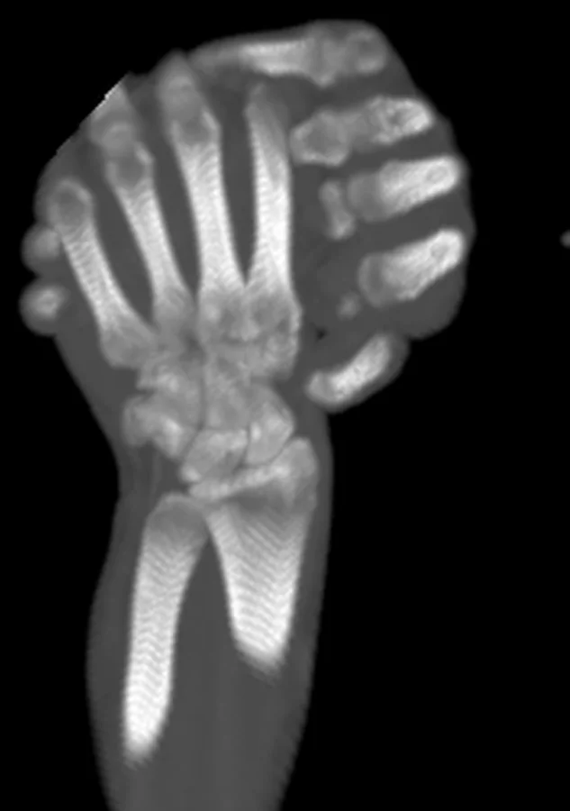
Özet
İntravenöz kontrast ekstravazasyonu, enjeksiyon sırasında iyotlu kontrastın damar dışına, çoğunlukla enjeksiyon yerinin cilt altı dokularına sızmasıdır. Tanısı büyük ölçüde kliniktir ve rutin görüntüleme gerekmez; enjeksiyon sırasında hastanın şikayetleri ve basınç uyarıları izlenmelidir. Görüntüleme, özellikle ciddi ekstravazasyonlarda yayılımın ve subfasyal ya da subkutan yerleşimin belgelenmesine yardımcı olabilir; cerrahiye yönlendirme kararı ekstravaze hacme değil klinik şiddete dayanır. Sıkı subfasyal kompartımanların tutulumu, daha gevşek subkutan dokuların tutulumuna göre daha fazla doku hasarına yol açabilir.
Okuma sırası
- antekubital ven. İncelemede, kontrastın beklenen damar içi opaklaşmayı oluşturup oluşturmadığını ve çevre yumuşak dokulara sızıp sızmadığını değerlendir.
- damar dışı kontrast birikimi. Görüntülerde kontrast ekstravazatının cilt altı dokularda mı yoksa fasya altı kompartımanlarda mı bulunduğunu değerlendir.
- önkol yumuşak doku kompartımanları. Kesitsel görüntülerde ekstravazatın yayılımını ve sınırlarını takip et.
- distal önkol ve el. Ekstravazatın cilt altı dokularda mı yoksa fasya altı kompartımanlarda mı olduğunu değerlendir.
Faz ve pencere
- Portal tanısal
- Enjeksiyon bölgesi tarama alanında ise, tomografi kesitsel görüntüler damar dışı kontrastın subkutan veya fasya altı kompartımanlara yayılımını ve ekstansiyonunu değerlendirmeye yarar.
- Kontrastsız
- Tanısal yaklaşımda kontrastsız fazın rolü kaynaklarda vurgulanmamaktadır; değerlendirme enjeksiyon sonrası klinik bulgular ve gerekirse post-enjeksiyon görüntüleme ile yapılır.
Önerilen pencereler G genişlik, M merkez; değerler Hounsfield birimi (HU)
- Yumuşak dokuG 400 / M +40
- AnjiyoG 600 / M +150
BT bulguları
- Periferik damar lümeni dışına sızan kontrast çevre yumuşak dokulara yayılır.
- Biriken kontrastın enjeksiyon yeri çevresindeki subkutan planlara veya kaslar arası fasyal aralıklara yayılması
- Ciddi ekstravazasyonlarda görüntüleme, ekstravazatın subfasyal mı subkutan mı olduğunu ve yayılımını değerlendirmeye yardımcı olabilir.
- Tanı için rutin görüntüleme önerilmez; enjeksiyon sırasında hastanın şikayetleri ve injeksiyon sistemindeki basınç uyarıları izlenmelidir.
- El bileği, önkol veya el gibi sınırlı kompartımanlarda derin yayılım ve nörovasküler yapılar çevresinde bası olasılığı
Normalde
Ekstravazasyonda intravenöz kontrast çevre yumuşak dokulara sızar; ciddi olgularda görüntüleme, ekstravazatın subfasyal mı subkutan mı olduğunu ve yayılımını değerlendirmeye yardımcı olabilir. Kıyaslamada damar dışı birikimin sınırını, doku şişliğini ve yayılımın el/önkol kompartımanlarına uzanıp uzanmadığını değerlendir.
Ayırıcı tanı
- Ekstravazasyon, intravenöz kontrastın çevre yumuşak dokulara sızmasıdır.
- Enfeksiyöz selülit ayırıcı tanısında, ekstravazasyonun enjeksiyon sırasında veya sonrasında ağrı ya da şişlikle fark edilebileceği ve tanısının çoğunlukla klinik olduğu dikkate alınır.
Tuzaklar
- Ekstremite tarama alanı dışında kalmışsa BT enjeksiyon yerini göstermeyebilir; kontrastın damar dışına kaçtığını görüntüden dışlama.
- Damar dışı kontrast miktarı BT'de çok yoğun görünse de görüntülenen hacim klinik şiddeti tek başına belirlemez; basınç, ağrı, dolaşım ve nörolojik muayene ayrıca değerlendirilir.
- Küçük hacimli kontrast ekstravazasyonları kayıtlara geçmemiş olabilir; tanı çoğunlukla kliniktir ve rutin görüntüleme önerilmez.
- İlk değerlendirmede etkilenen ekstremitede distal nabız ve duyu bulgularını değerlendir; nörovasküler kompromi, doku nekrozu veya kompartman sendromu varsa acil cerrahi değerlendirme gerekir.
Kendini dene
Kontrast ekstravazasyonunda cerrahiye yönlendirme kararı temel olarak neye dayanmalıdır?
Cevabı göster
Klinik şiddete ve nörovasküler duruma. Kaynaklar, cerrahiye yönlendirme kararının ekstravaze hacimden çok klinik şiddete dayanması gerektiğini belirtmektedir.
Ciddi kontrast ekstravazasyonlarında kesitsel görüntülemede hangi özellik değerlendirilmelidir?
Cevabı göster
Subkutan versus fasya altı (kompartıman) tutulumu. Ciddi ekstravazasyonlarda görüntüleme, ekstravazatın cilt altı dokularda mı yoksa fasya altı kompartımanlarda mı olduğunu ve yayılımını değerlendirmeye yardımcı olur.
İlgili konular
Kaynaklar
Bu sayfadaki 28 cümle ve 2 quiz sorusu aşağıdaki kaynaklardan alıntılarla otomatik olarak karşılaştırıldı; 21 cümle bu karşılaştırmada düzeltildi (2026-10-09).
- Intravenous contrast medium extravasation: systematic review and updated ESUR Contrast Media Safety Committee Guidelinespmc.ncbi.nlm.nih.gov
- MRI and CT contrast media extravasationpmc.ncbi.nlm.nih.gov
- Spontaneous ureteral rupture and review of the literature.pubmed.ncbi.nlm.nih.gov
- CT-guided nephrostomy-An expedient tool for complex clinical scenarios.pubmed.ncbi.nlm.nih.gov
- Renal trauma: imaging evaluation and implications for clinical management.pubmed.ncbi.nlm.nih.gov
- MRI and CT contrast media extravasation: A systematic review.pubmed.ncbi.nlm.nih.gov
- Computed Tomography Angiography With High Flow Rates: An In Vitro and In Vivo Feasibility Study.pubmed.ncbi.nlm.nih.gov
- Contrast Media Extravasation of Computed Tomography and Magnetic Resonance Imaging: Management Guidelines for the Radiologist.pubmed.ncbi.nlm.nih.gov
- Prospective study of access site complications of automated contrast injection with peripheral venous access in MDCT.pubmed.ncbi.nlm.nih.gov
- Imaging primer for CT angiography in peripheral vascular trauma.pubmed.ncbi.nlm.nih.gov
- A review of split-bolus single-pass CT in the assessment of trauma patients.pubmed.ncbi.nlm.nih.gov
- Contrast-enhanced ultrasound for evaluation of renal trauma during acute hemorrhagic shock: a canine model.pubmed.ncbi.nlm.nih.gov
- "Blush" on trauma computed tomography: not as bad as we think!pubmed.ncbi.nlm.nih.gov
- Contrast-enhanced CT for Colonic Diverticular Bleeding before Colonoscopy: A Prospective Multicenter Study.pubmed.ncbi.nlm.nih.gov
- Risk of contrast extravasation with vascular access in computed tomography.pubmed.ncbi.nlm.nih.gov
- Clinical guidelines for the management of cytostatic extravasation in children.pubmed.ncbi.nlm.nih.gov
- Integrating CT-Derived Body Composition and Real-Time Injection Pressure Monitoring for Predicting Contrast Media Extravasation: A Retrospective Cohort Study with Implications for Nurse-Led Risk Stratification.pubmed.ncbi.nlm.nih.gov
- Perioperative Intravenous Infiltration: From Recognition to Prevention.pubmed.ncbi.nlm.nih.gov
- Intra-articular Genicular Artery Pseudoaneurysm Mimicking Pigmented Villonodular Synovitis: A Case Report.pubmed.ncbi.nlm.nih.gov
- Retroperitoneal glass foreign body with renal parenchymal involvement after 25 years of silent retention: a case report.pubmed.ncbi.nlm.nih.gov
- Traumatic retropharyngeal hematoma successfully treated with n-butyl cyanoacrylate transarterial embolization: A case report and literature review.pubmed.ncbi.nlm.nih.gov
- The space of Retzius: anatomy, imaging features, and patterns of disease spread.pubmed.ncbi.nlm.nih.gov
- Osteitis Pubis in Athletes in the Era of Pubic-Related Groin Pain: Contemporary Concepts in Diagnosis, Rehabilitation, and Surgical Decision-Making.pubmed.ncbi.nlm.nih.gov
- Upper-Limb Compartment Syndrome Following Rapid Infusion Catheter Extravasation During Orthotopic Liver Transplantation: A Case Report.pubmed.ncbi.nlm.nih.gov
- Iatrogenic compartment syndrome: a comparative case report.pubmed.ncbi.nlm.nih.gov
- Computed tomography of renal emergencies: A comprehensive diagnostic guide for radiology residents.pubmed.ncbi.nlm.nih.gov
- Current Progress in the CT- and MRI-Based Detection and Evaluation of Acute Pancreatitis Complications.pubmed.ncbi.nlm.nih.gov
- Host-response trajectories in necrotizing fasciitis: host-pathogen interactions, immune evasion, and immunoparalysis.pubmed.ncbi.nlm.nih.gov
- Submucosal laryngeal lesions: A puzzling diagnostic conundrum.pubmed.ncbi.nlm.nih.gov
- A magnetic nanorobot with guided aggregation and thrombin-triggered coagulation for endovascular embolization.pubmed.ncbi.nlm.nih.gov
- Overcoming Biophysical Barriers in Melanoma Photomedicine: From Photodynamic Therapy to Smart Nanodelivery and Photoimmunotherapy.pubmed.ncbi.nlm.nih.gov
- Soft tissue hematomas on ultrasound: a case-based review and practical guide to diagnosis.pubmed.ncbi.nlm.nih.gov
- Ultrasound of the Cheek: Examination Technique, Anatomy, and Abnormal Findings.pubmed.ncbi.nlm.nih.gov
- Imaging of Abdominal Wall Masses, Masslike Lesions, and Diffuse Processes.pubmed.ncbi.nlm.nih.gov
- A Narrative Review of the Classification and Use of Diagnostic Ultrasound for Conditions of the Achilles Tendon.pubmed.ncbi.nlm.nih.gov
- Proliferative and non-proliferative lesions of the rat and mouse soft tissue, skeletal muscle and mesothelium.pubmed.ncbi.nlm.nih.gov
- Sacral CSF-venous fistula: a rare entity. Illustrative case.pubmed.ncbi.nlm.nih.gov
- Endovascular management of iliac vessel injury complicating lumbar disc surgery: a case report and review of literature.pubmed.ncbi.nlm.nih.gov
- Vascular complications in percutaneous biliary interventions.pubmed.ncbi.nlm.nih.gov
- Subcutaneous Pectoral Edema After Arthroscopic Labral Repair Despite Normal Irrigation Fluid Usage and Surgery Duration.pubmed.ncbi.nlm.nih.gov
- Magnetic resonance imaging of the wrist and hand.pubmed.ncbi.nlm.nih.gov
- Contrast-Enhanced CT Finding of Hot Spot Sign Secondary to Supra Hilar Mass Causing Superior Vena Cava Syndrome: A Case Report.pubmed.ncbi.nlm.nih.gov
- Interventional treatment of a bacterial embolus in the retrohepatic inferior vena cava secondary to invasive Klebsiella pneumoniae liver abscess syndrome: a case report and literature review.pubmed.ncbi.nlm.nih.gov
- Peribiliary Inflammatory Phlegmon Masquerading as Cholangiocarcinoma in Janus Kinase 2 V617F-positive Primary Myelofibrosis: A Multimodality Imaging Diagnosis.pubmed.ncbi.nlm.nih.gov
- Abdominal CT Scan Findings of Decompression Sickness: A Case Report.pubmed.ncbi.nlm.nih.gov
- Patterns of injury and violence in Yaoundé Cameroon: an analysis of hospital data.pubmed.ncbi.nlm.nih.gov
- A bicentric study on the application of dual energy CT for predicting hemorrhagic transformation post endovascular thrombectomy.pubmed.ncbi.nlm.nih.gov
- From congenital variants to critical clues: A radiologic review of inferior vena cava pathologies.pubmed.ncbi.nlm.nih.gov
- Photon counting CT vs. flat-panel CT in the evaluation of enhancement patterns in chronic subdural hematoma after middle meningeal artery embolization.pubmed.ncbi.nlm.nih.gov
- Imaging for Pediatric Head and Neck Emergencies: Critical Insights.pubmed.ncbi.nlm.nih.gov
- Strep pyogenes infection causing rapid onset orbital cellulitis, superior ophthalmic vein thrombosis, and necrotizing scleritis with dehiscence of extraocular muscle following strabismus surgery.pubmed.ncbi.nlm.nih.gov
- CT of Soft Tissue Infections: Current and Future Perspectives from Diagnosis to Treatment.pubmed.ncbi.nlm.nih.gov
- Advances in Fluorescent Adjuncts in Pediatric Surgery: A Comprehensive Review of Applications of Indocyanine Green Across Surgical Specialties.pubmed.ncbi.nlm.nih.gov
- Intraoperative technological advances and new frontiers in precision glioma surgery.pubmed.ncbi.nlm.nih.gov
- Amyloid-related imaging abnormalities (ARIA) in anti-amyloid therapies for Alzheimer's disease: An update from the Alzheimer's Association ARIA workgroup.pubmed.ncbi.nlm.nih.gov
- Haemorrhage, hypercoagulability and ischaemia: Evolution of brain injury after aneurysmal subarachnoid haemorrhage.pubmed.ncbi.nlm.nih.gov
- Ascites in acute pancreatitis: A window into disease severity.pubmed.ncbi.nlm.nih.gov
- Peripheral Intravenous Cannula Fracture With Intravascular Retention: Early Diagnosis and Management in a Case Report and Literature Review.pubmed.ncbi.nlm.nih.gov
- Challenging Retrieval of a Migrated Peripheral Venous Cannula Fragment in an Obstetric Patient: Case Report.pubmed.ncbi.nlm.nih.gov
- Nonpharmacological Interventions for Pain Relief During Peripheral Venous Cannulation: Implications for Practice.pubmed.ncbi.nlm.nih.gov
- Acute and chronic complications of cesarean delivery: An update for the general radiologist.pubmed.ncbi.nlm.nih.gov
- Cervical Necrotizing Fasciitis Across the COVID-19 Pandemic: A Single-Center Exploratory Retrospective Cohort Study.pubmed.ncbi.nlm.nih.gov
- The Gems and Pitfalls of Inflammatory Breast Disease: An Illustrated Review of Mastitis. What the Radiologist Should Know.pubmed.ncbi.nlm.nih.gov
- Adult renal lymphangioma misdiagnosed as a simple renal cyst: A case report.pubmed.ncbi.nlm.nih.gov
- Stabler Sign Secondary to Postoperative Breast Hematoma: CT Evidence of Extraperitoneal Blood Tracking.pubmed.ncbi.nlm.nih.gov
- Acute mediastinitis, mediastinal granuloma, and fibrosing mediastinitis: a narrative review.pubmed.ncbi.nlm.nih.gov
- Safety and Efficacy of Contrast Media Administration via Selected Vascular Access Devices in Computed Tomography.pubmed.ncbi.nlm.nih.gov
- Depicting Characteristic Staghorn Vessels in Solitary Fibrous Tumor of the Liver With Contrast-Enhanced Ultrasound and Ultrasound Localized Microscopy: A Case Report.pubmed.ncbi.nlm.nih.gov
- Primary malignant peripheral nerve sheath tumor of the liver: a case report and literature review.pubmed.ncbi.nlm.nih.gov
- Spontaneous pseudoaneurysm of a profunda femoris artery perforator in a pediatric patient.pubmed.ncbi.nlm.nih.gov
- Anaesthetic Challenges During Colonoscopy-Induced Intestinal Perforation in a Cat.pubmed.ncbi.nlm.nih.gov
- Optimising Autologous Breast Reconstruction: Pneumothorax and Pneumomediastinum-A Literature Review and First Reported Case of Pneumomediastinum Following Bilateral DIEP Flap Reconstruction.pubmed.ncbi.nlm.nih.gov
- Purulent Pericarditis and Tamponade From a Migrating Esophageal Foreign Body.pubmed.ncbi.nlm.nih.gov
- Case Report: Endovascular treatment of spontaneous iliac vein rupture complicated with lower extremity deep vein thrombosis.pubmed.ncbi.nlm.nih.gov
- Diagnosis and Treatment Approach for Giant Muscle Hematoma of the Thigh Due to a Post-traumatic Arteriovenous Fistula: A Case Report.pubmed.ncbi.nlm.nih.gov
- A Novel Perfused Full-Thickness Human Skin Microphysiological System for Modeling Injury, Regeneration, Tumor Pathophysiology, and Therapeutics Development.pubmed.ncbi.nlm.nih.gov
- Wearable Electronics for Precision Diagnosis Through Advanced Manufacturing and Integration.pubmed.ncbi.nlm.nih.gov
- Myoferlin contributes to invasiveness of human T-cell leukemia virus type 1-infected T cells.pubmed.ncbi.nlm.nih.gov
- A Complex Case of Unilateral Stasis Dermatitis With Id Reaction Misdiagnosed as Cellulitis: A Diagnostic Challenge.pubmed.ncbi.nlm.nih.gov
- Uterine Dehiscence and Abdominal Wall Necrotizing Fasciitis Following Cesarean Section: A Case Report.pubmed.ncbi.nlm.nih.gov
- Venous vessel-wall assessment with superb microvascular imaging in Behçet syndrome.pubmed.ncbi.nlm.nih.gov
- Case-control study of the characteristics and risk factors of hot clot artefacts on 18F-FDG PET/CT.pubmed.ncbi.nlm.nih.gov
- 14. Discogenic Low Back Pain.pubmed.ncbi.nlm.nih.gov
- Application of a portable sealed positive pressure infusion device in a porcine model of hemorrhagic shock.pubmed.ncbi.nlm.nih.gov
- Iatrogenic Dextrose Hyperosmolar Extravasation Injury in Suspected Endogenous Hyperinsulinism.pubmed.ncbi.nlm.nih.gov
- Contained rupture of a thoracic aortic aneurysm at the diaphragmatic hiatus: A case report.pubmed.ncbi.nlm.nih.gov
- Meleney's gangrene triggered by suprapubic catheter dislodgment and urine extravasation: A case report and review of management strategies.pubmed.ncbi.nlm.nih.gov
- Acoustic characterization of shell and size-engineered submicron contrast agents using attenuation measurements.pubmed.ncbi.nlm.nih.gov
- Acoustic immune reprogramming: a novel paradigm for spatiotemporally controlled immune regulation using ultrasound-responsive nanoplatforms.pubmed.ncbi.nlm.nih.gov
- Demystifying anti-inflammatory therapeutic strategies against pancreatitis and concomitant diseases: a 2025 perspective.pubmed.ncbi.nlm.nih.gov
- Contrast-Induced Neurotoxicity Following Mechanical Thrombectomy for Acute Left MCA Occlusion: A Reversible Radiological Mimic of Intracranial Haemorrhage.pubmed.ncbi.nlm.nih.gov
- Pitfall in necrotizing pancreatitis: Organized hematoma with arteriovenous fistula mimicking a splenic artery pseudoaneurysm.pubmed.ncbi.nlm.nih.gov
- Green-Synthesized Nanomaterials for Kidney Cancer: Current Progress and Future Perspectives.pubmed.ncbi.nlm.nih.gov
- Surgical interventions for pleural infection.pubmed.ncbi.nlm.nih.gov
- Neuroimaging Patterns of Intracranial Infections: Meningitis, Cerebritis, and Their Complications.pubmed.ncbi.nlm.nih.gov
- Conference Proceedings: Select Abstracts Presented at the 2025 Advocate Aurora Scientific Day.pubmed.ncbi.nlm.nih.gov
- Cross-Sectional Imaging of Third Molar-Related Abnormalities.pubmed.ncbi.nlm.nih.gov
- In ovo model with emu eggs as novel alternative to animal testing in preclinical imaging research.pubmed.ncbi.nlm.nih.gov
- Radiopharmaceutical administration practices-Are they best practice?pubmed.ncbi.nlm.nih.gov
- Radiation Dose Reduction Opportunities in Vascular Imaging.pubmed.ncbi.nlm.nih.gov
- Non-traumatic necrotizing fasciitis caused by Bacillus cereus in a child with acute lymphoblastic leukemia: a case report and proposed gastrointestinal-to-hematogenous pathway.pubmed.ncbi.nlm.nih.gov
- Imaging in scrub typhus - a pictorial review.pubmed.ncbi.nlm.nih.gov
- Acoustically activatable liposomes as a translational nanotechnology for site-targeted drug delivery and noninvasive neuromodulation.pubmed.ncbi.nlm.nih.gov
- Towards controlled drug delivery in brain tumors with microbubble-enhanced focused ultrasound.pubmed.ncbi.nlm.nih.gov
- Pneumothorax and Hemothorax: Practical Approaches for Radiologists.pubmed.ncbi.nlm.nih.gov
- Imaging in the postpartum period.pubmed.ncbi.nlm.nih.gov
- Spontaneous rectus sheath hematoma: Beyond hematoma size, volume, and treatment method - The dominant influence of comorbidity on prognosis and mortality: A single-center retrospective study.pubmed.ncbi.nlm.nih.gov
- Radiomitigators: Breakthroughs in Post-Radiation Recovery.pubmed.ncbi.nlm.nih.gov
- Cutaneous Hyalohyphomycosis and Its Atypical Clinical Presentations in Immunosuppressed Patients.pubmed.ncbi.nlm.nih.gov
- From mechanism to applications: Advanced microneedles for clinical medicine.pubmed.ncbi.nlm.nih.gov
- Mycotic aneurysms as a rare cause of subarachnoid hemorrhage.pubmed.ncbi.nlm.nih.gov
- Granulomatosis with polyangiitis: clinical characteristics and updates in diagnosis.pubmed.ncbi.nlm.nih.gov
Eğitim amaçlıdır; hasta başında tanı ve tedavi kararının yerine geçmez, tıbbi tavsiye değildir. Metinler yapay zekâ desteğiyle yazıldı ve otomatik bir adımda kaynak alıntılarıyla karşılaştırıldı; bu bir tıbbi doğrulama değildir ve içerik uzman radyolog incelemesinden geçmedi. Klinik kararlarda güncel kılavuzları, kurum protokollerini ve radyoloji raporunu esas alın.