Özet
Dalak inflamatuvar miyofibroblastik tümörü (IMT), inflamatuvar hücreler içeren iğsi miyofibroblastik hücrelerden oluşan, dalakta çok seyrek görülen solid bir neoplazidir. WHO yumuşak doku tümörü sınıflamasında orta biyolojik davranışlı, nadiren metastaz yapabilen tümörler arasındadır. BT'de lezyon çoğunlukla hipoattenüe ve hipoenfaze bir kitle olarak izlenir; santral yıldızsı kalsifikasyon varlığı tanıyı destekleyebilir ancak kesin tanının yerini tutmaz. Görüntüleme özellikleri özgül olmadığından kesin tanı patolojiye dayanır. Komşu organ invazyonu veya ek hastalık bulgularını atlamak tanısal değerlendirmeyi eksik bırakabilir.
Faz ve pencere
- Portal tanısal
- Lezyon kontrastlı BT'de hipoattenüe ve hipoenfaze görünür; MR görüntülerinde ise yavaş ve gecikmiş kontrastlanma gösterir.
- Kontrastsız
- Kitle dalak dokusuna göre düşük veya benzer atenüasyonda olabilir; santral yıldızsı kalsifikasyon varsa kontrast öncesi görüntülerde daha net seçilir.
Önerilen pencereler: Batın (G 400 / M 50), Yumuşak doku (G 400 / M 40).
BT bulguları
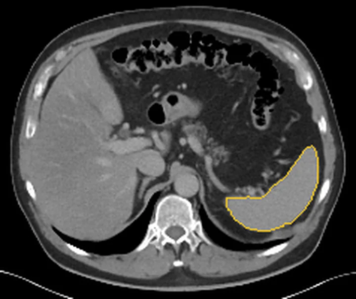
- Genellikle soliter, iyi sınırlı, solid ve dalak parankimine göre hipoattenüan kitle
- Lezyon genellikle hipoattenüe ve hipoenfazedir; MR'da yavaş ve gecikmiş kontrastlanma gösterir.
- Santral yıldızsı kalsifikasyon eşlik edebilir ve IMT olasılığını artırır
- Lezyonun komşu organlarla ilişkisi ve invazyon varlığı imajing ile kesin ayrım yapılamadığı için histopatolojik değerlendirme gereklidir.
- Dalak hilusu ve üst abdominal lenf nodları eşlik eden yayılım bulguları açısından taranır
- BT görünümü malign splenik kitlelerle örtüştüğünden doku tanısı olmadan kesin sınıflama yapılamaz
Ölçütler ve sınıflamalar
- WHO yumuşak doku tümörü sınıflamasında biyolojik davranış
- IMT, intermediate (orta biyolojik davranışlı; nadiren metastaz yapabilen) neoplazi olarak sınıflanır; görüntüleme histopatolojik tanının yerini tutmaz.
Normalde
Normal dalak portal venöz fazda homojen kontrastlanır; fokal solid kitle, parankim konturunda kabarıklık veya santral kalsifikasyon görülmez. Lezyonun dalak parankimi ile olan atenüasyon farkı, sınırlarının düzgünlüğü ve boyutu değerlendirilir.
Normal BT ile kıyasla
Aynı bölgenin, kaynak veri setinde patoloji içermediği belirtilen bir BT incelemesini kesit kesit kaydırın; organ sınırlarını açarak anatomiyi hasta görüntüsüyle karşılaştırın. Küçük rastlantısal bulgular tümüyle dışlanmamıştır.
Kaynak: TotalSegmentator v2.01 veri seti (Wasserthal ve ark., Radiology: Artificial Intelligence 2023), CC BY 4.0, vaka s0541; organ sınırları veri setinin otomatik segmentasyonundan, birleştirilerek sadeleştirildi. Görüntüler 2,5 mm kesit aralığına yeniden örneklendi.
Ayırıcı tanı
- Splenik lenfoma
- malignite ayırıcısında lenfadenopati ve bilinen primer malignite varlığı belirleyici olup, lezyonun imajing özellikleri IMT ile örtüşebilmektedir.
- Splenik hamartom
- çoğu kez iyi sınırlıdır ve arteriyel fazda belirgin kontrastlanabilir; portal faz örüntüsü değişkendir.
- Splenik anjiyosarkom
- dalakta belirsiz sınırlı, merkezi nekrotik alanlar içeren kompleks kitleler oluşturur ve sıkça eşlik eden metastazlarla birlikte yüksek derecede agresif bir maligniteyi düşündürür.
- Dalak metastazı
- bilinen altta yatan malignite varlığı malign lezyon için önemli bir prediktördür; imajing özellikleri diğer malignitelere benzer.
- Splenik tüberkülom
- difüz mikronodüler seyir daha tipik olmakla birlikte soliter kütle de yapabilir; tanı için klinik bilgi ve doku örneği gerekir.
Tuzaklar
- Santral yıldızsı kalsifikasyon BT'de IMT tanısını güçlü şekilde destekler ancak diğer lezyonları kesin olarak dışlamaz.
- İyi sınırlı olması kitlenin benign olduğunu kanıtlamaz; dalak IMT'si görüntüde lenfoma ve diğer neoplazileri taklit edebilir.
- IMT'nin imajing özellikleri lenfoma ve metastaz gibi malignitelere benzediğinden, ön tanı yalnızca görüntülemeye dayanarak konulmamalıdır.
Kendini dene
BT'de dalakta solid hipoattenüan kitle ve santral yıldızsı kalsifikasyon görülüyor. En olası tanı hangisidir?
Cevabı göster
Dalak inflamatuvar miyofibroblastik tümörü. Santral yıldızsı kalsifikasyon BT'de IMT tanısını güçlü biçimde destekler; kesin tanı histopatolojik ve immünohistokimyasal değerlendirmeyle konur.
BT'de malignite kuşkusu yaratan solid bir dalak kitlesinde kesin tanıyı sağlayan yöntem hangisidir?
Cevabı göster
Cerrahi rezeksiyon sonrası patolojik inceleme. Görüntüleme bulguları tanıya ipucu verebilir, ancak kesin tanı histopatolojik ve immünohistokimyasal değerlendirmeye dayanır.
Kaynaklar
Bu sayfadaki 35 cümle ve 2 quiz sorusu aşağıdaki kaynaklardan alıntılarla otomatik olarak karşılaştırıldı; 18 cümle bu karşılaştırmada düzeltildi (2026-10-09).
- The Spectrum of Solitary Benign Splenic Lesions—Imaging Clues for a Noninvasive Diagnosispmc.ncbi.nlm.nih.gov
- Common and Uncommon Splenic Lesions: A Reviewpmc.ncbi.nlm.nih.gov
- Sclerosing angiomatoid nodular transformation of the spleen in an 18-year-old man: a case reportpmc.ncbi.nlm.nih.gov
- Renal inflammatory myofibroblastic tumor: a case reportpmc.ncbi.nlm.nih.gov
- Clinicopathological Spectrum of EBV-Related Primary Splenic Tumors Identified by Splenectomy: A Case Seriespmc.ncbi.nlm.nih.gov
- [A Case of Primary Splenic Inflammatory Myofibroblastic Tumor].pubmed.ncbi.nlm.nih.gov
- Imaging features and radiologic-pathologic correlations of inflammatory pseudotumor-like follicular dendritic cell sarcoma.pubmed.ncbi.nlm.nih.gov
- Imaging features of inflammatory pseudotumor-like follicular dendritic cell sarcoma of the spleen.pubmed.ncbi.nlm.nih.gov
- Inflammatory Pseudotumor of the Pancreas Mimicking a Pancreatic Neoplasm.pubmed.ncbi.nlm.nih.gov
- Postoperative liver metastasis of primary inflammatory pseudotumorous follicular dendritic cell sarcoma of the spleen: a case report.pubmed.ncbi.nlm.nih.gov
- Inflammatory pseudotumor of the spleen: review of clinical presentation and diagnostic methods.pubmed.ncbi.nlm.nih.gov
- Metastatic Inflammatory Myofibroblastic Tumor of the Spleen: A Case Report and Review of the Literature.pubmed.ncbi.nlm.nih.gov
- Renal Pseudo-tumor Related to Renal Splenosis: Imaging Features.pubmed.ncbi.nlm.nih.gov
- A case of inflammatory pseudotumour of the gallbladder presenting as a big mass of uncertain behavior.pubmed.ncbi.nlm.nih.gov
- Surgical Treatment of Childhood Inflammatory Myofibroblastic Tumors.pubmed.ncbi.nlm.nih.gov
- Inflammatory myofibroblastic tumor: FDG PET/CT findings with pathologic correlation.pubmed.ncbi.nlm.nih.gov
- Laparoscopic splenectomy for inflammatory pseudotumor of the spleen.pubmed.ncbi.nlm.nih.gov
- Postchemotherapy Histiocyte-Rich Pseudotumor of the Spleen Simulating Residual Disease in a Patient With Non-Hodgkin Lymphoma on FDG PET/CT.pubmed.ncbi.nlm.nih.gov
- Inflammatory myofibroblastic tumor of the spleen: a case report.pubmed.ncbi.nlm.nih.gov
- Ectopic Splenic Tissue: Diagnostic Challenges in Limited Samples and Intraoperative Consultations.pubmed.ncbi.nlm.nih.gov
- Epstein-Barr Virus-Positive Inflammatory Follicular Dendritic Cell Sarcoma Occurring in Spleen of a Patient after Chemotherapy for Lung Carcinoma: A Case Report.pubmed.ncbi.nlm.nih.gov
- ALK-positive inflammatory myofibroblastic tumor in the pelvis of a child: a case report and literature review.pubmed.ncbi.nlm.nih.gov
- Primary Renal Mesenchymal Tumors: A Study from North India, Emphasizing Rare Entities and Molecular Profiles.pubmed.ncbi.nlm.nih.gov
- Molecularly guided management of pediatric endobronchial inflammatory myofibroblastic tumor: a report of two cases on surgical and targeted intervention.pubmed.ncbi.nlm.nih.gov
- Gastric inflammatory myofibroblastic tumor in adults: a case report and literature review.pubmed.ncbi.nlm.nih.gov
- An extremely rare case of malignant jejunal mesenteric inflammatory myofibroblastic tumor in a 61-year-old male patient: A case report and literature review.pubmed.ncbi.nlm.nih.gov
- Primary and secondary tumors of the peritoneum: key imaging features and differential diagnosis with surgical and pathological correlation.pubmed.ncbi.nlm.nih.gov
- Overlooked and Challenging Encounters-Inflammatory Pseudotumors in the Abdomen and Pelvis: A Pictorial Essay.pubmed.ncbi.nlm.nih.gov
- Case Report: a hypermetabolic mediastinal mass (SUVmax 16.7) mimicking malignancy with spontaneous resolution: an inflammatory myofibroblastic proliferation and diagnostic pitfall.pubmed.ncbi.nlm.nih.gov
- Diagnostic pitfalls in systemic juvenile idiopathic arthritis: insights from 6 misdiagnosed cases.pubmed.ncbi.nlm.nih.gov
- Pediatric Mesenteric Inflammatory Myofibroblastic Tumor Presenting as a Diagnostic Challenge: A Case Report and Literature Review.pubmed.ncbi.nlm.nih.gov
- Spleen metastasis as rare systemic manifestation of thymoma: a case report.pubmed.ncbi.nlm.nih.gov
- Acute gastrointestinal hemorrhage as the sole initial manifestation of gastric leiomyosarcoma: a case report highlighting the discrepancy between preoperative assessment and final pathology.pubmed.ncbi.nlm.nih.gov
- Inflammatory myofibroblastic tumor from molecular diagnostics to current treatment.pubmed.ncbi.nlm.nih.gov
- Plexiform fibromyxoma of the lesser curvature of the stomach in a 16-year-old adolescent: A case report and literature review.pubmed.ncbi.nlm.nih.gov
- A rare case of syphilitic hepatic inflammatory pseudotumor misdiagnosed as liver metastasis: Case report and mini-review.pubmed.ncbi.nlm.nih.gov
- Hepatic Epstein-Barr Virus-Positive Inflammatory Follicular Dendritic Cell Sarcoma: A Diagnostic Imaging Challenge - A Focused Review and Case Report.pubmed.ncbi.nlm.nih.gov
- Pictorial Review of Rare Pancreatic Tumors and Tumor-like Lesions: Radiologic-Pathologic Correlation.pubmed.ncbi.nlm.nih.gov
- Identification of CT Features to Differentiate Pulmonary Sarcoma from Carcinoma.pubmed.ncbi.nlm.nih.gov
- Uncommon liver tumors: Case report and literature review.pubmed.ncbi.nlm.nih.gov
- Concomitant Polycythemia Vera and Primary Splenic Diffuse Large B-Cell Lymphoma Complicated by Gastrosplenic Fistula: A Case Report.pubmed.ncbi.nlm.nih.gov
- The Spectrum of Solitary Benign Splenic Lesions-Imaging Clues for a Noninvasive Diagnosis.pubmed.ncbi.nlm.nih.gov
- Typical and Atypical Imaging Features of Malignant Lymphoma in the Abdomen and Mimicking Diseases.pubmed.ncbi.nlm.nih.gov
- Hepatic inflammatory pseudotumor presenting in an 8-year-old boy: A case report and review of literature.pubmed.ncbi.nlm.nih.gov
- Clinicopathological Features of Inflammatory Myofibroblastic Tumor in the Breast.pubmed.ncbi.nlm.nih.gov
- Hepatic Inflammatory Pseudotumor-Focusing on Its Heterogeneity.pubmed.ncbi.nlm.nih.gov
- Primary cardiac tumours: a single-center 41-year experience.pubmed.ncbi.nlm.nih.gov
- International Society of Paediatric Surgical Oncology (IPSO) Surgical Practice Guidelines.pubmed.ncbi.nlm.nih.gov
- Colonic pleomorphic undifferentiated sarcoma with a leukemoid reaction: case report.pubmed.ncbi.nlm.nih.gov
- Staged traditional Chinese medicine treatment for suspected pulmonary inflammatory pseudotumor with progressive lesion reduction: a case report and literature review.pubmed.ncbi.nlm.nih.gov
- Single-Site ALK-Positive Histiocytosis With TFG-ALK Fusion: Expanding the Clinical Spectrum of This Rare Entity.pubmed.ncbi.nlm.nih.gov
- Peribiliary Inflammatory Phlegmon Masquerading as Cholangiocarcinoma in Janus Kinase 2 V617F-positive Primary Myelofibrosis: A Multimodality Imaging Diagnosis.pubmed.ncbi.nlm.nih.gov
- Triple-Phase Computed Tomography for Differentiating Canine Cutaneous and Subcutaneous Mast Cell Tumors From Benign Lesions: A Size-Based Evaluation.pubmed.ncbi.nlm.nih.gov
- Intrapancreatic accessory spleen mimicking pancreatic malignancy: Diagnostic role of Tc-99m heat-damaged red blood cell scintigraphy.pubmed.ncbi.nlm.nih.gov
- Pelvic ALK-Positive Inflammatory Myofibroblastic Tumor Presenting 8 Years After Uterine Smooth Muscle Tumor of Uncertain Malignant Potential.pubmed.ncbi.nlm.nih.gov
- Inflammatory Myofibroblastic Tumor of the Urinary Bladder Presenting as a Radiological cT3 Mass: A Case Report and Narrative Review of 89 Cases.pubmed.ncbi.nlm.nih.gov
- Abdominal wall inflammatory myofibroblastic tumor initially misdiagnosed as leiomyoma: a rare case report.pubmed.ncbi.nlm.nih.gov
- Primary Pulmonary Epithelioid Inflammatory Myofibroblastic Sarcoma With SQSTM1::ALK Fusion.pubmed.ncbi.nlm.nih.gov
- Case Report: Single-cell transcriptomic profiling of a pediatric ALK-negative gastric inflammatory myofibroblastic tumor.pubmed.ncbi.nlm.nih.gov
- Sclerosing angiomatoid nodular transformation of the spleen in an 18-year-old man: a case report.pubmed.ncbi.nlm.nih.gov
- Rare Site of an Inflammatory Myofibroblastic Tumor at the Epididymis: A Case Report.pubmed.ncbi.nlm.nih.gov
- Bladder stromal tumor of uncertain malignant potential arising in ectopic prostatic tissue: Case report with MRI-pathology correlation.pubmed.ncbi.nlm.nih.gov
- Spleen EBV-positive inflammatory follicular dendritic cell sarcoma: a case report and literature review.pubmed.ncbi.nlm.nih.gov
- Uncommon granulomatous manifestation in Epstein-Barr virus-positive follicular dendritic cell sarcoma: a case report.pubmed.ncbi.nlm.nih.gov
- Non-odontogenic tumors of the facial bones in children and adolescents: role of multiparametric imaging.pubmed.ncbi.nlm.nih.gov
- Inflammatory Myofibroblastic Tumor: An Updated Review.pubmed.ncbi.nlm.nih.gov
- Inflammatory pseudotumor-like extranodal classic Hodgkin lymphoma manifesting as bowel perforation in acquired immunodeficiency syndrome patient with disseminated leishmaniasis: a case report and approach to differential diagnosis.pubmed.ncbi.nlm.nih.gov
- Prenatally Diagnosed Infantile Myofibroma of Sartorius Muscle-A Differential for Soft Tissue Masses in Early Infancy.pubmed.ncbi.nlm.nih.gov
- Calcifying fibrous tumor of the gastrointestinal tract: A clinicopathologic review and update.pubmed.ncbi.nlm.nih.gov
- Pseudotumor inflammatory of the spleen: A rare entity with diagnostic and therapeutic challenges: A case report.pubmed.ncbi.nlm.nih.gov
- Clinical Features and Factors Contributing to Preoperative Misdiagnosis of Gastric Gastrointestinal Stromal Tumors: A Retrospective Analysis of 27 Cases.pubmed.ncbi.nlm.nih.gov
- Spontaneous Massive Hemorrhage Caused by Inflammatory Myofibroblastic Tumor (IMT) of the Greater Omentum: A Case Report.pubmed.ncbi.nlm.nih.gov
- Common and Uncommon Splenic Lesions: A Review.pubmed.ncbi.nlm.nih.gov
- Imaging and pathological features of pediatric sclerosing angiomatoid nodular transformation of the spleen: a case series and hypothesis-generating perspective on non-operative management.pubmed.ncbi.nlm.nih.gov
- 18F-Fluorodeoxyglucose-Positron Emission Tomography/Computed Tomography (FDG PET/CT) in the Characterization of Massive Cardiac Infiltration by Peripheral T-cell Lymphoma, Not Otherwise Specified (PTCL-NOS).pubmed.ncbi.nlm.nih.gov
- Difficult Diagnosis of Interdigitating Dendritic Cell Sarcoma of the Retroperitoneum: A Case Report and A Brief Review of the Literature.pubmed.ncbi.nlm.nih.gov
- Gastric inflammatory myofibroblastic tumor: a case report.pubmed.ncbi.nlm.nih.gov
- Clinical diagnosis and treatment of abdominal inflammatory myofibroblastic tumors.pubmed.ncbi.nlm.nih.gov
- Clinicopathological Spectrum of EBV-Related Primary Splenic Tumors Identified by Splenectomy: A Case Series.pubmed.ncbi.nlm.nih.gov
- Primary ALK-positive anaplastic large cell lymphoma of the urinary bladder in a pediatric patient: a case report and review of literature.pubmed.ncbi.nlm.nih.gov
- Splenic inflammatory pseudotumor-like follicular dendritic cell sarcoma: a case report with imaging features and literature review.pubmed.ncbi.nlm.nih.gov
- Renal inflammatory myofibroblastic tumor: a case report.pubmed.ncbi.nlm.nih.gov
- Small Intestine Tumors: Diagnostic Role of Multiparametric Ultrasound.pubmed.ncbi.nlm.nih.gov
- Diagnostic Endoscopic Ultrasound (EUS) of the Luminal Gastrointestinal Tract.pubmed.ncbi.nlm.nih.gov
- Special Issue: EACR 2026 Congress: Innovative Cancer Science, 8-11 June 2026.pubmed.ncbi.nlm.nih.gov
- Inflammatory myofibroblastic tumour of the spleen: A diagnostic challenge.pubmed.ncbi.nlm.nih.gov
- Multimodality Imaging Features of Various Splenic Lesions: Clinical and Histopathologic Correlation.pubmed.ncbi.nlm.nih.gov
- Expanded Radiologic Characterization and Long-Term Imaging Follow-Up of Sclerosing Angiomatoid Nodular Transformation (SANT): A Case Report.pubmed.ncbi.nlm.nih.gov
- Case Report: Two rare cases of aggressive fibromatosis originating in the intestinal tract.pubmed.ncbi.nlm.nih.gov
- Primary splenic angiosarcoma presenting with multiple bone destruction as the initial manifestation: a case report and narrative review.pubmed.ncbi.nlm.nih.gov
- Leiomyosarcoma of the maxillary sinus presenting with epiphora: a case report.pubmed.ncbi.nlm.nih.gov
- Solitary colonic metastasis from hepatocellular carcinoma: A case report and synthesis of 15 published cases.pubmed.ncbi.nlm.nih.gov
- Late solitary pancreatic metastasis from clear-cell renal cell carcinoma: A case report.pubmed.ncbi.nlm.nih.gov
- Abdominal visceral tuberculosis: a malignancy mimic.pubmed.ncbi.nlm.nih.gov
- Comments and Illustrations of Ultrasound Findings in Extrapulmonary Tuberculosis Manifestations.pubmed.ncbi.nlm.nih.gov
- Extrapulmonary tuberculosıs: an old but resurgent problem.pubmed.ncbi.nlm.nih.gov
- A retrospective analysis of initial ultrasonographic misdiagnosis of fibrous hamartoma of infancy at the ultrasound center of a tertiary hospital in China.pubmed.ncbi.nlm.nih.gov
- Case Report: Fibromatosis-like metaplastic breast cancer: a case with detection of three genetic alterations, accompanied by local recurrence, lymph node metastasis, and distant metastasis.pubmed.ncbi.nlm.nih.gov
- Pulmonary Adenoleiomyomatous Hamartoma: Case Report of a Rare Entity With Comprehensive Literature Review.pubmed.ncbi.nlm.nih.gov
- Anaplastic Lymphoma Kinase (ALK)-Positive Large B-cell Lymphoma in Children: A Case Report and Review of Literature.pubmed.ncbi.nlm.nih.gov
- Case report: Diagnostic trap: metastatic endometrial stromal sarcoma with breast metastasis.pubmed.ncbi.nlm.nih.gov
- Utility of positron emission tomography-computed tomography in the evaluation of fever of unknown origin in a resource-limited tropical nation.pubmed.ncbi.nlm.nih.gov
- International Society of Paediatric Surgical Oncology (IPSO) Minimally Invasive Surgery (MIS) Guidelines.pubmed.ncbi.nlm.nih.gov
- Clinicopathological analysis of sclerosing haemangiomatoid nodular transformation of the spleen: analysis of three cases and a literature review.pubmed.ncbi.nlm.nih.gov
- Current Perspectives on Mesenchymal Dendritic Cell Neoplasms of Lymphoid Tissue: Insights into Ontogeny, Updates on Classification, and Clinicopathologic Characteristics.pubmed.ncbi.nlm.nih.gov
Eğitim amaçlıdır; hasta başında tanı ve tedavi kararının yerine geçmez, tıbbi tavsiye değildir. Metinler yapay zekâ desteğiyle yazıldı ve otomatik bir adımda kaynak alıntılarıyla karşılaştırıldı; bu bir tıbbi doğrulama değildir ve içerik uzman radyolog incelemesinden geçmedi. Klinik kararlarda güncel kılavuzları, kurum protokollerini ve radyoloji raporunu esas alın.