Özet
İnce barsak duvar içi hematomu spontan olarak, özellikle antikoagülasyon zemininde, ya da künt travma sonrasında gelişebilir. İntramural hematom şüphesinde kontrastsız BT, kontrastlı BT’den önce çekilmelidir; duvar çevresel kalınlaşıp lümen daralabilir. Kontrastlı BT’de duvar kontrastlanması ve aktif kanama bulguları değerlendirilebilir; ancak kontrastlanma hematomu maskeleyebileceği için öncesinde kontrastsız BT çekilmelidir. İntramural hematom lümen daralmasıyla ince barsak obstrüksiyonuna yol açabilir; iskemi veya perforasyon eşlik ederse bunlar komplikasyon olarak değerlendirilmelidir.
Faz ve pencere
- Kontrastsız tanısal
- Duvar içinde spontan yüksek atenüasyonlu, çoğunlukla çevresel veya asimetrik kalınlaşma ve buna bağlı lümen daralması görülür; bu patolojinin tanısal temel fazıdır.
- Portal
- Kontrastlı BT’de duvar kontrastlanması ve aktif kanama bulguları değerlendirilebilir; kontrastlanma hematomu maskeleyebileceğinden kontrastsız seri önce çekilmelidir.
Önerilen pencereler G genişlik, M merkez; değerler Hounsfield birimi (HU)
- BatınG 400 / M +50
- Yumuşak dokuG 400 / M +40
BT bulguları
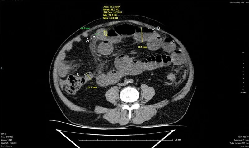
- Jejunumda sık görülen, kısa veya uzun bir segment boyunca konsantrik duvar kalınlaşması.
- Kontrastsız seride duvar katmanları içinde kendiliğinden hiperdens kan ürünleri.
- Submukozal genişleme ve duvar kalınlaşmasına bağlı lümen daralması; proksimal anslarda sıvı-gaz dilatasyonu gelişebilir.
- Komşu mezenterik yağda infiltrasyon görülebilir.
- Travma ilişkili barsak duvarı hematomunda perforasyon eşlik edebileceğinden barsak duvarı sürekliliği de değerlendirilmelidir.
- Duvarın belirgin kontrastlanmaması, pnömatozis veya serbest hava eşlik ederse yalnızca hematomla açıklanmayan komplikasyon olasılığı artar.
- Kontrastsız BT'de intramural atenüasyon artışı görülebilir; bir olgu serisinde ortalama yaklaşık 38 HU, odaksal değerler ise 70 HU'ya kadar bildirilmiştir.
Normalde
Normal jejunum ve ileumla karşılaştırmada, hematom düşünülen anslarda duvar kalınlaşması, artmış intramural atenüasyon ve lümen daralması aranır. Şüpheli ansı aynı düzeydeki komşu barsakla karşılaştırırken duvarın kalınlığını, spontan yoğunluğunu ve lümen çapını birlikte değerlendir; obstrüksiyon varsa proksimal ansların genişliği distaldeki çökmüş lümenle zıtlık gösterir.
Normal BT ile kıyasla
Aynı bölgenin, kaynak veri setinde patoloji içermediği belirtilen bir BT incelemesini kesit kesit kaydırın; organ sınırlarını açarak anatomiyi hasta görüntüsüyle karşılaştırın. Küçük rastlantısal bulgular tümüyle dışlanmamıştır.
Kaynak: TotalSegmentator v2.01 veri seti (Wasserthal ve ark., Radiology: Artificial Intelligence 2023), CC BY 4.0, vaka s0541; organ sınırları veri setinin otomatik segmentasyonundan, birleştirilerek sadeleştirildi. Görüntüler 2,5 mm kesit aralığına yeniden örneklendi.
Ayırıcı tanı
- İskemik duvar ödemi
- çoğunlukla kontrastlı görüntüde azalmış duvar boyanması, mezenterik ödem veya damar tıkanıklığıyla desteklenir.
- İnfeksiyöz enterit, ince barsak duvar kalınlaşmasının ayırıcı tanısında yer alabilir; bu görüntüleme bulgusu tek başına hematom tanısı koydurmaz.
- İnce barsak lenfoması tümöral ayırıcı tanıdadır; ince barsak tümörlerinde duvar kalınlaşması ve lümen darlığı görülebilir, bu bulgular hematoma özgü değildir.
- İnce barsak adenokarsinomu tümöral ayırıcı tanıdadır; ince barsak tümörlerinde duvar kalınlaşması, lümen darlığı ve proksimal segmentlerde genişleme görülebilir.
- Duvar içi ödem ve hematom
- kontrastlı seride her ikisi de katmanlanma veya hedef benzeri görünüm oluşturabilir; ancak hematomda bu katmanlar artmış atenüasyona sahiptir.
Tuzaklar
- Yalnız kontrastlı BT’de hematom boyanmış duvar gibi algılanabilir; kontrastsız görüntüde aynı duvarın zaten yüksek atenüasyonlu olup olmadığını kontrol et.
- Kollabe ans fizyolojik olarak kalın görünebilir; hematomda duvar içi yoğunluk artışı ve lümen daralması ardışık kesitlerde sürer.
- İntramural kanama ile eşlik eden iskemi birlikte bulunabilir; damar açıklığı ve ans duvarı kontrastlanmasını ayrıca incele.
- Komşu mezenterik yağ infiltrasyonu hematoma eşlik edebilir; tanıda kontrastsız BT'deki intramural hiperdensite, duvar kalınlaşması ve lümen daralması birlikte değerlendirilir.
Kendini dene
Antikoagülan kullanan hastada kusma ve karın ağrısı var. Kontrastsız BT’de jejunum çevresel kalın, spontan hiperdens ve lümen dar. Bu bulgularla en olası tanı nedir?
Cevabı göster
İntramural hematom. Kan ürünleri kontrast verilmeden önce de duvar içinde parlak görünür ve ansın geçiş kanalını sıkıştırabilir. İskemiyi ayırmada bağırsak duvarı kontrastlanmasının azalması veya kaybolması yardımcı olabilir.
Kontrastsız BT’de ince barsak duvarı kalınlaşmış ve lümen daralmış, ancak duvar içi atenüasyon artışı yok. Hematom tanısı bu bulgularla desteklenir mi?
Cevabı göster
Hayır; duvar içi atenüasyon artışı da aranır. İntramural hematomun karakteristik BT bulguları duvar kalınlaşması, artmış duvar içi atenüasyon ve lümen daralmasını içerir; yalnız kalınlaşma ve daralma hematom tanısını desteklemek için yeterli değildir.
Kaynaklar
Bu sayfadaki 37 cümle ve 2 quiz sorusu aşağıdaki kaynaklardan alıntılarla otomatik olarak karşılaştırıldı; 16 cümle bu karşılaştırmada düzeltildi (2026-10-09).
- Intestinal Spontaneous Intramural Hematoma Secondary to Anticoagulation Therapy: A Case Reportpmc.ncbi.nlm.nih.gov
- Warfarin Poisoning and Blunt Abdominal Trauma: A Rare Cause of Small Bowel Obstructionpmc.ncbi.nlm.nih.gov
- When the colon takes a hit: a rare case of acute colonic intramural hematoma after blunt abdominal traumapmc.ncbi.nlm.nih.gov
- Spontaneous intramural small-bowel hematoma secondary to coagulopathy in patients receiving vitamin K antagonists: A case series reportpmc.ncbi.nlm.nih.gov
- Blocked by a clot: subserosal cecal hematoma causing bowel obstruction – a case reportpmc.ncbi.nlm.nih.gov
- Small bowel intramural hematoma caused by warfarin: case report and literature review.pubmed.ncbi.nlm.nih.gov
- Small bowel intramural hematoma secondary to abdominal massage.pubmed.ncbi.nlm.nih.gov
- Treatment of brodifacoum overdose with prothrombin complex concentrate.pubmed.ncbi.nlm.nih.gov
- Intestinal Spontaneous Intramural Hematoma Secondary to Anticoagulation Therapy: A Case Report.pubmed.ncbi.nlm.nih.gov
- Spontaneous intramural hematoma of the small bowel in anticoagulated patients-Insights into management: A case report.pubmed.ncbi.nlm.nih.gov
- Anatomic Suitability for Transcaval Access Based on Computed Tomography.pubmed.ncbi.nlm.nih.gov
- Multidetector Computed Tomography Findings of Adhesive Internal Hernias.pubmed.ncbi.nlm.nih.gov
- Transabdominal ultrasonography of the small bowel.pubmed.ncbi.nlm.nih.gov
- Apixaban-induced haematoma causing small bowel intussusception.pubmed.ncbi.nlm.nih.gov
- MDCT of primary, locally recurrent, and metastatic duodenal gastrointestinal stromal tumours (GISTs): a single institution study of 25 patients with review of literature.pubmed.ncbi.nlm.nih.gov
- Intramural hematoma of colon: Case report of 2 cases.pubmed.ncbi.nlm.nih.gov
- Intramural duodenal haematoma after endoscopic biopsy: case report and review of the literature.pubmed.ncbi.nlm.nih.gov
- Gastrointestinal tract metastasis presenting as intussusception in invasive lobular carcinoma of the breast: A case report.pubmed.ncbi.nlm.nih.gov
- Spontaneous Intramural Intestinal Hemorrhage versus Acute Mesenteric Ischemia by CT Evaluation.pubmed.ncbi.nlm.nih.gov
- Bad Blood: An Unusual Case of Small Bowel Obstruction From Hematoma-Induced Intussusception.pubmed.ncbi.nlm.nih.gov
- Management and outcomes of blunt and penetrating traumatic aortic injuries in children.pubmed.ncbi.nlm.nih.gov
- Non-traumatic Intramural Duodenal Hematoma Secondary to Alcoholic Pancreatitis.pubmed.ncbi.nlm.nih.gov
- Intramural Hematoma of Gastrointestinal Tract in People with Hemophilia A and B.pubmed.ncbi.nlm.nih.gov
- Radiological Evaluation of Bowel Ischemia.pubmed.ncbi.nlm.nih.gov
- A literature review and case report: Traumatic appendicitis in an elderly patient with spinal cord injury and medical comorbidities.pubmed.ncbi.nlm.nih.gov
- Evaluation of isolated abdominal visceral artery dissection with multi-scale spiral computed tomography: a retrospective case series.pubmed.ncbi.nlm.nih.gov
- Trends in nonoperative management of traumatic injuries - A synopsis.pubmed.ncbi.nlm.nih.gov
- Spontaneous intramural small-bowel hematoma secondary to coagulopathy in patients receiving vitamin K antagonists: A case series report.pubmed.ncbi.nlm.nih.gov
- Pediatric blunt duodenal injury: a 9-year retrospective single-center series from Vietnam.pubmed.ncbi.nlm.nih.gov
- Small Intestine Tumors: Diagnostic Role of Multiparametric Ultrasound.pubmed.ncbi.nlm.nih.gov
- Gastrointestinal Involvement in Adult IgA Vasculitis: A Comprehensive Review.pubmed.ncbi.nlm.nih.gov
- Predictive Value of Preoperative CT Findings for Bowel Ischemia in Patients with Blunt Mesenteric Injury: A Retrospective Cohort Study.pubmed.ncbi.nlm.nih.gov
- The Diagnostic Value of D-Dimer Testing in the Differential Diagnosis of Acute Abdomen in the Emergency Department.pubmed.ncbi.nlm.nih.gov
- Case Report: Delayed intra-abdominal hemorrhage in a patient with a duodenal stump tumor after Billroth II gastrectomy.pubmed.ncbi.nlm.nih.gov
- A pictorial essay on cross-sectional imaging findings of pathologies in the second (D2) segment of the duodenum in adults.pubmed.ncbi.nlm.nih.gov
- Conservative Management of a Jejuno-Jejunal Intussusception in a Patient with Severe Haemophilia A.pubmed.ncbi.nlm.nih.gov
- Findings of Abdominal Imaging in Patients with COVID-19 - Part 1: Hollow Organs.pubmed.ncbi.nlm.nih.gov
- Gallbladder hemorrhage in a patient on apixaban: A rare cause of acute abdominal pain.pubmed.ncbi.nlm.nih.gov
- Cross-sectional imaging of acute gynaecologic disorders: CT and MRI findings with differential diagnosis-part I: corpus luteum and haemorrhagic ovarian cysts, genital causes of haemoperitoneum and adnexal torsion.pubmed.ncbi.nlm.nih.gov
- A Case of Direct-Acting Oral Anticoagulant-Induced Intramural Colon Hematoma Successfully Treated by Laparoscopic Surgery.pubmed.ncbi.nlm.nih.gov
- Diagnostic Role of Multi-Detector Computed Tomography in Acute Mesenteric Ischemia.pubmed.ncbi.nlm.nih.gov
- Contained Juxtarenal Aortic Perforation in the Setting of Severe Warfarin-Associated Coagulopathy.pubmed.ncbi.nlm.nih.gov
- Duodenal Trauma: Mechanisms of Injury, Diagnosis, and Management.pubmed.ncbi.nlm.nih.gov
- Delayed small bowel obstruction due to blunt abdominal trauma with mesenteric injury: a case report.pubmed.ncbi.nlm.nih.gov
- Spontaneous superior mesenteric vein tear.pubmed.ncbi.nlm.nih.gov
- Pneumoperitoneum without significant bowel perforation in patients with blunt trauma: a systematic review and meta-analysis.pubmed.ncbi.nlm.nih.gov
- Gastrointestinal complications of cocaine use: a narrative literature review.pubmed.ncbi.nlm.nih.gov
- Colonic Ischemia in a Patient With Pemphigus Vulgaris: Coincidence or Causation.pubmed.ncbi.nlm.nih.gov
- Hepatic portal venous gas in severe neurocritical illness after intracranial surgery: Two case reports and literature review.pubmed.ncbi.nlm.nih.gov
- Chronic intestinal pseudo-obstruction secondary to intestinal leiomyositis in a ragdoll cat.pubmed.ncbi.nlm.nih.gov
- Computed tomography in the vivo anatomy of rabbit kidneys (<i>Oryctolagus cuniculus</i>).pubmed.ncbi.nlm.nih.gov
- Computed Tomographic Findings of Eosinophilic Enteritis in Eight Cats: Case Series.pubmed.ncbi.nlm.nih.gov
- Large vessel vasculitis combined with inflammatory bowel disease: epidemiology, shared pathogenesis, and therapeutic advances.pubmed.ncbi.nlm.nih.gov
- Optimal treatment strategies for hepatic portal venous gas: A retrospective assessment.pubmed.ncbi.nlm.nih.gov
- Patterns of injury and violence in Yaoundé Cameroon: an analysis of hospital data.pubmed.ncbi.nlm.nih.gov
- Stricturing Diseases of the Gastrointestinal Tract-Current Clinical Practice.pubmed.ncbi.nlm.nih.gov
- The Usefulness and Limitations of Radiologic Findings for Diagnosis of Infectious Diseases: A Call for Antimicrobial Stewardship.pubmed.ncbi.nlm.nih.gov
- Small intestine choriocarcinoma with brain, lung and liver metastases in a male patient: a case report.pubmed.ncbi.nlm.nih.gov
- Craniocervical arterial dissections with cerebral infarction: a five-case series with diagnostic application of vessel wall MRI.pubmed.ncbi.nlm.nih.gov
- AI-based diagnosis of acute aortic syndrome from noncontrast CT.pubmed.ncbi.nlm.nih.gov
- Clinical Imaging and Innovations in Aortic Aneurysm and Dissection.pubmed.ncbi.nlm.nih.gov
- Gastrointestinal perforation: clinical and MDCT clues for identification of aetiology.pubmed.ncbi.nlm.nih.gov
- CT of Penetrating Abdominopelvic Trauma.pubmed.ncbi.nlm.nih.gov
- Sonography of acute appendicitis and its mimics in children.pubmed.ncbi.nlm.nih.gov
- Mesenteric Ischemia: A Comprehensive Review of the Role of Radiology in Diagnosis.pubmed.ncbi.nlm.nih.gov
- Emergency in pediatric rheumatology: a narrative review.pubmed.ncbi.nlm.nih.gov
- Case Report: A case of melanosis coli complicated by pneumatosis intestinalis-induced volvulus.pubmed.ncbi.nlm.nih.gov
- Endovascular Coil Embolization of a Ruptured Gastroduodenal Artery Aneurysm: A Case Report.pubmed.ncbi.nlm.nih.gov
- CT angiography of acute aortic syndrome in patients with chronic kidney disease.pubmed.ncbi.nlm.nih.gov
- Case report: Duodenal obstruction caused by gastroduodenal artery pseudoaneurysm with hematoma: an unusual case and literature review.pubmed.ncbi.nlm.nih.gov
- Acute and chronic complications of cesarean delivery: An update for the general radiologist.pubmed.ncbi.nlm.nih.gov
- Spontaneous Isolated Superior Mesenteric Artery Dissection Presenting as Chronic Postprandial Abdominal Pain: Conservative Management of a Yun Type IIb Case.pubmed.ncbi.nlm.nih.gov
- ESR Essentials: Gynaecological causes of acute pelvic pain in women: a primer for emergent evaluation-practice recommendations by the European Society of Emergency Radiology.pubmed.ncbi.nlm.nih.gov
- Conservative Management of Idiopathic Spontaneous Intraperitoneal Hemorrhage.pubmed.ncbi.nlm.nih.gov
- Spontaneous Suburothelial Hemorrhage: The Crucial Role of Radiology in Preventing Unnecessary Interventions.pubmed.ncbi.nlm.nih.gov
- Computed Tomographic Features of Bezoars and Other Gastrointestinal Foreign Bodies in Dogs and Cats: A Comparative Analysis.pubmed.ncbi.nlm.nih.gov
- Post-operative complications of cholecystectomy: what the radiologist needs to know.pubmed.ncbi.nlm.nih.gov
- Crohn's Disease: Radiological Answers to Clinical Questions and Review of the Literature.pubmed.ncbi.nlm.nih.gov
- Imaging Evaluation of Ovarian Masses in a Pediatric Population: A Comprehensive Overview.pubmed.ncbi.nlm.nih.gov
- Case Report: Retained gallstone migrating to the chest wall after cholecystectomy presenting as chronic abscess, sinus tract, and inflammatory pseudotumor.pubmed.ncbi.nlm.nih.gov
- Odontogenic Maxillary Sinusitis With Sequestrum-Like Bone Formation Mimicking Fungal Sinusitis: A Case Report.pubmed.ncbi.nlm.nih.gov
- Case Report: Pediatric Erdheim-Chester disease with cerebral ventricular system infiltration and epiphyseal involvement.pubmed.ncbi.nlm.nih.gov
- Successful Conservative Management of a Traumatic Intramural Duodenal Hematoma Using a Double Elementary Diet Tube.pubmed.ncbi.nlm.nih.gov
- When the colon takes a hit: a rare case of acute colonic intramural hematoma after blunt abdominal trauma.pubmed.ncbi.nlm.nih.gov
- Blocked by a clot: subserosal cecal hematoma causing bowel obstruction - a case report.pubmed.ncbi.nlm.nih.gov
- When Histology Is Unknown: Surgical Decision-Making in Small Bowel Tumors-From Preoperative Imaging to Intraoperative Findings: A Narrative Review.pubmed.ncbi.nlm.nih.gov
- Early-onset colorectal cancer in patients under 30: a case series from Tanzania highlighting an emerging epidemiological shift.pubmed.ncbi.nlm.nih.gov
- Ileal tuberculosis presenting with severe unexplained anemia and unusual pseudo-tumoral imaging features: A case report.pubmed.ncbi.nlm.nih.gov
- Relative colon wall attenuation - distinguishing ischemia from other colon wall changes.pubmed.ncbi.nlm.nih.gov
- Computed Tomographic Findings and Clinical Manifestations of Acute Severe Hypersensitivity to a Non-Ionic Iodinated Contrast Agent in a Castrated Male Dog: A Case Report.pubmed.ncbi.nlm.nih.gov
- Advancing precision triage in strangulated small bowel obstruction: from static scores to dynamic multiparametric models.pubmed.ncbi.nlm.nih.gov
- CT of acute abdomen in the elderly.pubmed.ncbi.nlm.nih.gov
- Jejunal Ectopic Pancreas: A Case Report and Literature Review.pubmed.ncbi.nlm.nih.gov
- Evaluation of imaging findings in gastrointestinal anisakiasis in emergency CT and ultrasound.pubmed.ncbi.nlm.nih.gov
- Diffuse cystic lung diseases-a primer for radiologists.pubmed.ncbi.nlm.nih.gov
- Multifaceted Imaging of Renal Lesions With an Emphasis on Cross-Sectional Imaging.pubmed.ncbi.nlm.nih.gov
- Clinical Applications of Dual-Energy CT.pubmed.ncbi.nlm.nih.gov
- Adenocarcinoma of the Duodenojejunal Flexure in a Patient With Intestinal Nonrotation.pubmed.ncbi.nlm.nih.gov
- Lupus enteritis and peritonitis presenting as initial manifestations of systemic lupus erythematosus: diagnostic challenge in tuberculosis-endemic regions - case report.pubmed.ncbi.nlm.nih.gov
- Eosinophilic Duodenitis and Jejunitis with Ascites and Peripheral Eosinophilia: A Diagnostic Challenge-Case Report.pubmed.ncbi.nlm.nih.gov
- Lupus Enteritis Presenting as Acute Abdomen with Clear Gelatinous Stools in a Woman with Systemic Lupus Erythematosus: A Case Report and Literature Review.pubmed.ncbi.nlm.nih.gov
- The Many Faces of the Angry Peritoneum.pubmed.ncbi.nlm.nih.gov
- Clinics in diagnostic imaging (201). Small bowel intramural haematoma induced by anticoagulation therapy with associated reactive ileus.pubmed.ncbi.nlm.nih.gov
- Duodenal imaging on the spotlight: from A to Z.pubmed.ncbi.nlm.nih.gov
- Long-term safety and effectiveness of the "OptEase" vena cava filter.pubmed.ncbi.nlm.nih.gov
- Negative surgical exploration for CT-proven pneumoperitoneum: a diagnostic pitfall in suspected gastrointestinal perforation-a six-case series.pubmed.ncbi.nlm.nih.gov
- Intracranial venous thrombosis imaging by anatomical location: an educational review with mimics and diagnostic pitfalls.pubmed.ncbi.nlm.nih.gov
- Median Arcuate Ligament Syndrome (MALS) With a Spontaneous Dissecting Aneurysm of the Celiac Axis.pubmed.ncbi.nlm.nih.gov
- Spontaneous mesenteric hemorrhage following a sneezing episode associated with an intestinal pseudodiverticulum: a case report.pubmed.ncbi.nlm.nih.gov
- An atypical presentation of mesenteric bucket-handle injury following direct trauma during football: a case report.pubmed.ncbi.nlm.nih.gov
- A Case of Concomitant Inferior Epigastric Artery Injury and Superior Vesical Artery Pseudoaneurysm after Cesarean Section Treated with Transarterial and Transvaginal Embolization.pubmed.ncbi.nlm.nih.gov
- Severe celiac trunk stenosis following blunt trauma in a patient with diabetic ketoacidosis: a diagnostic dilemma of traumatic injury versus median arcuate ligament syndrome.pubmed.ncbi.nlm.nih.gov
- Medico-Legal Considerations of Seat Belt Syndrome With Severe Abdominal Trauma: A Case Report and Literature Review.pubmed.ncbi.nlm.nih.gov
- Omental infarction as an unusual manifestation of decompensated chronic left heart failure: a rare case report.pubmed.ncbi.nlm.nih.gov
- Delayed Recognition of Multiple Small Bowel Perforations and Mesenteric Lacerations Following Combined Blunt and Penetrating Lumbar Trauma: A Case Report.pubmed.ncbi.nlm.nih.gov
Eğitim amaçlıdır; hasta başında tanı ve tedavi kararının yerine geçmez, tıbbi tavsiye değildir. Metinler yapay zekâ desteğiyle yazıldı ve otomatik bir adımda kaynak alıntılarıyla karşılaştırıldı; bu bir tıbbi doğrulama değildir ve içerik uzman radyolog incelemesinden geçmedi. Klinik kararlarda güncel kılavuzları, kurum protokollerini ve radyoloji raporunu esas alın.