
Özet
Hızlı çok kesitli BT’de, özellikle erişkin ince barsağında, kısa süreli entero-enterik invajinasyon klinik olarak sessiz ve kendiliğinden düzelen bir bulgu olarak saptanabilir. Kısa entero-enterik invajinasyon, çekici odak ve proksimal obstrüksiyon bulguları olmadan görülebilir; segment uzunluğu tek başına lezyonun kalıcılığını güvenilir biçimde öngörmez. Kalıcı veya çekici odaklı invajinasyonu geçici varyantla karıştırmak gereksiz cerrahiye yol açabilir; obstrüksiyon ya da iskemi bulgularını göz ardı etmek ise komplike invajinasyonun ve perforasyon riskinin atlanmasına neden olabilir.
Faz ve pencere
- Portal tanısal
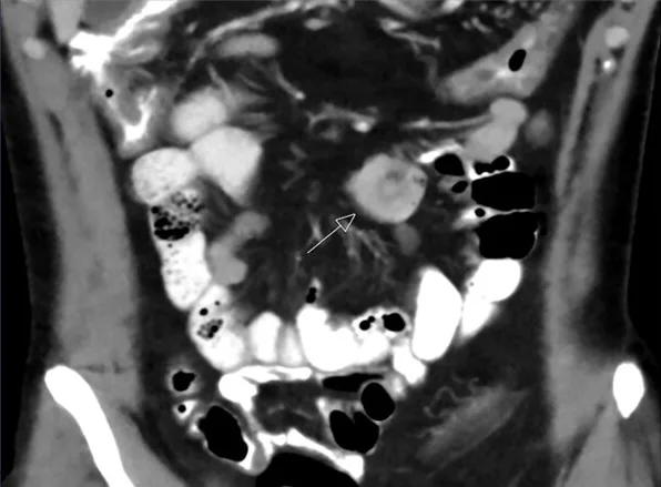
- İç içe duvarların kısa bir bölümde oluşturduğu hedef/sosis görünümü; invagine mezenter, çekici odak, proksimal ans genişlemesi ve duvar kontrastlanmasıyla birlikte incelenir.
Önerilen pencereler: Batın (G 400 / M 50), Yumuşak doku (G 400 / M 40).
BT bulguları
- Kısa entero-enterik segmentte eşmerkezli hedef görünümü veya uzun eksende küçük sosis biçimli iç içe ans.
- İnvagine duvarlar arasında az miktarda mezenter yağı/damar bulunabilir; eşlik eden belirgin mezenterik ödem beklenmez.
- İnvajinasyon ucunda belirgin polipoid ya da kitle biçimli çekici odak saptanmaz.
- Proksimal ince barsak anslarında geçiş noktasına bağlı belirgin genişleme ve distal kollaps yoktur.
- Duvar kalınlaşması, duvar kontrastlanma kaybı, pnömatozis veya çevre sıvı gibi iskemi bulguları eşlik etmez.
- Önceki/sonraki incelemede görünümün kaybolması veya yeri değişmesi geçici olma olasılığını destekler; tek zamanlı BT dinamizmi kanıtlamaz.
Ölçütler ve sınıflamalar
- Kısa segment için çalışmada bildirilen olasılık eşiği
- ≤3,5 cm uzunluk, bir erişkin serisinde kendiliğinden düzelen olgularla ilişkiliydi; tek başına kesin dışlama kuralı değildir.
Normalde
İnvajinasyonda iç içe geçmiş bağırsak segmentleri ve aralarındaki mezenterik yağ, BT’de “bağırsak içinde bağırsak” görünümünü oluşturur. Kısa invajinasyonda iç içe geçmiş bağırsak duvarları ve aralarındaki mezenterik yağ izlenir; ayrıca proksimal anslarda genişleme ile duvar kalınlaşması veya kontrastlanma değişikliği gibi komplikasyon bulguları değerlendirilir.
Normal BT ile kıyasla
Aynı bölgenin, kaynak veri setinde patoloji içermediği belirtilen bir BT incelemesini kesit kesit kaydırın; organ sınırlarını açarak anatomiyi hasta görüntüsüyle karşılaştırın. Küçük rastlantısal bulgular tümüyle dışlanmamıştır.
Kaynak: TotalSegmentator v2.01 veri seti (Wasserthal ve ark., Radiology: Artificial Intelligence 2023), CC BY 4.0, vaka s0119; organ sınırları veri setinin otomatik segmentasyonundan, birleştirilerek sadeleştirildi. Görüntüler 2,5 mm kesit aralığına yeniden örneklendi.
Ayırıcı tanı
- Çekici odaklı ince barsak invajinasyonu
- uzamış/persistan hedef, lümene uzanan kitle veya geçiş noktasında obstrüksiyon görülebilir.
- İnce barsak tümörü
- fokal duvar ya da lümen içi kitle vardır; iç içe duvarların ve taşınmış mezenterin yokluğu ayırt ettirir.
- Fekalize ince barsak içeriği
- obstrüksiyon yoksa proksimal anslarda belirgin dilatasyon veya hava-sıvı seviyesi beklenmez, konsantrik duwar katları görülmez.
- İnce barsak volvulusu
- BT’de mezenter ekseni çevresinde dönmüş anslar ve geçiş noktası görülebilir; obstrüksiyonla birlikte iskemi bulguları varsa komplike invajinasyon da değerlendirilmelidir.
Tuzaklar
- 3,5 cm eşiği sınırlı bir erişkin çalışmasından gelir; daha uzun segmentler de kendiliğinden düzelebilir ve kısa segmentte klinik risk bulguları varsa eşik güvence sağlamaz.
- Aksiyel kesitte görüntüleme açısına bağlı olarak hedef benzeri görünümler değişebilir; gerçek invajinasyonda iç içe duvarlar ve taşınmış mezenter çoklu düzlemlerde takip edilebilir.
- Tek BT, kendiliğinden redüksiyonu göstermez; rapor dilinde gözlenen kısa invajinasyonu kesin olarak ‘geçici’ ilan etme.
- Obstrüksiyon, çekici lezyon, duvar kontrastlanmasında azalma veya mezenterik sıvı varsa sessiz varyant varsayımı uygun değildir.
Kendini dene
BT’de kısa entero-enterik hedef görünümü var; proksimal obstrüksiyon ve belirgin çekici odak saptanmıyor. En uygun yorum hangisidir?
Cevabı göster
Geçici entero-enterik invajinasyon olasılığı. Çekici odak ve obstrüksiyon bulguları olmayan entero-enterik invajinasyon geçici olabilir. Tek BT incelemesi zaman içindeki kendiliğinden düzelmeyi göstermez; bu nedenle görünümün geçici olduğunu kesinleştirmez.
Kısa invajinasyon uzunluğu hakkında doğru yorum hangisidir?
Cevabı göster
≤3,5 cm bir çalışmada düzelmeyle ilişkiliydi. 3,5 cm ve altı bir erişkin serisinde kendiliğinden düzelen olgularla ilişkili bulunmuştur; eşik kesin tanı ya da tedavi kuralı değildir. Daha uzun segmentlerin bir kısmı da kendiliğinden düzelmiştir.
İlgili konular
Kaynaklar
Bu sayfadaki 33 cümle ve 2 quiz sorusu aşağıdaki kaynaklardan alıntılarla tek tek karşılaştırıldı; 8 cümle bu karşılaştırmada düzeltildi (2026-10-09).
- Intussusception in Adults: The Role of MDCT in the Identification of the Site and Cause of Obstructionpmc.ncbi.nlm.nih.gov
- Malignancy predictors and treatment strategies for adult intestinal intussusception.pubmed.ncbi.nlm.nih.gov
- Clinical course and predictive factors of incidentally detected adult intussusceptions on CT: a retrospective cohort study.pubmed.ncbi.nlm.nih.gov
- Transient intussusception - What the sonographer needs to know.pubmed.ncbi.nlm.nih.gov
- A Rare Presentation of Adult Intussusception.pubmed.ncbi.nlm.nih.gov
- Intussusception as the Primary Presentation of a Cecal Tumor.pubmed.ncbi.nlm.nih.gov
- Inflammatory Fibroid Polyp or Vanek Tumor as an Uncommon Cause of Adult Intussusception: Diagnostic and Surgical Considerations.pubmed.ncbi.nlm.nih.gov
- Adult Intussusception and Ischemic Bowel Potentially Associated with CurQD Supplementation: A Case Report of A Diagnosis Driven by Point-of-care Ultrasound.pubmed.ncbi.nlm.nih.gov
- Early postoperative ileoileal intussusception following adhesiolysis and enterouterine fistula resection in an adult: a rare case report.pubmed.ncbi.nlm.nih.gov
- Multiple Transient Idiopathic Small Bowel Intussusceptions in an Adult: A Rare Intraoperative Finding.pubmed.ncbi.nlm.nih.gov
- A rare case of duodenojejunal intussusception causing biliary obstruction during pregnancy.pubmed.ncbi.nlm.nih.gov
- The Many Faces of Intestinal Tumors in Adults, Including the Primary Role of CT Imaging in Emergencies and the Important Role of Cross-Sectional Imaging: A Pictorial Review.pubmed.ncbi.nlm.nih.gov
- Peutz-Jeghers syndrome revealed by CT finding of acute small bowel intussusception: A case report.pubmed.ncbi.nlm.nih.gov
- Adult ileocecocolic intussusception with mucocele of the appendix: a rare presentation.pubmed.ncbi.nlm.nih.gov
- Pedunculated colonic lipoma causing adult colo-colic intussusception: A case report and literature review.pubmed.ncbi.nlm.nih.gov
- Adult ileo-ileal intussusception from primary small bowel leiomyosarcoma: MRI diagnostic insights with corroborative histopathology.pubmed.ncbi.nlm.nih.gov
- Double intussusception: a rare case report and review of the literature.pubmed.ncbi.nlm.nih.gov
- A Rare Adult Ileal Intussusception Caused by Perineurioma.pubmed.ncbi.nlm.nih.gov
- Proximal Roux-en-Y Limb Intussusception After Pediatric Living Donor Liver Transplantation for Biliary Atresia: A Rare Surgical Emergency.pubmed.ncbi.nlm.nih.gov
- Presumed septic shock from small-bowel obstruction due to a vitellointestinal remnant in a 12-year-old: successful surgical management - a case report.pubmed.ncbi.nlm.nih.gov
- A rare cause of small intestinal obstruction in children: a case report of calcifying fibrous tumor.pubmed.ncbi.nlm.nih.gov
- Role of magnetic resonance in bowel pathologies: Beyond inflammatory bowel disease.pubmed.ncbi.nlm.nih.gov
- Adult ileocolic intussusception caused by a terminal ileal lipoma: A case report.pubmed.ncbi.nlm.nih.gov
- Mesenteric Ischemia: A Comprehensive Review of the Role of Radiology in Diagnosis.pubmed.ncbi.nlm.nih.gov
- Idiopathic intestinal intussusception causing intestinal obstruction in an adult: a case report and review of the literature.pubmed.ncbi.nlm.nih.gov
- Pediatric tuina combined with glycerin enema in the management of pediatric intussusception: an observational study.pubmed.ncbi.nlm.nih.gov
- Pathological lead points in pediatric secondary intussusception: A single-center retrospective observational study of five common etiologies.pubmed.ncbi.nlm.nih.gov
- mTOR pathway diseases: challenges and opportunities from bench to bedside and the mTOR node.pubmed.ncbi.nlm.nih.gov
- Rare Adult Ileo-Ileo-Colic Intussusception Caused by an Ileal Lipoma with Spontaneous Ileal Rupture and Rectal Hemorrhage: A Case Report with a Hypothesis-Generating Discussion of Mesenteric Anatomical Variability.pubmed.ncbi.nlm.nih.gov
- Twist in the cecum: A case series of mobile cecum, cecal volvulus and cecal bascule.pubmed.ncbi.nlm.nih.gov
- Elective Repair of Atypical Internal Hernia Following Roux-En-Y Gastric Bypass in a Patient With Recurrent Obstructive Symptoms: A Case Report.pubmed.ncbi.nlm.nih.gov
- A Collision Tumour Phenomenon: Dual Metastatic Deposits From Melanoma and Adrenal Carcinoma Causing Small Bowel Intussusception.pubmed.ncbi.nlm.nih.gov
- Small Bowel Metastasis of Osteosarcoma Presenting with Intussusception: A Case Report.pubmed.ncbi.nlm.nih.gov
- Adult Ileo-Ileal Intussusception Secondary to a 9 cm Pedunculated Submucosal Lipoma: Clinical, Radiologic, and Pathologic Correlation.pubmed.ncbi.nlm.nih.gov
- Ileal leiomyoma disguised as intussusception: Imaging pitfalls in a 28-year-old female.pubmed.ncbi.nlm.nih.gov
- Imaging Modalities for Evaluation of Intestinal Obstruction.pubmed.ncbi.nlm.nih.gov
- Abdominal ultrasonography of the pediatric gastrointestinal tract.pubmed.ncbi.nlm.nih.gov
- Double Trouble in the Abdomen: Waugh Syndrome Presenting as Left-Sided Ileocolic Intussusception in an Infant.pubmed.ncbi.nlm.nih.gov
- When It Is Not NEC: Recognizing Mimics of Necrotizing Enterocolitis in Preterm Infants.pubmed.ncbi.nlm.nih.gov
- Complication imaging after laparoscopic Roux-en-Y gastric bypass: clues to the diagnosis and pitfalls.pubmed.ncbi.nlm.nih.gov
- Pediatric emergency medicine point-of-care ultrasound: summary of the evidence.pubmed.ncbi.nlm.nih.gov
- Patterns of injury and violence in Yaoundé Cameroon: an analysis of hospital data.pubmed.ncbi.nlm.nih.gov
- Imaging Acute Non-Traumatic Abdominal Pathologies in Pediatric Patients: A Pictorial Review.pubmed.ncbi.nlm.nih.gov
- Commentary: an eye on PET quantification.pubmed.ncbi.nlm.nih.gov
- Spontaneous onset and resolution of gastroesophageal intussusception during contrast-enhanced computed tomography.pubmed.ncbi.nlm.nih.gov
- Benign lesions prolapsing through the anus: a differential diagnostic algorithm and evidence-based surgical management-a narrative review.pubmed.ncbi.nlm.nih.gov
- Intussusception of the Left Atrial Appendage: A Systematic Review.pubmed.ncbi.nlm.nih.gov
- Adult intussusception in a tropical setting: real-world surgical diagnosis in low-resource settings.pubmed.ncbi.nlm.nih.gov
- Mucinous adenocarcinoma presenting as adult colonic intussusception: A case report.pubmed.ncbi.nlm.nih.gov
- Postoperative intussusception: a rare but critical complication in adult patients with Crohn's disease - case report and literature review.pubmed.ncbi.nlm.nih.gov
- Recurrent intussusception as initial manifestation of primary intestinal melanoma: Case report and literature review.pubmed.ncbi.nlm.nih.gov
- International Society of Paediatric Surgical Oncology (IPSO) Surgical Practice Guidelines.pubmed.ncbi.nlm.nih.gov
- Insights into canine reproductive health: ultrasonographic evaluation of the uterus-a review.pubmed.ncbi.nlm.nih.gov
- Society for Cardiovascular Magnetic Resonance 2024 Cases of SCMR case series.pubmed.ncbi.nlm.nih.gov
- Common, Less Common, and Unexpected Complications after Bariatric Surgery: A Pictorial Essay.pubmed.ncbi.nlm.nih.gov
- Gastrointestinal perforation: clinical and MDCT clues for identification of aetiology.pubmed.ncbi.nlm.nih.gov
- Diagnostic procedures for submucosal tumors in the gastrointestinal tract.pubmed.ncbi.nlm.nih.gov
- Adult Colonic Intussusception: A Focused Review of Diagnostic and Management Strategies.pubmed.ncbi.nlm.nih.gov
- Contemporary diagnosis and management of patients with adhesive small bowel obstruction.pubmed.ncbi.nlm.nih.gov
- Small bowel lymphatic malformation: Clinical presentation and a comprehensive literature review.pubmed.ncbi.nlm.nih.gov
- Comprehensive Management of Bowel Endometriosis: Surgical Techniques, Outcomes, and Best Practices.pubmed.ncbi.nlm.nih.gov
- Long-Term Survival in 241 Cases of Intussusception in Cattle and Factors Associated with Mortality.pubmed.ncbi.nlm.nih.gov
- Small Bowel Imaging from Stepchild of Roentgenology to MR Enterography, Part II: The Reliable Disclosure of Crohn's Disease and Non-Inflammatory Small Bowel Disorder Plot through MRI Findings.pubmed.ncbi.nlm.nih.gov
- Impact of gastric and bowel surgery on gastrointestinal drug delivery.pubmed.ncbi.nlm.nih.gov
- Idiopathic double-site jejunoileal intussusception with volvulus and transmural necrosis in a young adult: case report.pubmed.ncbi.nlm.nih.gov
- Expectant management of a pediatric patient with traumatic intussusception: Case report with literature review.pubmed.ncbi.nlm.nih.gov
- Giant paraumbilical hernia causing gastric outlet obstruction in a young man with class III obesity: a case report and literature review.pubmed.ncbi.nlm.nih.gov
- Solitary fibrous tumors from A to Z: a pictorial review with radiologic-pathologic correlation.pubmed.ncbi.nlm.nih.gov
- Giant intraperitoneal non-pancreatic pseudocyst: a case report.pubmed.ncbi.nlm.nih.gov
- Clinical, imaging and surgical outcomes of secondary intussusceptions in children: a retrospective analysis based on the benign and malignant pathologic lead points.pubmed.ncbi.nlm.nih.gov
- Diagnostic Utility of CT Findings as Indicators for Bowel Resection in Strangulated Small Bowel Obstruction.pubmed.ncbi.nlm.nih.gov
- Ileosigmoid knot: A case report of the lethal twist.pubmed.ncbi.nlm.nih.gov
- A rare case report of ileosigmoid knots that cause intestinal obstruction during Ramadan fasting.pubmed.ncbi.nlm.nih.gov
- Pediatric Spigelian Hernia and Spigelian-Cryptorchidism Syndrome: An Integrative Review.pubmed.ncbi.nlm.nih.gov
- Lymphatic Endothelial Cells in Health and Disease.pubmed.ncbi.nlm.nih.gov
- Duodenal imaging on the spotlight: from A to Z.pubmed.ncbi.nlm.nih.gov
- Semaglutide-Induced Small Bowel Pseudo-Obstruction and Ileitis in a Patient With Type 2 Diabetes: A Case Report.pubmed.ncbi.nlm.nih.gov
- Meckel's diverticulum: A challenging diagnosis.pubmed.ncbi.nlm.nih.gov
- Small bowel volvulus due to a mesenteric lipoma: a case report.pubmed.ncbi.nlm.nih.gov
- Primary Distal Ileal Volvulus Presenting As Acute Intestinal Obstruction in an Adult: A Case Report.pubmed.ncbi.nlm.nih.gov
- Incomplete reversed midgut rotation with 540° volvulus in an adult: a case report.pubmed.ncbi.nlm.nih.gov
- One patient with synchronous multiple primary colon cancers which is easy to miss: a case report.pubmed.ncbi.nlm.nih.gov
- Endoscopic drainage and one-stage titanium clip closure for appendiceal root abscess mimicking cecal submucosal tumor: a case report.pubmed.ncbi.nlm.nih.gov
- Bezoar-Induced Small Bowel Obstruction in a Patient With a Remote History of Gastric Bypass: A Case Report.pubmed.ncbi.nlm.nih.gov
- Imaging Characteristics of Pylorogastric, Duodenogastric, and True-Gastrogastric Intussusception in 25 Dogs and One Cat.pubmed.ncbi.nlm.nih.gov
- Chronic intussusception revealing primary diffuse large B-cell lymphoma of the small intestine: A case report.pubmed.ncbi.nlm.nih.gov
- Small bowel obstruction due to <i>Ascaris lumbricoides</i> in a child: A case report from Northwest Ethiopia.pubmed.ncbi.nlm.nih.gov
- Phytobezoar Impaction Within Meckel's Diverticulum Causing Distal Small Bowel Obstruction: A Case Report With Characteristic Computed Tomography Small Bowel Feces Sign.pubmed.ncbi.nlm.nih.gov
Eğitim amaçlıdır; hasta başında tanı ve tedavi kararının yerine geçmez. İçerik yapay zekâ destekli bir süreçle güncel literatüre göre cümle cümle kaynaklarla karşılaştırıldı, ancak uzman radyolog incelemesinden geçmedi. Klinik kararlarda güncel kılavuzları, kurum protokollerini ve radyoloji raporunu esas alın.