Özet
İnce barsak adenokarsinomu duodenum, jejunum veya ileumdan köken alabilen malign bir tümördür; hastalar belirsiz karın yakınmaları, obstrüksiyon, kanama veya anemiyle başvurabilir. BT enterografi, ince barsak tümörlerinin tanı ve evrelemesinde; lezyonların, barsak dışı yayılımın ve lenfatik ya da uzak metastazların değerlendirilmesinde kullanılır. Konvansiyonel BT'de barsak distansiyonunun yetersizliği küçük mukozal lezyonları örtebilir; şüphe sürerse hedefe yönelik enterografi önem kazanır. Fokal stenozun veya metastatik yayılımın atlanması hastalığın evrelemesini ve obstrüksiyon nedeninin saptanmasını eksik bırakabilir.
Okuma sırası
- ince barsak lümeni ve geçiş noktası. Koronal seride duodenumdan terminal ileuma kadar ince barsak anslarını sırayla takip et ve lümenin aniden daraldığı tek bir kısa segment bul.
- fokal barsak duvarı. Daralma düzeyinde duvarın bir tarafta kalınlaşıp kalınlaşmadığını, konturun omuzlanmasını ve lümene uzanan yumuşak dokuyu değerlendir.
- proksimal ve distal anslar. Proksimal ans çapını distal kollabe ansla karşılaştır; geçiş noktasına kadar biriken sıvı ve gazın obstrüksiyonla uyumunu göster.
- mezenter, karaciğer ve komşu organlar. Son olarak komşu mezenterik lenf düğümlerini, karaciğer parankimini, peritonu ve tümörün pankreas ya da damarlarla temasını araştır.
Faz ve pencere
- Portal tanısal
- Portal veya enterik faz kontrastlı BT'de kısa segmentli, düzensiz duvar kalınlaşması ve ani lümen daralmasını ara; intravenöz kontrast sonrası orta derecede heterojen kontrastlanma görülebilir. Duodenal adenokarsinom polipoid ve iyi sınırlı lezyon şeklinde; jejunal ve ileal adenokarsinom ise ani başlayan konsantrik veya düzensiz kenarlı annüler daralma şeklinde görülebilir.
- Kontrastsız
- Kontrastsız BT obstrüksiyonun proksimal dilatasyonunu ve kitle etkisini gösterebilir, ancak tümörün kontrastlanmasını, duvar katmanlarını ve damar/organ yayılımını güvenle karakterize etmez.
Önerilen pencereler: Batın (G 400 / M 50), Yumuşak doku (G 400 / M 40).
BT bulguları
- Kısa barsak segmentinde asimetrik veya konsantrik düzensiz mural kalınlaşma
- Ani omuzlanmalı, düzensiz lümen daralması ve proksimal ince barsak dilatasyonu
- Jejunum veya ileumda tek odaklı lezyon; atlayan çoklu segmentler Crohn hastalığını düşündürür
- Duodenumda polipoid veya kitle benzeri yumuşak doku ve komşu pankreas/mezenterik damar ilişkisi
- Tümör çevresindeki yağ dokuda infiltrasyon bulguları BT’de ekstramural yayılım açısından değerlendirilir.
- Bölgesel mezenterik lenf düğümleri, karaciğer odakları veya peritoneal nodüller
- Tümör düzeyinde ülserasyon, kanama bulgusu, perforasyon ya da obstrüksiyona bağlı staz
- Duodenal adenokarsinomda rutin kontrastlı BT ile nodal evreleme sınırlıdır; bir çalışmada patolojik referansa göre N evresi hastaların %42’sinde doğru belirlenmiş ve lenf nodu metastazlarını saptama duyarlılığı %24 bulunmuştur.
Normalde
Barsak distansiyonunun yetersizliği mukozal detayları gizleyebileceğinden, güvenilir duvar değerlendirmesi için nötral oral kontrastla enterografi yapılması önerilir. Lezyon şüphesinde aynı segmentin proksimal ve distal devamlılığını izleyip duvar simetrisini, lümen çapını, ani geçiş noktasını ve komşu yağ planı/lenf düğümlerini normal anslarla karşılaştır.
Normal BT ile kıyasla
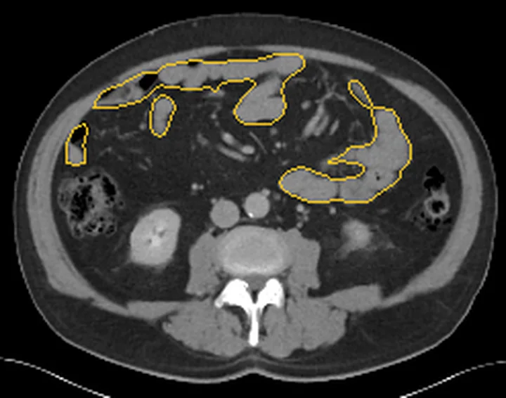
Aynı bölgenin, kaynak veri setinde patoloji içermediği belirtilen bir BT incelemesini kesit kesit kaydırın; organ sınırlarını açarak anatomiyi hasta görüntüsüyle karşılaştırın. Küçük rastlantısal bulgular tümüyle dışlanmamıştır.
Kaynak: TotalSegmentator v2.01 veri seti (Wasserthal ve ark., Radiology: Artificial Intelligence 2023), CC BY 4.0, vaka s0541; organ sınırları veri setinin otomatik segmentasyonundan, birleştirilerek sadeleştirildi. Görüntüler 2,5 mm kesit aralığına yeniden örneklendi.
Ayırıcı tanı
- İnce barsak lenfoması
- daha uzun segmentli, homojen veya anevrizmatik genişleme yapan tutulum ve çoklu lenf düğümleri görülebilir.
- Gastrointestinal stromal tümör
- çoğu kez ekzofitik kitle oluşturur ve lümen darlığına rağmen obstrüksiyon daha az belirgin olabilir.
- İnce barsak nöroendokrin tümörü
- küçük primer odağa eşlik eden belirgin kontrastlanan mezenterik desmoplastik kitle ve kalsifikasyon olabilir.
- Crohn hastalığında tarak belirtisi (comb sign) ve atlanmış (skip) duvar kalınlaşması alanları görülebilir; adenokarsinomdan ayrımda tek fokal lezyon yerine çoklu atlanmış alanların bulunması Crohn hastalığını destekler.
- İnce barsak adenomu, adenokarsinomun ayırıcı tanısında yer alır.
- İnce barsak adenokarsinomu kısa segmentli düzensiz duvar kalınlaşması veya annüler daralma şeklinde görülebilir; lenfomada uzun ya da çoklu segment tutulumu ve anevrizmatik lümen genişlemesi, GIST’te ise ekzenterik büyüme görülebilir.
Tuzaklar
- Barsak distansiyonunun yetersizliği mukozal detayları gizleyebileceğinden, şüpheli bulguların güvenilir değerlendirilmesi için nötral oral kontrastla enterografi yapılması önerilir.
- Adenokarsinomu lenfoma ile karıştırma; adenokarsinom daha sık kısa stenoz ve obstrüksiyon yaparken lenfoma anevrizmatik genişleme gösterebilir.
- Duodenumun retroperitoneal konumu ve komşu organlara yakınlığı nedeniyle, duodenumdaki primer veya ikincil süreçlerin ve komşu organlarla ilişkilerinin çok düzlemli BT rekonstrüksiyonlarında değerlendirilmesi yararlıdır.
- Barsak distansiyonu olmadan küçük ince barsak tümörleri BT'de görülmeyebilir; klinik şüphe sürüyorsa BT enterografi gibi distansiyon sağlayan incelemeler lezyonların saptanmasına yardımcı olabilir.
Kendini dene
Enterografi BT'de tek bir jejunal kısa segmentte düzensiz omuzlanmalı stenoz ve proksimal ans dilatasyonu görülüyor. En olası tanı hangisidir?
Cevabı göster
Adenokarsinom. Fokal düzensiz stenoz ve obstrüksiyon adenokarsinom için tipiktir. Lenfoma daha uzun segmentli genişleme; Crohn skip inflamasyon; GIST ise çoğunlukla ekzofitik kitle paterni oluşturur.
İnce barsak adenokarsinomunda intravenöz kontrast sonrası genellikle hangi kontrastlanma paterni görülür?
Cevabı göster
Orta derecede heterojen kontrastlanma. İnce barsak adenokarsinomunda intravenöz kontrast sonrası genellikle orta derecede heterojen kontrastlanma bildirilir.
İlgili konular
Kaynaklar
Bu sayfadaki 38 cümle ve 2 quiz sorusu aşağıdaki kaynaklardan alıntılarla otomatik olarak karşılaştırıldı; 12 cümle bu karşılaştırmada düzeltildi (2026-10-09).
- Trending of small bowel adenocarcinoma research from 2000 to 2022: A bibliometric analysispmc.ncbi.nlm.nih.gov
- Management of peritoneal metastases from colorectal cancer and small bowel adenocarcinoma in patients with inflammatory bowel diseasepmc.ncbi.nlm.nih.gov
- The Many Faces of Intestinal Tumors in Adults, Including the Primary Role of CT Imaging in Emergencies and the Important Role of Cross-Sectional Imaging: A Pictorial Reviewpmc.ncbi.nlm.nih.gov
- BRCA2 ‐Related Hereditary Cancer Syndrome‐Associated Small Bowel Adenocarcinoma With Multiple BRCA2 Mutations: A Case Report and Review of the Literaturepmc.ncbi.nlm.nih.gov
- Small bowel adenocarcinoma: An overviewpmc.ncbi.nlm.nih.gov
- Small Bowel Adenocarcinoma.pubmed.ncbi.nlm.nih.gov
- Updates in the diagnosis and management of small-bowel tumors.pubmed.ncbi.nlm.nih.gov
- Small bowel adenocarcinoma: An overview.pubmed.ncbi.nlm.nih.gov
- Enteroscopy and radiology for the management of celiac disease complications: Time for a pragmatic roadmap.pubmed.ncbi.nlm.nih.gov
- Gastrointestinal cancers in inflammatory bowel disease: An update with emphasis on imaging findings.pubmed.ncbi.nlm.nih.gov
- Primary small-bowel malignancy: update in tumor biology, markers, and management strategies.pubmed.ncbi.nlm.nih.gov
- Carcinomas in inflammatory bowel disease: a narrative review on diagnostic imaging techniques.pubmed.ncbi.nlm.nih.gov
- Small bowel adenocarcinoma of the jejunum: a case report and literature review.pubmed.ncbi.nlm.nih.gov
- CT and MR Enterography in the Evaluation of Celiac Disease.pubmed.ncbi.nlm.nih.gov
- Small bowel adenocarcinoma: French intergroup clinical practice guidelines for diagnosis, treatments and follow-up (SNFGE, FFCD, GERCOR, UNICANCER, SFCD, SFED, SFRO).pubmed.ncbi.nlm.nih.gov
- Role of computed tomography angiography in detection and staging of small bowel carcinoid tumors.pubmed.ncbi.nlm.nih.gov
- Abdominal wall infiltration by small bowel adenocarcinoma in a patient with Crohn's disease--a case report.pubmed.ncbi.nlm.nih.gov
- Imaging manifestations of malignancy in inflammatory bowel disease: patterns, pitfalls and pearls.pubmed.ncbi.nlm.nih.gov
- Multimodality imaging features of small bowel cancers complicating Crohn's disease: a pictorial review.pubmed.ncbi.nlm.nih.gov
- When Histology Is Unknown: Surgical Decision-Making in Small Bowel Tumors-From Preoperative Imaging to Intraoperative Findings: A Narrative Review.pubmed.ncbi.nlm.nih.gov
- Advanced local jejunal carcinoma treated with cytoreductive surgery (CRS) and hyperthermic intraperitoneal chemotherapy (HIPEC): a case report and review of the literature.pubmed.ncbi.nlm.nih.gov
- Ileal tuberculosis presenting with severe unexplained anemia and unusual pseudo-tumoral imaging features: A case report.pubmed.ncbi.nlm.nih.gov
- Epidemiological and clinical trends of malignant small-bowel tumors in Asia.pubmed.ncbi.nlm.nih.gov
- Simultaneous DOTATATE uptake in three non-neuroendocrine pathologies: Follicular lymphoma, Crohn's disease, and vertebral hemangioma.pubmed.ncbi.nlm.nih.gov
- Pleomorphic rhabdomyosarcoma of the left lower extremity with synchronous gastric and small intestinal metastases presenting as intussusception: a case report.pubmed.ncbi.nlm.nih.gov
- Immune checkpoint inhibitor-related intestinal pseudo-obstruction and multisystem immune-related adverse events in a post-gastrectomy patient: a case report and literature review.pubmed.ncbi.nlm.nih.gov
- The Many Faces of Intestinal Tumors in Adults, Including the Primary Role of CT Imaging in Emergencies and the Important Role of Cross-Sectional Imaging: A Pictorial Review.pubmed.ncbi.nlm.nih.gov
- High-Frequency Miniprobe Endoscopic Ultrasonography Across the Gastrointestinal Tract.pubmed.ncbi.nlm.nih.gov
- Duodenal imaging on the spotlight: from A to Z.pubmed.ncbi.nlm.nih.gov
- Cholangiocarcinoma: Recent Advances in Molecular Pathobiology and Therapeutic Approaches.pubmed.ncbi.nlm.nih.gov
- Gastrointestinal perforation: clinical and MDCT clues for identification of aetiology.pubmed.ncbi.nlm.nih.gov
- An abdominal giant lipoma that was surgically resected: case report.pubmed.ncbi.nlm.nih.gov
- Artificial intelligence models: transforming early diagnosis and precise treatment of gastrointestinal cancers.pubmed.ncbi.nlm.nih.gov
- Pericardial Diseases: International Position Statement on New Concepts and Advances in Multimodality Cardiac Imaging.pubmed.ncbi.nlm.nih.gov
- The Role of Imaging in Inflammatory Bowel Diseases: From Diagnosis to Individualized Therapy.pubmed.ncbi.nlm.nih.gov
- Case Report: Masquerading doughnut: a case of misdiagnosed ileocecal tumor-induced intussusception.pubmed.ncbi.nlm.nih.gov
- Early and Late Complications After Sleeve Gastrectomy and Roux-en-Y Gastric Bypass: A Narrative Review.pubmed.ncbi.nlm.nih.gov
- Large Bowel Obstruction.pubmed.ncbi.nlm.nih.gov
- Patterns of injury and violence in Yaoundé Cameroon: an analysis of hospital data.pubmed.ncbi.nlm.nih.gov
- British Society of Gastroenterology consensus guidelines on the management of inflammatory bowel disease in adults.pubmed.ncbi.nlm.nih.gov
- Application of imaging technology for the diagnosis of malignancy in the pancreaticobiliary duodenal junction (Review).pubmed.ncbi.nlm.nih.gov
- Gastroenteropancreatic Neuroendocrine Tumors-Current Status and Advances in Diagnostic Imaging.pubmed.ncbi.nlm.nih.gov
- CT and MR in peritoneal malignancies: pearls and pitfalls at preoperative examination.pubmed.ncbi.nlm.nih.gov
- Research progress on the application of dual energy CT in gastric diseases.pubmed.ncbi.nlm.nih.gov
- The Many Faces of the Angry Peritoneum.pubmed.ncbi.nlm.nih.gov
- Early-onset colorectal cancer in patients under 30: a case series from Tanzania highlighting an emerging epidemiological shift.pubmed.ncbi.nlm.nih.gov
- Presumptive Ewing Sarcoma of the Jejunum Presenting with Metastatic Disease: A Case Report and Focused Narrative Review.pubmed.ncbi.nlm.nih.gov
- Stenosing Ileal Angiosarcoma Presenting as Subocclusive Syndrome: A Rare Case Report and Literature Review.pubmed.ncbi.nlm.nih.gov
- Small intestine choriocarcinoma with brain, lung and liver metastases in a male patient: a case report.pubmed.ncbi.nlm.nih.gov
- Acute lower gastrointestinal bleeding from Meckel's diverticulum in a young adult: a case report on surgical management.pubmed.ncbi.nlm.nih.gov
- CT-Based Reference Ranges for Canine Pyloric Thickness: Influence of Body Size and Normalization With Aortic Diameter.pubmed.ncbi.nlm.nih.gov
- Challenges in the initial diagnosis of colon carcinoma from symptoms of a bladder and colon fistula: a case report.pubmed.ncbi.nlm.nih.gov
- Usefulness of ultrasound in diagnosing constipation in children: a narrative review.pubmed.ncbi.nlm.nih.gov
- Clinical applications of 4D flow magnetic resonance imaging in hepatic vascular disorders: practical biomarkers, protocol design, and translational implementation.pubmed.ncbi.nlm.nih.gov
- Assessing differences in growth and shape between symptomatic and asymptomatic abdominal aortic aneurysms.pubmed.ncbi.nlm.nih.gov
- Diffeomorphic Surface Modeling for MRI-Based Characterization of Gastric Anatomy and Motility.pubmed.ncbi.nlm.nih.gov
- The forgotten appearance of metastatic melanoma in the small bowel.pubmed.ncbi.nlm.nih.gov
- Role of contrast-enhanced ultrasound in evaluation of the bowel.pubmed.ncbi.nlm.nih.gov
- Ileal Gastrointestinal Stromal Tumor Presenting With Subacute Intestinal Obstruction and Synchronous Peritoneal Metastases: A Case Report.pubmed.ncbi.nlm.nih.gov
- Cholecystogastric fistula masquerading as gastrointestinal stromal tumour: Unusual presentation of gallbladder adenomyomatosis - a case report.pubmed.ncbi.nlm.nih.gov
- Mesenteric Fat Necrosis Mimicking a Malignant Mesenteric Tumor: A Case Report.pubmed.ncbi.nlm.nih.gov
- 불분명한 소아 복부 종괴: 영상 기반 진단 접근법.pubmed.ncbi.nlm.nih.gov
- Multivalvular Cardiac Involvement from Giant Hepatic Metastases of an Ileal Neuroendocrine Tumor.pubmed.ncbi.nlm.nih.gov
- Diagnostic Utility of Endoscopic Features and Endoscopic Ultrasonography for Ulcerative Colitis-Associated Neoplasia: A Retrospective Study on the Role of Endoscopic Submucosal Dissection as a Total Biopsy.pubmed.ncbi.nlm.nih.gov
- Brunner's gland hamartoma in the setting of a duodenal diverticulum: A case report.pubmed.ncbi.nlm.nih.gov
- Gastric Adenocarcinoma Associated With CTLA-4 Haploinsufficiency in the Background of Common Variable Immunodeficiency: A Rare Case.pubmed.ncbi.nlm.nih.gov
- Long-term safety and effectiveness of the "OptEase" vena cava filter.pubmed.ncbi.nlm.nih.gov
- Correlation between imaging and histology in benign solitary retroperitoneal nerve sheath tumors: a pictorial review.pubmed.ncbi.nlm.nih.gov
- Pancreatic Lipomatosis: An Extensive Pictorial Review.pubmed.ncbi.nlm.nih.gov
- Advances in CT Imaging of GI Malignancies.pubmed.ncbi.nlm.nih.gov
- From companion animals to patients: interspecies lessons in neuroendocrine oncology.pubmed.ncbi.nlm.nih.gov
- CT colonography: revisited after 30 years.pubmed.ncbi.nlm.nih.gov
- Imaging Approach to Diverticular Disease of the Small Bowel.pubmed.ncbi.nlm.nih.gov
- SAR Consensus Recommendations for Defining Small Bowel Crohn Disease Strictures at CT and MR Enterography.pubmed.ncbi.nlm.nih.gov
- A comprehensive dataset of magnetic resonance enterography images with intestinal segment annotations.pubmed.ncbi.nlm.nih.gov
- Duropathies as Unifying Concept-Part Two: A Narrative Overview of Clinical and Neuroradiological Features.pubmed.ncbi.nlm.nih.gov
- Lymph Node Assessment with Multiparametric Ultrasound: Normal Values, Morphologic Patterns, and Diagnostic Algorithms.pubmed.ncbi.nlm.nih.gov
- Dental-craniofacial manifestation and treatment of rare diseases.pubmed.ncbi.nlm.nih.gov
- The role of enteroscopy in the diagnosis and interventional management of small intestine diseases.pubmed.ncbi.nlm.nih.gov
- Nonocclusive mesenteric ischemia secondary to organophosphorus poisoning: A case report.pubmed.ncbi.nlm.nih.gov
- Faecolith-induced small bowel obstruction: A rare acute surgical emergency - A case series.pubmed.ncbi.nlm.nih.gov
- International Society of Paediatric Surgical Oncology (IPSO) Minimally Invasive Surgery (MIS) Guidelines.pubmed.ncbi.nlm.nih.gov
- Update on the Role of Imaging in the Diagnosis, Staging, and Prognostication of Gallbladder Cancer.pubmed.ncbi.nlm.nih.gov
- A narrative review of imaging for pancreas adenocarcinoma: staging, surgical considerations, and surveillance.pubmed.ncbi.nlm.nih.gov
- Small intestinal metastasis of triple-negative breast cancer mimicking lymphoma: imaging findings and case report.pubmed.ncbi.nlm.nih.gov
- Ruptured jejunal gastrointestinal stromal tumor (GIST) presenting with acute hemoperitoneum in a young adult with autosomal dominant polycystic kidney disease (ADPKD).pubmed.ncbi.nlm.nih.gov
- A Rare Presentation of Benign Prostatic Hyperplasia With Pure Stromal Hyperplasia as a Large Isolated Pelvic Mass With Normal Prostate Imaging.pubmed.ncbi.nlm.nih.gov
- An Unusual Case Report of Duodenojejunal Flexure Tumor With a Systematic Review of the Literature.pubmed.ncbi.nlm.nih.gov
- Small Bowel Imaging from Stepchild of Roentgenology to MR Enterography: Part I: Guidance in Performing and Observing Normal and Abnormal Imaging Findings.pubmed.ncbi.nlm.nih.gov
- Small Bowel Imaging from Stepchild of Roentgenology to MR Enterography, Part II: The Reliable Disclosure of Crohn's Disease and Non-Inflammatory Small Bowel Disorder Plot through MRI Findings.pubmed.ncbi.nlm.nih.gov
- Trans-hiatal small-bowel herniation after laparoscopic radical total gastrectomy for gastric cancer: a case report and literature review.pubmed.ncbi.nlm.nih.gov
- Adenocarcinoma of the Duodenojejunal Flexure in a Patient With Intestinal Nonrotation.pubmed.ncbi.nlm.nih.gov
- Gut decisions based on the liver: prediction of colorectal neoplasia using AI-based liver analysis of routine CT scans.pubmed.ncbi.nlm.nih.gov
- Preoperative <sup>18</sup>F-FDG PET/CT as a surgical triage tool for synchronous neoplasm detection in acute left-sided obstructive colorectal cancer: a two-institution retrospective cohort study.pubmed.ncbi.nlm.nih.gov
- Advances in Artificial Intelligence for Gastrointestinal Endoscopy: 2026 Update.pubmed.ncbi.nlm.nih.gov
- Role of magnetic resonance in bowel pathologies: Beyond inflammatory bowel disease.pubmed.ncbi.nlm.nih.gov
- An unusual cause of a vesical mass: Small bowel diverticulum presenting with a bladder fistula: A case report and review of the literature.pubmed.ncbi.nlm.nih.gov
- Pediatric Hereditary Polyposis Syndromes: Diagnosis, Surveillance, and Management Across Evolving Guidelines.pubmed.ncbi.nlm.nih.gov
- Recurrent intussusception caused by giant ileal gastric heterotopia in a patient with ileocecectomy: A rare case report and literature review.pubmed.ncbi.nlm.nih.gov
- Gastrointestinal Tract Polyp Detection, Tracking, and Surveillance and the Role of Artificial Intelligence.pubmed.ncbi.nlm.nih.gov
- Small-Bowel Perforation Caused by Multiple Metastases from Lung Adenocarcinoma Preceded by Obscure Gastrointestinal Bleeding: A Case Report.pubmed.ncbi.nlm.nih.gov
- Synchronous Vaginal and Liver Metastases in Rectosigmoid Cancer: Case Report and Literature Review.pubmed.ncbi.nlm.nih.gov
- Primary duodenal carcinoma presenting as subacute intestinal obstruction: Diagnostic and surgical challenge.pubmed.ncbi.nlm.nih.gov
- Diagnostic accuracy of imaging modalities for primary small-bowel tumors: a systematic review and diagnostic test accuracy meta-analysis.pubmed.ncbi.nlm.nih.gov
- Signet-Ring Cell Carcinoma Masquerading as Crohn's Disease: A Diagnostic Challenge in Multifocal Colonic Strictures with Ascites.pubmed.ncbi.nlm.nih.gov
- Obstructing colonic involvement by gastric-origin adenocarcinoma during adjuvant chemotherapy after curative-intent gastrectomy: a case report with literature review.pubmed.ncbi.nlm.nih.gov
- Ileal malignant melanoma presenting with severe iron-deficiency anemia and active small-bowel bleeding: primary intestinal melanoma versus metastasis - a case report and narrative review.pubmed.ncbi.nlm.nih.gov
- Seminal vesicle fistula: a rare pathology in complicated Crohn's disease.pubmed.ncbi.nlm.nih.gov
- Crohn's disease Activity: Abdominal Computed Tomography Histopathology Correlation.pubmed.ncbi.nlm.nih.gov
- Imaging findings of intestinal tuberculosis.pubmed.ncbi.nlm.nih.gov
- Endoscopic "Two-Step Method" for resection of a large tumor at the major duodenal papilla: A case report (with video).pubmed.ncbi.nlm.nih.gov
- Cross-sectional imaging features of duodenal obstruction: A case series highlighting etiologic diversity.pubmed.ncbi.nlm.nih.gov
- Clinical Manifestations and Outcomes of Lung Cancer Patients With Appendiceal Metastasis.pubmed.ncbi.nlm.nih.gov
- Routine contrast-enhanced CT is insufficient for TNM-staging of duodenal adenocarcinoma.pubmed.ncbi.nlm.nih.gov
- Metastatic appendiceal mucinous adenocarcinoma presenting as bilateral ovarian masses mimicking advanced ovarian cancer: a case report.pubmed.ncbi.nlm.nih.gov
Eğitim amaçlıdır; hasta başında tanı ve tedavi kararının yerine geçmez, tıbbi tavsiye değildir. Metinler yapay zekâ desteğiyle yazıldı ve otomatik bir adımda kaynak alıntılarıyla karşılaştırıldı; bu bir tıbbi doğrulama değildir ve içerik uzman radyolog incelemesinden geçmedi. Klinik kararlarda güncel kılavuzları, kurum protokollerini ve radyoloji raporunu esas alın.