Özet
İnce barsak vasküler lezyonları nadirdir ve açıklanamayan gastrointestinal kanama ya da demir eksikliği anemisiyle ortaya çıkabilir. Eski literatürde bazı lezyonlar “kavernöz hemanjiom” olarak adlandırılmıştır; güncel ISSVA sınıflaması vasküler tümörlerle vasküler malformasyonları ayrı gruplar olarak ele alır, bu nedenle bu terimler BT görünümüne dayanarak eş anlamlı kullanılmamalıdır. Kontrastlı BT’de hemanjiomlar hipervasküler görünebilir; arteriyel fazda periferik ve kesintili, portal fazda santral veya homojen kontrastlanma gösterebilir ve lezyon komşuluğunda mezenterik damar dolgunluğu görülebilir; BT ve diğer incelemeler küçük lezyonları saptamayabilir. Tek bir incelemenin negatif sonucuna dayanmak yanıltıcı olabilir; ince barsak lezyonlarının tanısı güç olabilir.
Okuma sırası
- Jejunal duvar odağı. Jejunal anslarda nodüler veya polipoid odağı ara; bulursan kontrastsız seride lezyon yoğunluğu ve flebolitleri, kontrastlı incelemede arteriyel ve portal faz kontrastlanma örüntüsünü karşılaştır. Erken arteriyel boyanmanın silik olması yavaş akımlı malformasyonu dışlamaz.
- jejunal lümen ve duvar. Odağın duvar içinde mi kaldığını, submukozal kabarıklıkla lümene taşıp taşmadığını aksiyel kesitlerde kontrol et.
- lezyon içi damar ağı. Lezyon içi kıvrımlı damar kanallarını ve olası flebolitleri ara; kontrastsız seride kalsifikasyonu, kontrastlı serilerde damar devamlılığını doğrula.
- lezyon komşuluğu ve periton. Son olarak lümen içi yeni kontrast odağı, proksimal dilatasyon, serbest sıvı ve ans duvarı perfüzyonunu tara.
Faz ve pencere
- Arteriyel tanısal
- Kavernöz hemanjiom olarak tanımlanan lezyonlarda arteriyel fazda periferik ve kesintili kontrastlanma görülebilir. Aktif ekstravazyon, arteriyel fazda lümene geçen damar yoğunluğunda yeni bir kontrast odağı olarak izlenir.
- Portal tanısal
- Lezyonun portal fazda santrale doğru dolması veya homojenleşmesi görülebilir; duvar kalınlaşması, lümene uzanan bileşen ve komşu damarlarla ilişkisi değerlendirilir. Aktif ekstravazyon, arteriyel fazda lümene geçen damar yoğunluğunda yeni bir kontrast odağı olarak izlenir.
- Kontrastsız
- Flebolitleri ve spontan hiperdens pıhtıyı saptamak için kullanılır; tek başına hemanjiomun vasküler doğasını göstermez.
Önerilen pencereler: Batın (G 400 / M 50), Yumuşak doku (G 400 / M 40), Anjiyo (G 600 / M 150).
BT bulguları
- Kavernöz hemanjiomlar BT’de heterojen yoğunlukta nodüler veya polipoid lezyonlar şeklinde görülebilir ve genellikle lümene doğru uzanır.
- Lümene polipoid uzanım; büyük lezyonda ans lümeninde daralma veya obstrüksiyon.
- Damar yapılı lezyon çevresinde kıvrımlı besleyici ya da drene edici damarlar.
- Duvar içinde veya lezyon komşuluğunda yuvarlak flebolitler; kontrastsız seride kalsifik odak olarak seçilir.
- Arteriyel görüntülerde lümene doğru genişleyen kontrast odağı aktif ekstravazasyon lehinedir.
- İnce barsak hemanjiomları tek veya çoklu olabilir; çoklu olgularda deri veya karaciğer gibi başka organlarda da benzer lezyonlar bulunabilir.
Normalde
Kavernöz hemanjiomlarda BT’de nodüler veya polipoid lezyon, flebolitler ve lezyon komşuluğunda mezenterik damar dolgunluğu görülebilir.
Normal BT ile kıyasla
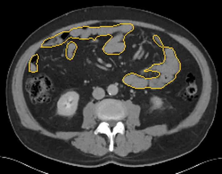
Aynı bölgenin, kaynak veri setinde patoloji içermediği belirtilen bir BT incelemesini kesit kesit kaydırın; organ sınırlarını açarak anatomiyi hasta görüntüsüyle karşılaştırın. Küçük rastlantısal bulgular tümüyle dışlanmamıştır.
Kaynak: TotalSegmentator v2.01 veri seti (Wasserthal ve ark., Radiology: Artificial Intelligence 2023), CC BY 4.0, vaka s0541; organ sınırları veri setinin otomatik segmentasyonundan, birleştirilerek sadeleştirildi. Görüntüler 2,5 mm kesit aralığına yeniden örneklendi.
Ayırıcı tanı
- İnce barsak GIST’i çoğunlukla ekzofitik büyüyen solid bir kitle şeklindedir ve heterojen kontrastlanma gösterebilir.
- İnce barsak nöroendokrin tümörlerinde mezenterik nodal metastazlara bağlı desmoplastik reaksiyon, barsaklarda çekilme, lümen daralması ve obstrüksiyona yol açabilir.
- İnce barsak angiodisplazisi kanamaya yol açabilir; aktif kanama dinamik kontrastlı BT anjiyografide kontrast ekstravazasyonu olarak görülebilir.
- Ektopik ince barsak varisleri portal hipertansiyonla ilişkili olabilir; kesitsel görüntüleme tanıda ve tedavi planlamasında rol oynar.
- İntramural ince barsak hematomu BT’de belirgin jejunal duvar kalınlaşması şeklinde görülebilir.
Tuzaklar
- Non-kontrastlı seride heterojen yoğunluk ve flebolitler görülebilir; lezyonun kistik/solid bileşenleri ve kalsifikasyonları bu seride ayırt edilir.
- Flebolitler, venöz malformasyonların yavaş akımlı kanalları içindeki kalsifiye trombüsleri temsil eder; görüntülemede lezyonla ilişkileri değerlendirilmelidir.
- İnce barsak venöz malformasyonu kontrastlı BT’de homojen kontrastlanma ve portal venöz fazda kalıcı kontrastlanma gösterebilir.
- Kesitsel görüntüleme ve kapsül endoskopi ince barsak hemanjiomunu saptamayabilir; balon destekli enteroskopi lezyonun yerini belirlemeye yardımcı olabilir.
Kendini dene
GİS kanaması olan hastada jejunum duvarında kontrastlanan odak ve birkaç duvar içi flebolit görülüyor. En olası tanı nedir?
Cevabı göster
Venöz malformasyon (eski literatürde kavernöz hemanjiom). Duvarla ilişkili vasküler odak ve aynı yerdeki birden çok flebolit venöz malformasyonu destekler; eski yayınlarda benzer lezyonlar kavernöz hemanjiom olarak adlandırılmıştır. GIST genellikle solid doku kitlesi, nöroendokrin tümör ise mezenterik desmoplazi veya metastatik odaklar eşliğinde değerlendirilir. BT tek başına vasküler lezyonun histolojik sınıfını belirlemez.
Anjiyografide kontrastın jejunal bir dal üzerinden ince barsak lümenine geçmesi aşağıdakilerden hangisini gösterir?
Cevabı göster
Aktif kanama (ekstravazasyon). Anjiyografide kontrastın jejunal bir daldan ince barsak içine ekstravazasyonu aktif kanamayı gösterir.
Kaynaklar
Bu sayfadaki 38 cümle ve 2 quiz sorusu aşağıdaki kaynaklardan alıntılarla tek tek karşılaştırıldı; 21 cümle bu karşılaştırmada düzeltildi (2026-10-09).
- Practical Lessons from the Small Bowel Bleeding Lesions: A Case Report on Small Bowel Cavernous Hemangiomapmc.ncbi.nlm.nih.gov
- Cavernous Hemangioma: A Rare Cause of Massive Lower Gastrointestinal Bleedingpmc.ncbi.nlm.nih.gov
- Small Intestinal Cavernous Hemangioma Presenting with Recurrent Gastrointestinal Bleeding: A Case Report and Narrative Reviewpmc.ncbi.nlm.nih.gov
- Laparoscopic resection of a small intestinal venous malformation presenting as acute bowel obstruction without hemorrhage in a child: a case reportpmc.ncbi.nlm.nih.gov
- Klippel–Trénaunay Syndrome With Pelvic/Rectal and Urinary Bladder Involvement: A Rare Case Reportpmc.ncbi.nlm.nih.gov
- Small Intestinal Cavernous Hemangioma Presenting with Recurrent Gastrointestinal Bleeding: A Case Report and Narrative Review.pubmed.ncbi.nlm.nih.gov
- Intestinal Lymphangioma With Extensive Secondary Lymphangiectasia Causing Life-Threatening Hemorrhage: First Case Requiring Small Bowel Transplantation.pubmed.ncbi.nlm.nih.gov
- Gastrointestinal Klippel-Trénaunay Syndrome Mimicking Ulcerative Colitis: A Case Report.pubmed.ncbi.nlm.nih.gov
- Electrochemotherapy in pediatrics: a systematic review.pubmed.ncbi.nlm.nih.gov
- Mesenteric Venous Malformation Case Report in an Adolescent With Midgut Volvulus.pubmed.ncbi.nlm.nih.gov
- Trp53 loss drives the neoplastic transformation of Pik3caH1047R-induced vascular malformation in a mouse model.pubmed.ncbi.nlm.nih.gov
- Giant rectal vascular malformation: a rare differential diagnosis in rectal bleeding.pubmed.ncbi.nlm.nih.gov
- Bleeding per rectum in pediatric population: A pictorial review.pubmed.ncbi.nlm.nih.gov
- Spectrum of bowel wall thickening on ultrasound with pathological correlation in children.pubmed.ncbi.nlm.nih.gov
- Immune checkpoint inhibitor-related intestinal pseudo-obstruction and multisystem immune-related adverse events in a post-gastrectomy patient: a case report and literature review.pubmed.ncbi.nlm.nih.gov
- Meckel's diverticulum complicated by a congenital intestinal adhesive band: a case report and literature review.pubmed.ncbi.nlm.nih.gov
- Case Report: Diagnostic and therapeutic reflections on one case of greater omental adhesion misdiagnosed as peritoneal metastasis following rectal cancer surgery.pubmed.ncbi.nlm.nih.gov
- Laparoscopic resection of a small intestinal venous malformation presenting as acute bowel obstruction without hemorrhage in a child: a case report.pubmed.ncbi.nlm.nih.gov
- Klippel-Trénaunay Syndrome With Pelvic/Rectal and Urinary Bladder Involvement: A Rare Case Report.pubmed.ncbi.nlm.nih.gov
- Intradural extramedullary cavernous malformation: A common lesion in an uncommon location.pubmed.ncbi.nlm.nih.gov
- Case Report: Progressive acquired ileal mesenteric venous malformation after right hemicolectomy: serial imaging evolution leading to delayed gastrointestinal bleeding.pubmed.ncbi.nlm.nih.gov
- An unusual superior mesenteric arteriovenous malformation causing obscure gastrointestinal bleeding: a case report.pubmed.ncbi.nlm.nih.gov
- Anal and Perianal Masses: The Common, the Uncommon, and the Rare.pubmed.ncbi.nlm.nih.gov
- Ischemic colitis due to a mesenteric arteriovenous malformation in a patient with a connective tissue disorder.pubmed.ncbi.nlm.nih.gov
- Imaging and percutaneous treatment of vascular anomalies.pubmed.ncbi.nlm.nih.gov
- Blue rubber bleb nevus syndrome: our experience and new endoscopic management.pubmed.ncbi.nlm.nih.gov
- Multimodality Imaging Review of Anorectal and Perirectal Diseases with Clinical, Histologic, Endoscopic, and Operative Correlation, Part II: Infectious, Inflammatory, Congenital, and Vascular Conditions.pubmed.ncbi.nlm.nih.gov
- Gastrointestinal Blue Rubber Bleb Nevus Syndrome in Children: A Case Series Across Endoscopic, Surgical, and Medical Management.pubmed.ncbi.nlm.nih.gov
- Mesenteric Venous Malformation With Subocclusive Symptoms: A Case Report.pubmed.ncbi.nlm.nih.gov
- Jejunojejunal intussusception induced by a gastrointestinal stromal tumor: a case report and literature review.pubmed.ncbi.nlm.nih.gov
- Case Report: Delayed coil migration after endovascular embolization of an inferior mesenteric arteriovenous malformation.pubmed.ncbi.nlm.nih.gov
- Superselective Transcatheter Embolization of a Small-bowel Arteriovenous Malformation Using N-butyl Cyanoacrylate-lipiodol: A Case Report and Literature Review.pubmed.ncbi.nlm.nih.gov
- Slow-flow, high-impact: radiologic features in venous malformations of the female genital tract.pubmed.ncbi.nlm.nih.gov
- Transarterial Embolization for Malignancy-Related Gastrointestinal Bleeding: Outcomes with and Without CT Extravasation.pubmed.ncbi.nlm.nih.gov
- Rare Delayed Complications of Laparoscopic Adjustable Gastric Band Erosion: Contained Gastric Perforation and Bleeding Pseudoaneurysm.pubmed.ncbi.nlm.nih.gov
- Life-Threatening Melena from Deep Ileal Angiodysplasia Successfully Treated by Intraoperative Enteroscopy-Guided Segmental Resection: A Case Report.pubmed.ncbi.nlm.nih.gov
- Ileal tuberculosis presenting with severe unexplained anemia and unusual pseudo-tumoral imaging features: A case report.pubmed.ncbi.nlm.nih.gov
- Synchronous Low-Grade Alimentary Lymphoma and Gastrointestinal Mast Cell Tumor with a Collision Pattern in a Cat.pubmed.ncbi.nlm.nih.gov
- Hiding in Plain Sight: A Case of Intestinal Tuberculosis Masquerading as Benign Lower GI Bleeding.pubmed.ncbi.nlm.nih.gov
- Dynamic contrast-enhanced computed tomography for small bowel bleeding: Two cases of arteriovenous malformation and gastrointestinal stromal tumor.pubmed.ncbi.nlm.nih.gov
- Small bowel lymphatic malformation: Clinical presentation and a comprehensive literature review.pubmed.ncbi.nlm.nih.gov
- When Histology Is Unknown: Surgical Decision-Making in Small Bowel Tumors-From Preoperative Imaging to Intraoperative Findings: A Narrative Review.pubmed.ncbi.nlm.nih.gov
- Giant jejunal gastrointestinal stromal tumor presenting as refractory anemia: a case report of complex R0 resection.pubmed.ncbi.nlm.nih.gov
- Cholecystogastric fistula masquerading as gastrointestinal stromal tumour: Unusual presentation of gallbladder adenomyomatosis - a case report.pubmed.ncbi.nlm.nih.gov
- Subcentimeter Ileal Neuroendocrine Tumor Resection for Obscure Gastrointestinal Bleeding Followed by Pacemaker-Treated Sick Sinus Syndrome: A Case Report.pubmed.ncbi.nlm.nih.gov
- Incidental cystic pancreatic neuroendocrine neoplasm and differential diagnosis.pubmed.ncbi.nlm.nih.gov
- Multiparametric Abdominal Ultrasound as a Source of Imaging Biomarkers in Predictive and Personalized Medicine: A Narrative Review.pubmed.ncbi.nlm.nih.gov
- Imipenem/Cilastatin Sodium as Reabsorbable Embolic Agent in Acute Lower Gastrointestinal Bleeding: A Multicentric Prospective Study.pubmed.ncbi.nlm.nih.gov
- Intraluminal hematoma of an ileal conduit causing postrenal acute kidney injury 14 years after radical cystectomy: A case report.pubmed.ncbi.nlm.nih.gov
- Splenic Artery Embolization, A Therapy that Can be Life-Saving for Small Bowel Variceal Bleeding.pubmed.ncbi.nlm.nih.gov
- Narrative review of the current management of ectopic varices: an update.pubmed.ncbi.nlm.nih.gov
- Recurrent Peristomal Variceal Hemorrhage Secondary to Portal Hypertension: A Diagnostic Pitfall in Metastatic Rectal Cancer.pubmed.ncbi.nlm.nih.gov
- Bad Blood: An Unusual Case of Small Bowel Obstruction From Hematoma-Induced Intussusception.pubmed.ncbi.nlm.nih.gov
- Spontaneous intramural small bowel hematoma: unusual cause of intestinal subocclusion in an anticoagulated patient-a case report.pubmed.ncbi.nlm.nih.gov
- Spontaneous intramural hematoma of the small bowel in anticoagulated patients-Insights into management: A case report.pubmed.ncbi.nlm.nih.gov
- Post-operative complications of cholecystectomy: what the radiologist needs to know.pubmed.ncbi.nlm.nih.gov
- A Narrative Review on Toxidromes in the Psychiatric Population: Implications for Overdose Prevention.pubmed.ncbi.nlm.nih.gov
- Intramural Hematoma of Gastrointestinal Tract in People with Hemophilia A and B.pubmed.ncbi.nlm.nih.gov
- Long-term safety and effectiveness of the "OptEase" vena cava filter.pubmed.ncbi.nlm.nih.gov
- Common and Uncommon Splenic Lesions: A Review.pubmed.ncbi.nlm.nih.gov
- Splenoses and Other Ectopic and Heterotopic Splenic Tissue: The Use of Long-Lasting Enhancement in Contrast-Enhanced Ultrasound to Avoid Unnecessary Intervention.pubmed.ncbi.nlm.nih.gov
- Role of MRI for assessment of GI bleeding: a pictorial review of indications, technique and performance.pubmed.ncbi.nlm.nih.gov
- Trends and Changes in Endoscopic Management and Clinical Outcomes of Colonic Diverticular Bleeding during the Coronavirus Disease-2019 Pandemic.pubmed.ncbi.nlm.nih.gov
- Expert consensus on perioperative management for liver tumors treated with co-ablation system therapy.pubmed.ncbi.nlm.nih.gov
- Aspirin Eugenol Ester Alleviates Gastric Injury by Inhibiting Ferroptosis and Oxidative Stress.pubmed.ncbi.nlm.nih.gov
- Acute intestinal bleeding due to enteric duplication cyst (EDC) and visceral malrotation in a young male adult: A case report.pubmed.ncbi.nlm.nih.gov
- Intestinal Malrotation in Adults: A Retrospective Cross-Sectional Study Based on Computed Tomography Findings.pubmed.ncbi.nlm.nih.gov
- Imaging Approach to Diverticular Disease of the Small Bowel.pubmed.ncbi.nlm.nih.gov
- Excision of a long-standing low-flow venous malformation of the fourth digit in a 30-year-old male: a case report.pubmed.ncbi.nlm.nih.gov
- Imaging Characteristics of Pylorogastric, Duodenogastric, and True-Gastrogastric Intussusception in 25 Dogs and One Cat.pubmed.ncbi.nlm.nih.gov
- Imaging features and clinical management of mesenteric cavernous lymphangioma: A case report and literature review.pubmed.ncbi.nlm.nih.gov
- Diagnostic value of CT enterography combined with inflammatory indicators in active Crohn's disease.pubmed.ncbi.nlm.nih.gov
- Case Report: Eccrine spiradenoma of the shoulder mimicking a vascular lesion on ultrasonography and CT.pubmed.ncbi.nlm.nih.gov
- Abdominal tuberculosis presenting as a massive lower GI bleed in a cerebral palsy patient-a case report.pubmed.ncbi.nlm.nih.gov
- Endoscopic Management of Post-Bariatric Surgery Complications: Diagnostic Work-Up and Innovative Approaches for Leak, Fistula, and Stricture Management.pubmed.ncbi.nlm.nih.gov
- Lupus Enteritis Presenting as Acute Abdomen with Clear Gelatinous Stools in a Woman with Systemic Lupus Erythematosus: A Case Report and Literature Review.pubmed.ncbi.nlm.nih.gov
- Small Bowel Gastrointestinal Stromal Tumors: A 15-Year Cohort Study Focusing on Jejuno-Ileal Site-Specific Outcomes and Prognostic Factors.pubmed.ncbi.nlm.nih.gov
- Abdominal apoplexy complicating ulcerative colitis: a case report of venous mesenteric hemorrhage.pubmed.ncbi.nlm.nih.gov
- Massive Psoas Muscle Bleeding from the Contralateral Lumbar Artery Obscured by a Lumbar Vertebra Fracture: A Case Report.pubmed.ncbi.nlm.nih.gov
- Cross-sectional imaging after pancreatic surgery: The dialogue between the radiologist and the surgeon.pubmed.ncbi.nlm.nih.gov
- MRI of uncommon lesions of the large bowel: a pictorial essay.pubmed.ncbi.nlm.nih.gov
- Microcystic lymphatic malformation of the small intestine presenting as acute abdominal pain in a 5-year-old boy: a case report and review of the literature.pubmed.ncbi.nlm.nih.gov
- Eosinophilic Duodenitis and Jejunitis with Ascites and Peripheral Eosinophilia: A Diagnostic Challenge-Case Report.pubmed.ncbi.nlm.nih.gov
- Role of magnetic resonance in bowel pathologies: Beyond inflammatory bowel disease.pubmed.ncbi.nlm.nih.gov
- Comprehensive Imaging Evaluation and Staging of Crohn's Disease: When and Why to Use Intestinal Ultrasound, MRE, or CTE: Current Guidelines and Future Directions.pubmed.ncbi.nlm.nih.gov
- Imaging in inflammatory bowel disease 2025: ECCO-ESGAR-ESP-IBUS diagnostic and monitoring recommendations with MRI and intestinal ultrasound in treat-to-target strategies.pubmed.ncbi.nlm.nih.gov
- A Grade 1, Metastatic, Well-Differentiated Neuroendocrine Tumor Presenting as Closed-Loop Small Bowel Obstruction With Ischemic Bowel.pubmed.ncbi.nlm.nih.gov
- Laparoscopic resection of small bowel neuroendocrine tumors: A long-term survival analysis.pubmed.ncbi.nlm.nih.gov
- Endometriosis MR mimickers: T1-hyperintense lesions.pubmed.ncbi.nlm.nih.gov
- What a difference a delay makes! CT urogram: a pictorial essay.pubmed.ncbi.nlm.nih.gov
- Renal stones on portal venous phase contrast-enhanced CT: does intravenous contrast interfere with detection?pubmed.ncbi.nlm.nih.gov
- The Importance of Magnetic Resonance Enterography in Monitoring Inflammatory Bowel Disease: A Review of Clinical Significance and Current Challenges.pubmed.ncbi.nlm.nih.gov
- A Study of Small and Large Bowel Wall Thickness Using Computed Tomography and Its Histopathological Correlation.pubmed.ncbi.nlm.nih.gov
- Computed tomography enterography-based radiomics nomograms to predict inflammatory activity for ileocolonic Crohn's disease: a preliminary single-center retrospective study.pubmed.ncbi.nlm.nih.gov
- Current insights into the diagnosis and management of small bowel bleeding.pubmed.ncbi.nlm.nih.gov
- Jejunal Diverticulosis as a Rare Cause of Severe Obscure Gastrointestinal Bleeding in an Elderly Patient: A Case Report.pubmed.ncbi.nlm.nih.gov
- Computed Tomographic Features of Bezoars and Other Gastrointestinal Foreign Bodies in Dogs and Cats: A Comparative Analysis.pubmed.ncbi.nlm.nih.gov
- Evaluating luminal and post-operative Crohn's disease activity on magnetic resonance enterography: A review of radiological disease activity scores.pubmed.ncbi.nlm.nih.gov
- Imaging of Strictures in Crohn's Disease.pubmed.ncbi.nlm.nih.gov
- Gastrointestinal bleeding in children: diagnostic approach.pubmed.ncbi.nlm.nih.gov
- Patterns of injury and violence in Yaoundé Cameroon: an analysis of hospital data.pubmed.ncbi.nlm.nih.gov
Eğitim amaçlıdır; hasta başında tanı ve tedavi kararının yerine geçmez. İçerik yapay zekâ destekli bir süreçle güncel literatüre göre cümle cümle kaynaklarla karşılaştırıldı, ancak uzman radyolog incelemesinden geçmedi. Klinik kararlarda güncel kılavuzları, kurum protokollerini ve radyoloji raporunu esas alın.